Underwater endoscopic resection for flat lesions in the stomach often results in immediate bleeding?
Xiaojing Quan, Ying Zheng, Hongbo Wu, Lingzhi Qin, Baicang Zou, Bin Qin

Abstract
Genes, proteins, chemicals, diseases, species, mutations and cell lines named across the full text — each resolved to its canonical identifier and authoritative record.
Click any figure to enlarge with its caption.
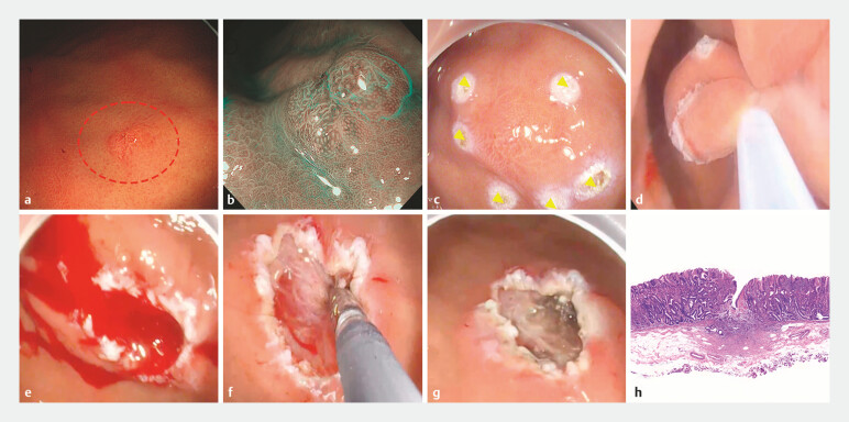 Fig. 1
Peer Reviews
No public reviews on file for this paper yet. If you reviewed it on a platform where reviews are public (OpenReview, ICLR, NeurIPS, ICML), you can paste yours below so the community can read it here.
Videos
No videos yet. Explain this paper in a talk, walkthrough, or lecture? Add one.
Taxonomy
TopicsGastrointestinal disorders and treatments · Gastrointestinal Bleeding Diagnosis and Treatment · Gastrointestinal Tumor Research and Treatment
Conventional endoscopic mucosal resection (EMR) often presents challenges when treating flat lesions in the stomach. In this study, we successfully performed underwater endoscopic mucosal resection (UEMR) in three cases of flat pre-cancerous lesions located in the gastric body ( Video 1 ).
Underwater endoscopic mucosal resection (UEMR) in three cases of flat pre-cancerous lesions located in the gastric body.Video 1
The first case involved a 71-year-old woman who underwent an outpatient magnified gastroscopy screening for monitoring a flat lesion in the gastric body. During the procedure, a 6 mm flat elevated lesion was identified on the anterior wall of the middle gastric body ( Fig. 1 a, b ). Given the economic and time-efficiency advantages over endoscopic submucosal dissection (ESD), UEMR was chosen for this procedure. Despite the use of anesthesia, the patient’s respiratory movements caused significant gastric wall activity. To address this, the lesion was marked with a snare tip ( Fig. 1 c ), intraluminal gas was evacuated, and the gastric lumen was immersed in normal saline ( Fig. 1 d ). This strategy effectively reduced gastric activity, allowing for easier lesion grasping with the snare. The lesion was successfully resected without perforation, though significant arterial bleeding occurred ( Fig. 1 e ), which was promptly managed with electrocautery ( Fig. 1 f, g ).
Images of the first case resected via UEMR. a Flat elevated lesion on the middle gastric body. b Observation of the lesion under chromoendoscopy. c Marking of the lesion with a snare tip. d Resection of the lesion. e Significant arterial bleeding following resection. f Immediate control of bleeding using electrocautery. g Postoperative wound following successful hemostasis. h Postoperative pathology confirming complete resection of the lesion. Abbreviation: UEMR, underwater endoscopic mucosal resection.
The other two cases involved flat lesions located on the greater curvature of the gastric body, each approximately 5 mm in size. Both lesions were successfully resected using UEMR; however, immediate postoperative bleeding occurred in both cases, which was effectively controlled using electrocautery. Pathological examination confirmed adenomas with negative surgical margins in all three cases ( Fig. 1 h ).
In summary, UEMR demonstrates an efficient and rapid alternative to ESD for the removal of flat gastric lesions. However, the occurrence of immediate postoperative bleeding highlights the necessity for careful monitoring and prompt intervention.
Endoscopy_UCTN_Code_CPL_1AH_2AZ_3AC