Intrachannel anterograde snare traction combined with a clip-and-line system for polyps in the appendix cavity
Zhiying Gao, Zhenguo Pan, Jian Zhao, Feng Pan

Abstract
Genes, proteins, chemicals, diseases, species, mutations and cell lines named across the full text — each resolved to its canonical identifier and authoritative record.
Click any figure to enlarge with its caption.
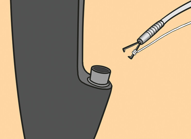 Fig. 1
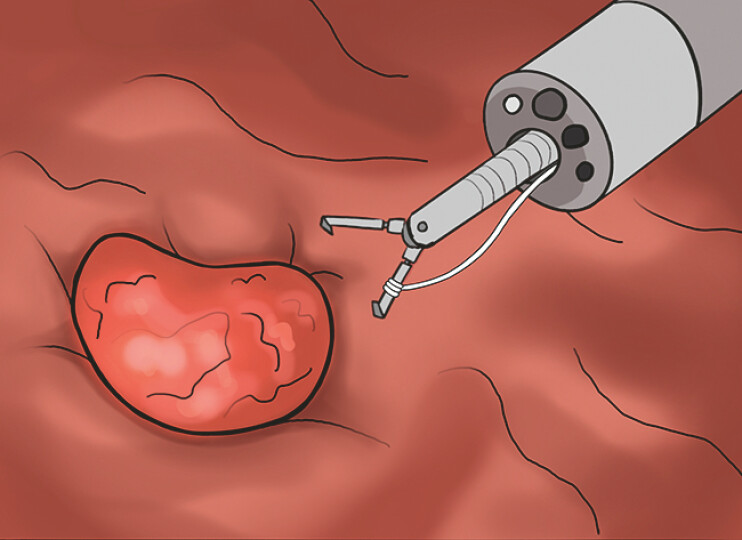 Fig. 2
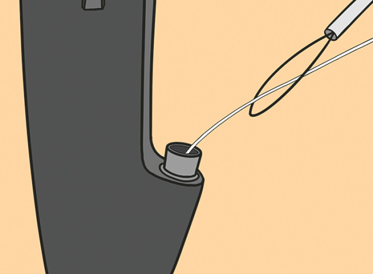 Fig. 3
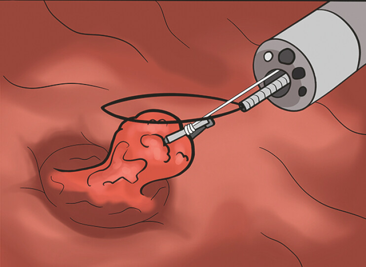 Fig. 4
- —Jiangsu Provincial Medical Key Discipline Cultivation Unit
Peer Reviews
No public reviews on file for this paper yet. If you reviewed it on a platform where reviews are public (OpenReview, ICLR, NeurIPS, ICML), you can paste yours below so the community can read it here.
Videos
No videos yet. Explain this paper in a talk, walkthrough, or lecture? Add one.
Taxonomy
TopicsIntraperitoneal and Appendiceal Malignancies · Gastrointestinal disorders and treatments · Minimally Invasive Surgical Techniques
A-57-year-old man with no symptoms underwent a colonoscopy due to a history of colon polyp. The procedure revealed a 1.5-cm polypoid eminence with an indistinct boundary to the base in the appendix cavity. Abdominal computed tomography (CT) showed a normal appendix. After obtaining informed consent, endoscopic removal of the appendix lesion was performed ( Video 1 ).
An improved operation approach involved a clip-and-line system and a snare device pierced by floss in the instrument channel which was used to excise the ileocecal lesion.Video 1
First, a single clip-and-line traction system by the instrument channel was applied to the lesion, for adequate exposure of the submucosal layer ( Fig. 1 , Fig. 2 ). Second, after the submucosal injection, a snare device was used to excise entirely the ileocecal lesion, which was pierced by floss in the instrument channel ( Fig. 3 ). Finally, the lesion was extracted from the intestinal lumen using a snare device ( Fig. 4 ). The procedure was successfully performed within 10 minutes.
A single clip-and-line system through the instrument channel approaches the lesion.
The ileocecal lesion was exposed adequately by the single clip-and-line system.
A snare device was pierced by floss through the instrument channel.
Anterograde snare traction combined with a clip-and-line system for polyps in the appendix cavity excised entirely the ileocecal lesion.
Postoperative histopathological analysis confirmed the presence of sessile serrated lesions (SSLs) in the ileocecal lesion. The patient made a swift recovery and was discharged 5 days after the procedure.
When confined entirely within the appendiceal lumen, these lesions are nearly undetectable by conventional colonoscopy. When the lesion is completely confined to the appendix, removal of the polyp is often performed along with removal of the appendix 1 . Our team performed a super minimally invasive procedure to remove the lesion completely while preserving the appendix that considered as an important immune organ. This case highlights the security and effectiveness of a super minimally invasive procedure to the appendix lesion when such lesions are identified in the colon.
Endoscopy_UCTN_Code_TTT_1AQ_2AD_3AB
The reference list from the paper itself. Each links out to its DOI / PubMed record.