Intrahepatic bile duct stone removal using a tapered-tip sheath system
Fumioki Toyoda, Yuya Muramoto, Tomoaki Matsumori, Masataka Yokode, Hiroshi Seno

Abstract
Genes, proteins, chemicals, diseases, species, mutations and cell lines named across the full text — each resolved to its canonical identifier and authoritative record.
Click any figure to enlarge with its caption.
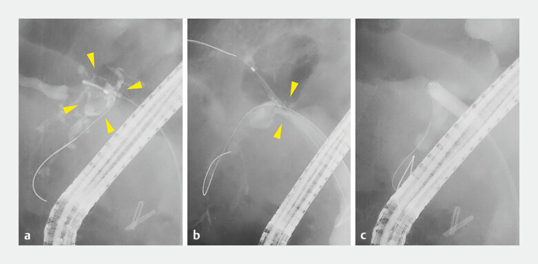 Fig. 1
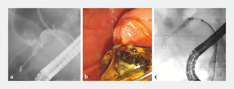 Fig. 2
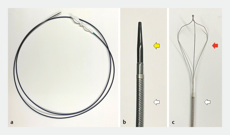 Fig. 3
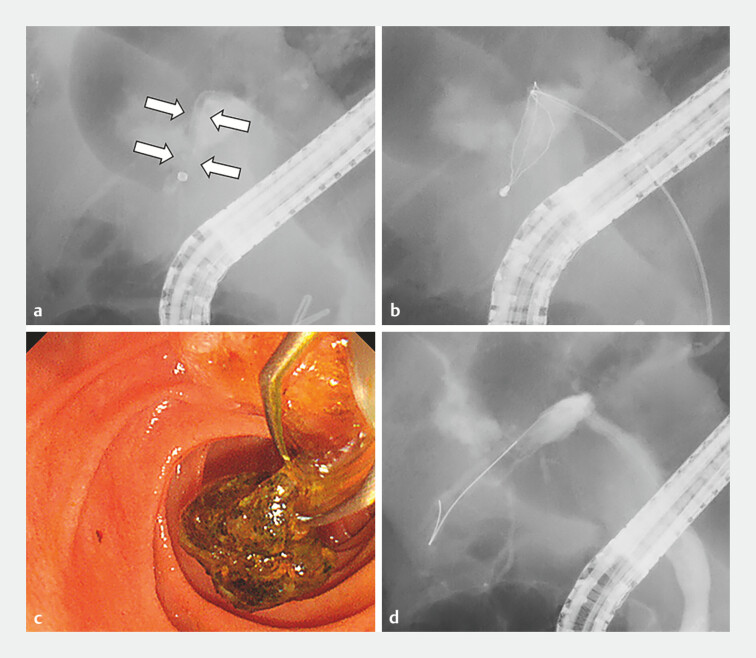 Fig. 4
Peer Reviews
No public reviews on file for this paper yet. If you reviewed it on a platform where reviews are public (OpenReview, ICLR, NeurIPS, ICML), you can paste yours below so the community can read it here.
Videos
No videos yet. Explain this paper in a talk, walkthrough, or lecture? Add one.
Taxonomy
TopicsGallbladder and Bile Duct Disorders · Pediatric Hepatobiliary Diseases and Treatments · Pancreatic and Hepatic Oncology Research
A 64-year-old man with hilar biliary strictures following liver transplantation was admitted for a scheduled biliary stent exchange. During initial endoscopic retrograde cholangiopancreatography (ERCP), a filling defect on the peripheral side of the B6 branch stricture was observed ( Fig. 1 a, b ), indicating intrahepatic bile duct (IHBD) stones. The stricture was dilated using a 6-mm dilation balloon catheter (REN; Kaneka Co., Inc.) ( Fig. 1 c ), and 5 Fr endoscopic nasobiliary drainage tubes (SilkyPass J type, Boston Scientific Co.) were inserted. Stones were removed during the second ERCP session. A basket catheter (Medi-Globe 8-Wire Nitinol Basket; Medico’s Hirata Inc) was inserted over the guidewire (EndoSelector, Boston Scientific Co.) ( Fig. 2 a ); however, not all of the stones were removed ( Fig. 2 b ). A three-layered mechanical lithotripter (Xemex Crusher Catheter LBMT320; Zeon Medical) was used for the larger stones but failed to pass through the stricture ( Fig. 2 c ). Therefore, a tapered tip sheath system (EndoSheather, Piolax), composed of a tapered inner catheter and an outer sheath ( Fig. 3 a, b ), was advanced over the guidewire and positioned at the periphery of the remaining filling defects. The inner sheath and guidewire were withdrawn ( Fig. 4 a ), and the three-layered inner basket of the lithotripter was inserted through the outer sheath ( Fig. 4 b ). The remaining stones were captured ( Fig. 4 c ), achieving complete stone removal ( Fig. 4 d , Video 1 ).
The images of the initial endoscopic retrograde cholangiopancreatography (ERCP) procedure. A cholangiography image showing a filling defect ( a yellow arrowheads) on the peripheral side of the B6 branch stricture ( b yellow arrowheads). c The B6 branch stricture was dilated using a 6-mm dilation balloon catheter (REN; Kaneka Co., Inc.).
The images of the second ERCP procedure. a A basket catheter (Medi-Globe 8-Wire Nitinol Basket; Medico Hirata Inc.) was inserted into the B6 branch. b Some stones were successfully removed using a basket catheter. c A three-layered mechanical lithotripsy basket (Xemex Crusher Catheter LBMT320; Zeon Medical) failed to pass through the stricture or angulation of the B6 branch.
Images of the tapered-tip sheath system. a Overview of the tapered-tip sheath system (EndoSheather, Piolax). b The tapered-tip inner catheter tip (a yellow arrow) and an outer sheath (a white arrow). c Tip of the inner basket of the lithotripter (Xemex Crusher Catheter LBMT320; Zeon Medical) (a red arrow) and an outer sheath (a white arrow).
Stone removal using the tapered-tip sheath system. a The tapered-tip sheath system was inserted over the guidewire to the peripheral side of the remaining filling defects. Thereafter, the inner sheath and the guidewire were withdrawn (white arrows). b The three-layered inner basket was inserted through the outer sheath. c The basket successfully recovered the remaining stones. d Complete removal of the B6 stones was achieved.
This video shows the efficient procedure for intrahepatic bile duct stone removal using a tapered-tip sheath system.Video 1
Biliary strictures are common complications of liver transplantation leading to IHBD stone formation distal to the stricture 1 . Endoscopic transpapillary stone removal can be challenging because of bile duct angulation and strictures 2 . The tapered-tip sheath system, originally developed for bile duct biopsy 3 , enabled the deployment of the basket along the same axis as the bile duct, facilitating efficient stone retrieval and improving the efficiency of IHBD stone removal.
Endoscopy_UCTN_Code_TTT_1AR_2AH
The reference list from the paper itself. Each links out to its DOI / PubMed record.
- 1Boeva I Karagyozov PI Tishkov I Post-liver transplant biliary complications: Current knowledge and therapeutic advances. World J Hepatol 202113667910.4254/wjh.v 13.i 1.66PMC 785686833584987 · doi ↗ · pubmed ↗
- 2Yasuda I Itoi T Recent advances in endoscopic management of difficult bile duct stones Dig Endosc 20132537638510.1111/den.1211823650878 · doi ↗ · pubmed ↗
- 3Matsumori T Uza N Shiokawa M Clinical impact of a novel device delivery system in the diagnosis of bile duct lesions: A single-center experience J Gastroenterol Hepatol 2022371360136610.1111/jgh.1586635434844 · doi ↗ · pubmed ↗