Author Correction: Transcription factor FoxO1 regulates myoepithelial cell diversity and growth
Rino Tokumasu, Rika Yasuhara, Seya Kang, Takahiro Funatsu, Kenji Mishima

Abstract
Genes, proteins, chemicals, diseases, species, mutations and cell lines named across the full text — each resolved to its canonical identifier and authoritative record.
Click any figure to enlarge with its caption.
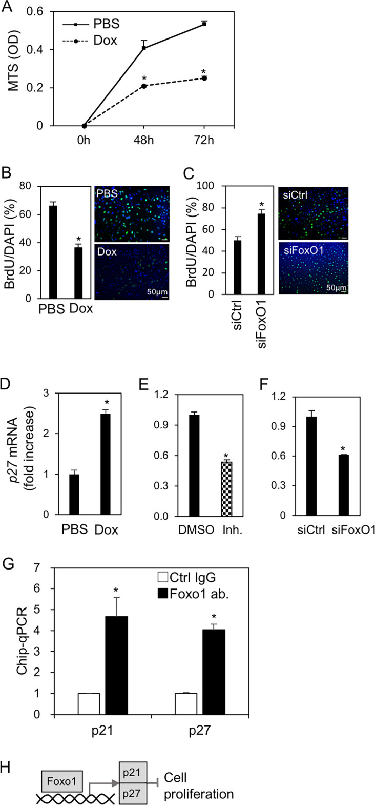 Figure 4
Peer Reviews
No public reviews on file for this paper yet. If you reviewed it on a platform where reviews are public (OpenReview, ICLR, NeurIPS, ICML), you can paste yours below so the community can read it here.
Videos
No videos yet. Explain this paper in a talk, walkthrough, or lecture? Add one.
Taxonomy
TopicsFOXO transcription factor regulation
Correction to: Scientific Reports 10.1038/s41598-024-51619-1, published online 11 January 2024
The original version of this Article contained an error in Fig. 4B, where, as a result of an error during figure assembly, a duplicate figure had been uploaded.
Fig. 4. FoxO1 suppressed ME cell proliferation via cell cycle arrest. (A) Viability of ME^PB-FoxO1^ cells treated with and without Dox (2 µg/mL) at the indicated time-points. (B,C) Cell proliferation rates were measured by BrdU incorporation assay. BrdU positive/DAPI (%, left) with and without Dox (2 µg/mL) for 24 h (B) or with and without transfection of siRNA for FoxO1 for 48 h (C). Immunofluorescent images were showed on the right (BrdU; green, DAPI; blue). (D–F) Expression of p27(KIP1) in ME^PB-FoxO1^ cells. Cells were treated with and without Dox (2 µg/mL) (D), pretreated with and without FoxO1 inhibitor (Inh.; AS1842856, 1 μM) (E) and transfected with siRNA for FoxO1 (F) in the presence of Dox (2 µg/mL) for 48h. The expression data of p21(CIP/WAF1) were shown in Fig. S4. (G) Chromatin immunoprecipitation-quantitative real-time PCR (ChIP-qPCR) analysis of the DNA binding activity of FoxO1 in ME cells. DNA sample was prepared from ME^PB-FoxO1^ cells treated with Dox (2 µg/mL) for 72 h. The associated DNA at the promoter regions of p21^CIP/WAF1^ (− 1722 to − 1712) and p27^KIP1^ (− 1036 to − 1026), after incubation with FoxO1 antibody-conjugated protein G beads, were immunoprecipitated and analyzed by qPCR. *P < 0.05. n = 3. All data were representative of three independent experiments. (H) A schematic for FoxO1-induced cell growth inhibition. See also Supplementary Fig. S4.
The original Article has been corrected.