Elapsed time changes of the brain radiopharmaceutical accumulation of the amyloid PET examination using 18F-flutemetamol
Shota Takemoto, Koji Onuki, Keiko Tanimoto, Masaho Taniguchi, Takako Suero, Mio Okamoto, Satoshi Kimura, Monami Osawa, Haruka Takeshige-Amano, Noriko Nishikawa, Shigeki Aoki, Ryohei Kuwatsuru, Nobutaka Hattori, Koji Murakami

TL;DR
This study shows that the amount of a brain imaging tracer decreases over time after injection, affecting key measurements used in amyloid PET scans.
Contribution
The study demonstrates that radiopharmaceutical washout over time affects quantitative amyloid PET indices, which is novel in clinical imaging timing research.
Findings
Pixel values in PET images decreased with elapsed time after radiopharmaceutical administration.
SUVr and Centiloid Scale values changed due to radiopharmaceutical washout.
Quantitative indices are affected by the timing of PET image acquisition.
Abstract
The purpose of this study was to examine how the radiopharmaceutical accumulation in the brain changes with the elapsed time between the administration of 18F-flutemetamol and the start of imaging, and to determine its effect on quantitative indices. The study population consisted of 25 subjects who agreed to participate in the study. After visual evaluation by the radiologist, 14 subjects tested negative for Aβ accumulation, and 11 subjects tested positive as well. The study population was treated with 18F-flutemeamol, and list mode acquisition was performed for 50 min starting at 60 min after the time of administration. From the acquired list data, five PET images were extracted at 10-min intervals from the start to the end of acquisition, a PET image corresponding to 20 min of acquisition from 60 min after administration, and a PET image corresponding to 20 min of acquisition from…
Genes, proteins, chemicals, diseases, species, mutations and cell lines named across the full text — each resolved to its canonical identifier and authoritative record.
Click any figure to enlarge with its caption.
 Figure 10
 Figure 11
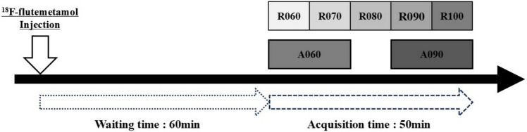 Figure 1
 Figure 2
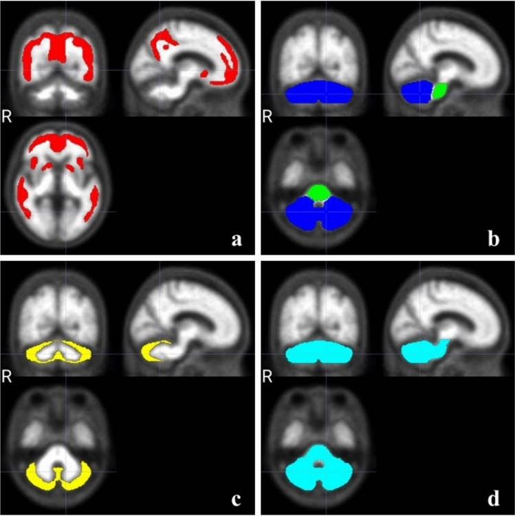 Figure 3
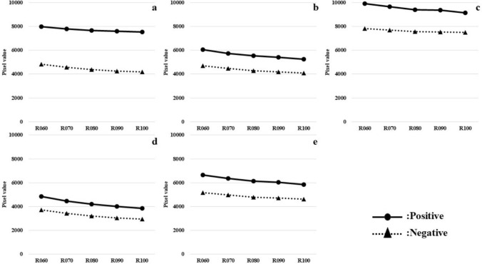 Figure 4
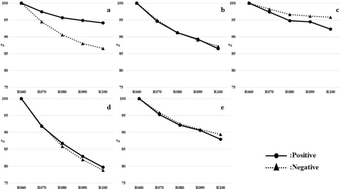 Figure 5
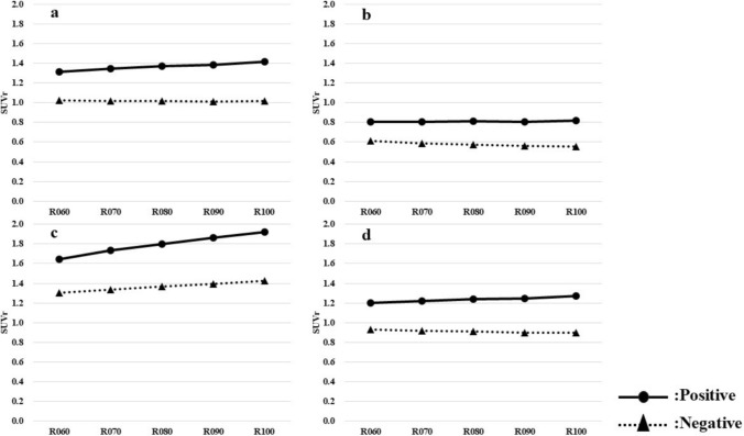 Figure 6
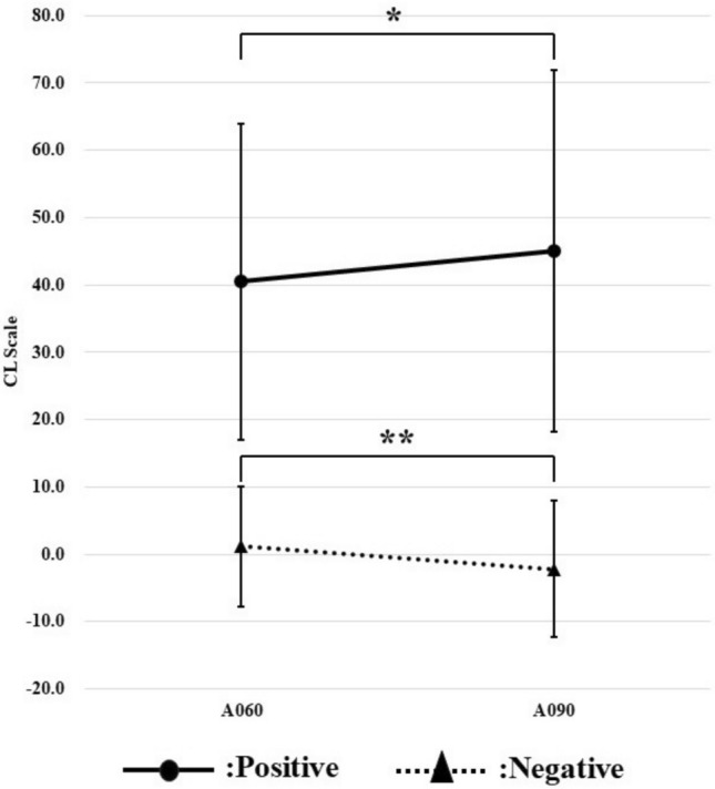 Figure 7
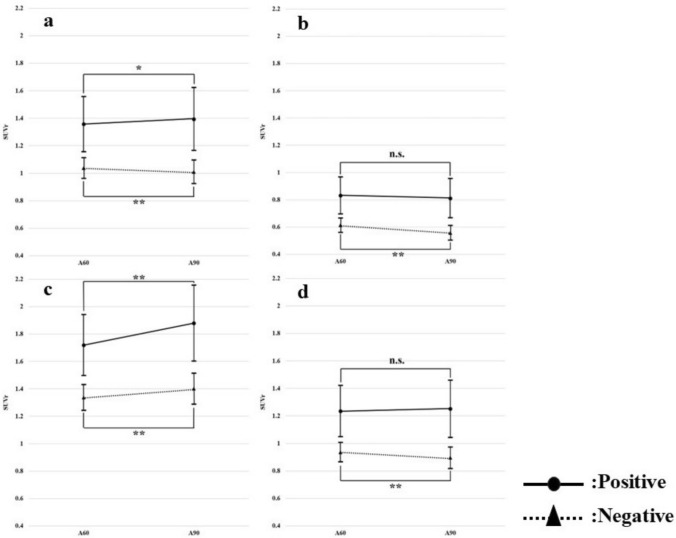 Figure 8
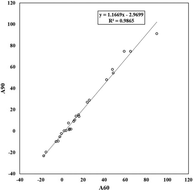 Figure 9
Peer Reviews
No public reviews on file for this paper yet. If you reviewed it on a platform where reviews are public (OpenReview, ICLR, NeurIPS, ICML), you can paste yours below so the community can read it here.
Videos
No videos yet. Explain this paper in a talk, walkthrough, or lecture? Add one.
Taxonomy
TopicsMedical Imaging Techniques and Applications · Lanthanide and Transition Metal Complexes · Radiopharmaceutical Chemistry and Applications