Validation of a semi-automated method to quantify lesion volume changes in multiple sclerosis on 2D proton-density-weighted scans based on image subtraction
Rozemarijn M. Mattiesing, Serena Stel, Alysha S. Mangroe, Iman Brouwer, Adriaan Versteeg, Ronald A. van Schijndel, Bernard M.J. Uitdehaag, Frederik Barkhof, Hugo Vrenken, Joost P.A. Kuijer

TL;DR
Researchers developed a reliable method to track changes in brain lesions in multiple sclerosis patients using 2D scans over up to five years.
Contribution
A semi-automated method for quantifying lesion volume changes using 2D proton-density-weighted images and image subtraction.
Findings
The method showed excellent reproducibility with ICCtrans values between 0.90 and 0.96.
Accuracy was good, with ICCacc values ranging from 0.67 to 0.86.
Higher lesion volume and atrophy were associated with reduced reproducibility and accuracy.
Abstract
The detection and quantification of changes in white matter lesions in the brain is important to monitor treatment effects in patients with multiple sclerosis (MS). Existing automatic tools predominantly require FLAIR images as input which are not always available, or only focus on new/enlarging activity. Therefore, we developed and validated a semi-automated method to quantify lesion volume changes based on 2D proton-density (PD)-weighted images and image subtraction. This semi-automated method provides insight in both “positive” activity (defined as new and enlarging lesions) and “negative” activity (disappearing and shrinking lesions). Yearly MRI scans of patients with early MS from the REFLEX/REFLEXION studies were used. The maximum follow-up period was 5 years. Two PD-weighted images were normalized, registered to a common halfway-space, intensity-matched, and subsequently…
Genes, proteins, chemicals, diseases, species, mutations and cell lines named across the full text — each resolved to its canonical identifier and authoritative record.
Click any figure to enlarge with its caption.
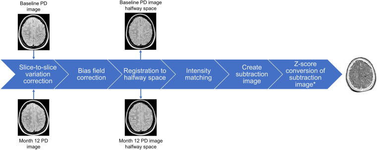 Figure 1
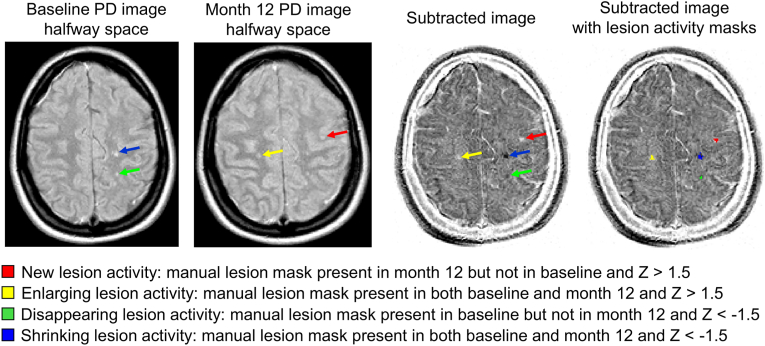 Figure 2
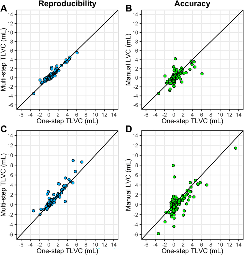 Figure 3
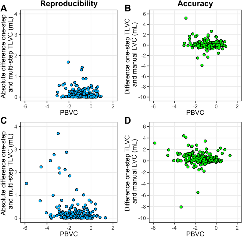 Figure 4
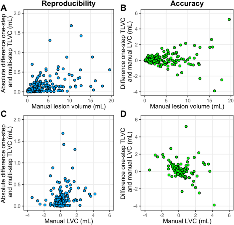 Figure 5
Peer Reviews
No public reviews on file for this paper yet. If you reviewed it on a platform where reviews are public (OpenReview, ICLR, NeurIPS, ICML), you can paste yours below so the community can read it here.
Videos
No videos yet. Explain this paper in a talk, walkthrough, or lecture? Add one.
Taxonomy
TopicsAdvanced MRI Techniques and Applications · RNA regulation and disease · Heat shock proteins research