The genome sequence of the Asparagus Beetle, Crioceris asparagi (Linnaeus, 1758)
Michael F. Geiser, Freya Read, Maxwell V.L. Barclay, Terrence Sylvester, Chenyang Cai, Doga Cedden

TL;DR
This paper presents the genome sequence of the Asparagus Beetle, including its chromosomes and mitochondrial DNA, with over 26,000 protein-coding genes identified.
Contribution
The study provides the first genome assembly of the Asparagus Beetle, including chromosomal scaffolding and gene annotation.
Findings
The genome assembly is 639.30 megabases long, with 93.04% scaffolded into 9 chromosomal pseudomolecules.
The mitochondrial genome is 15.76 kilobases in length and has been fully assembled.
Gene annotation identified 26,673 protein-coding genes using the Ensembl platform.
Abstract
We present a genome assembly from an individual male specimen of Crioceris asparagi (Asparagus Beetle; Arthropoda; Insecta; Coleoptera; Chrysomelidae). The genome sequence has a total length of 639.30 megabases. Most of the assembly (93.04%) is scaffolded into 9 chromosomal pseudomolecules, including the X and Y sex chromosomes. The mitochondrial genome has also been assembled and is 15.76 kilobases in length. Gene annotation of this assembly on Ensembl identified 26,673 protein-coding genes.
Genes, proteins, chemicals, diseases, species, mutations and cell lines named across the full text — each resolved to its canonical identifier and authoritative record.
Click any figure to enlarge with its caption.
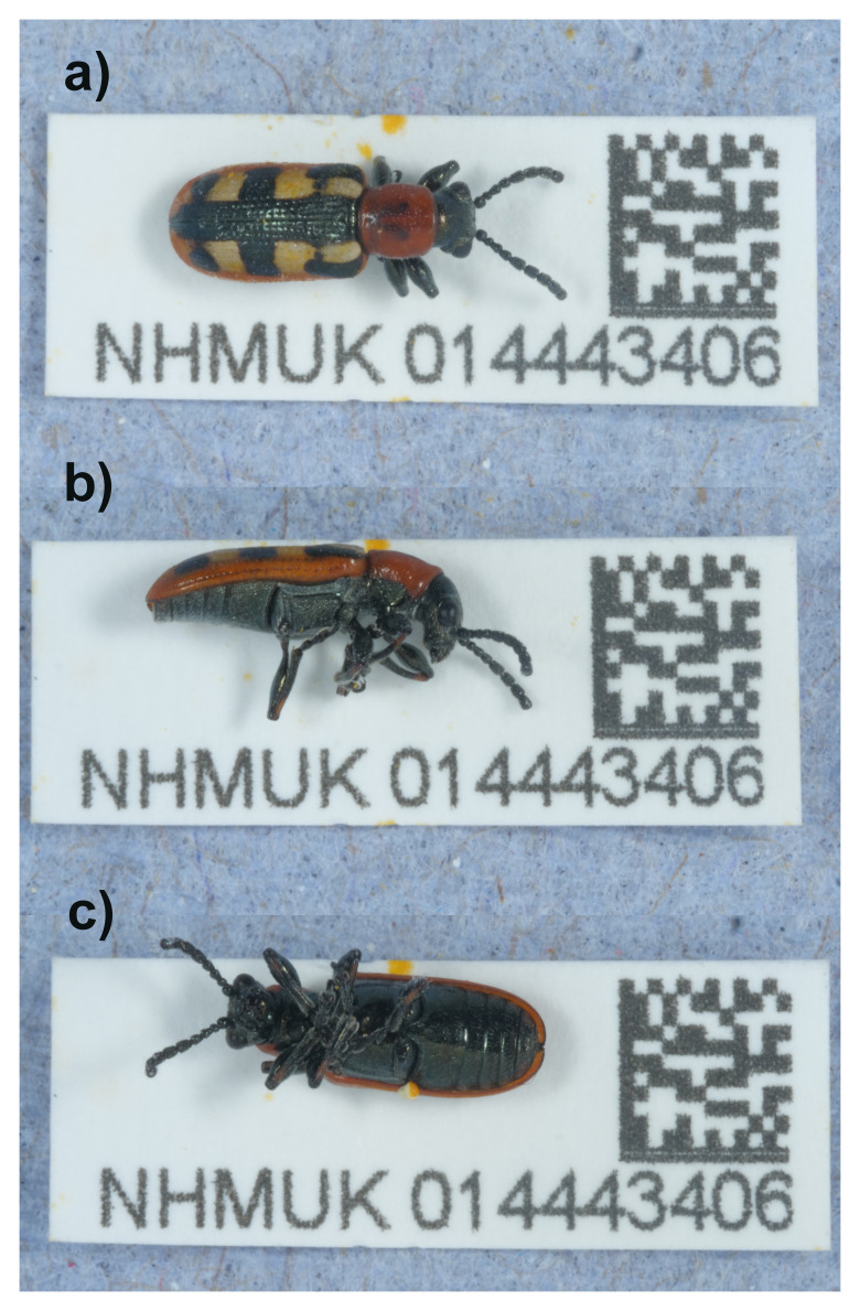 Figure 1
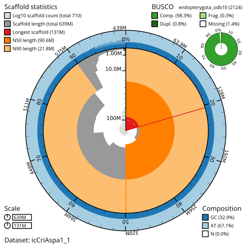 Figure 2
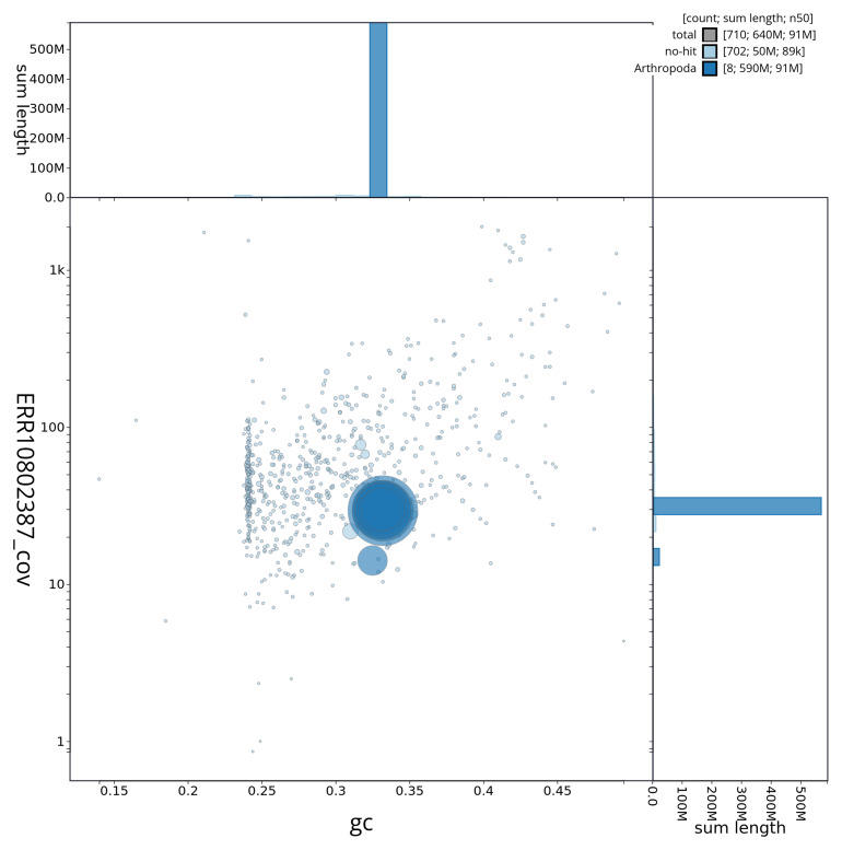 Figure 3
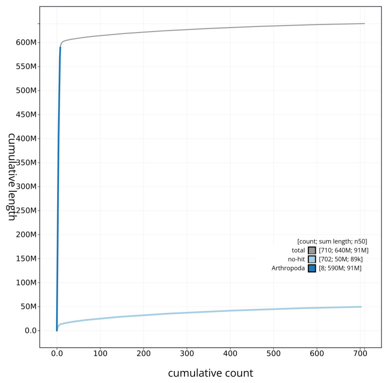 Figure 4
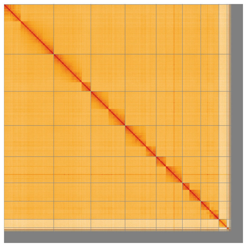 Figure 5
| Project information | |||
|---|---|---|---|
|
| Crioceris asparagi (common asparagus beetle) | ||
|
| PRJEB59077 | ||
|
|
| ||
|
| SAMEA111458050 | ||
|
| 131627 | ||
| Specimen information | |||
|
|
|
|
|
|
| icCriAspa1 | SAMEA111458352 | abdomen |
|
| icCriAspa1 | SAMEA111458147 | Head and thorax |
| Sequencing information | |||
|
|
|
|
|
|
| ERR10802453 | 1.09e+09 | 164.92 |
|
| ERR10802387 | 2.11e+06 | 25.24 |
| Genome assembly | ||
|---|---|---|
| Assembly name | icCriAspa1.1 | |
| Assembly accession | GCA_958507055.1 | |
|
|
| |
| Span (Mb) | 639.30 | |
| Number of contigs | 871 | |
| Number of scaffolds | 709 | |
| Longest scaffold (Mb) | 131.42 | |
| Assembly metrics
|
| |
| Contig N50 length (Mb) | 5.5 |
|
| Scaffold N50 length (Mb) | 90.6 |
|
| Consensus quality (QV) | 58.8 |
|
|
| primary: 90.19%; alternate:
|
|
| BUSCO
| C:98.4%[S:97.6%,D:0.8%],
|
|
| Percentage of assembly mapped
| 93.04% |
|
| Sex chromosomes | XY |
|
| Organelles | Mitochondrial genome: 15.76 kb |
|
| Genome annotation of assembly GCA_958507055.1 at Ensembl | ||
| Number of protein-coding genes | 26,673 | |
| Number of gene transcripts | 26,898 | |
| INSDC
| Name | Length
| GC% |
|---|---|---|---|
| 1 | 131.42 | 33.0 | |
| 2 | 97.98 | 33.0 | |
| 3 | 90.62 | 33.0 | |
| 4 | 82.3 | 33.0 | |
| 5 | 69.17 | 33.0 | |
| 6 | 48.75 | 33.0 | |
| 7 | 47.7 | 33.0 | |
| X | 21.83 | 32.5 | |
| Y | 5.05 | 31.0 | |
| MT | 0.02 | 24.0 |
| Software tool | Version | Source |
|---|---|---|
| BlobToolKit | 4.2.1 |
|
| BUSCO | 5.3.2 |
|
| bwa-mem2 | 2.2.1 |
|
| Cooler | 0.8.11 |
|
| FastK | 427104ea91c78c3b8b8b49f1a7d6bbeaa869ba1c |
|
| Gfastats | 1.3.6 |
|
| Hifiasm | 0.16.1-r375 |
|
| HiGlass | 44086069ee7d4d3f6f3f0012569789ec138f42b84
|
|
| Merqury.FK | d00d98157618f4e8d1a9190026b19b471055b2
|
|
| MitoHiFi | 2 |
|
| Nextflow | 23.04.0-5857 |
|
| PretextView | 0.2 |
|
| purge_dups | 1.2.3 |
|
| sanger-tol/ascc | - |
|
| sanger-tol/genomenote | v1.0 |
|
| sanger-tol/readmapping | 1.1.0 |
|
| Singularity | 3.9.0 |
|
| YaHS | 1.2a |
|
- —Wellcome Trust
Peer Reviews
No public reviews on file for this paper yet. If you reviewed it on a platform where reviews are public (OpenReview, ICLR, NeurIPS, ICML), you can paste yours below so the community can read it here.
Videos
No videos yet. Explain this paper in a talk, walkthrough, or lecture? Add one.
Taxonomy
TopicsGenomics and Phylogenetic Studies · Insect Resistance and Genetics · Chromosomal and Genetic Variations
Species taxonomy
Eukaryota; Opisthokonta; Metazoa; Eumetazoa; Bilateria; Protostomia; Ecdysozoa; Panarthropoda; Arthropoda; Mandibulata; Pancrustacea; Hexapoda; Insecta; Dicondylia; Pterygota; Neoptera; Endopterygota; Coleoptera; Polyphaga; Cucujiformia; Chrysomeloidea; Chrysomelidae; Criocerinae; Crioceris; Crioceris asparagi (Linnaeus, 1758) (NCBI:txid131627)
Background
Crioceris asparagi (Linnaeus, 1758), commonly known as the “Asparagus Beetle” is one of about 250 species of the leaf beetle family (Chrysomelidae) found on the British Isles ( Duff, 2018). Chrysomelidae are one of the largest families of Coleoptera, with over 40,000 currently valid species. Crioceris Geoffroy, 1762 is the type genus of the subfamily Criocerinae, represented by seven species in Britain ( Duff, 2018) and about 1500 worldwide ( Monrós, 1960; Vencl & Leschen, 2014). Crioceris is native to the Palaearctic, Oriental and Afrotropical regions, counting about 30 described species ( Bezděk & Sekerka, 2024; Heinze, 1962; Kimoto & Gressitt, 1979; Monrós, 1960). C. asparagi is currently the only Crioceris on the British checklist, though occasional records of two more species, C. duodecimpunctata (Linnaeus, 1758) and C. macilenta Weise, 1881, exist. The latter two are considered non-established accidental introductions ( Duff, 2018).
Adults and larvae of C. asparagi feed externally and conspicuously on the leaves and stems of wild, and more often cultivated, asparagus ( Asparagus spp.: Asparagaceae) and can be a pest ( Rheinheimer & Hassler, 2018). Pupation takes place in the soil at the base of the plant. Immature stages are described by Steinhausen ( 1994; 2001).
The native distribution of C. asparagi includes almost all of Europe except the far North, the Middle East, Central Asia and Siberia ( Bezděk & Sekerka, 2024). It has been an important non-native pest of asparagus in the USA and Canada for more than a century ( Chittenden, 1917).
Cox (2007) shows it to be widespread in England, but with only scarce records towards the North and West. He mentions a few scattered records in Wales, but considers it absent from Scotland and Northern Ireland. He also observes that its British distribution much more closely matches the range of cultivated asparagus, than the more coastal distribution of the uncommon wild asparagus. NBN Atlas Partnership (2024) shows that C. asparagi has apparently increased both its range and abundance, now also occurring in a few places in Scotland, but remains very scarce in Wales. Whether it is indigenous to the UK is still a matter of speculation, but Fowler (1890) believed it to be, so if it is an introduction, it is a long-standing one.
C. asparagi cannot easily be confused with any other British leaf beetle. Its cylindrical shape is somewhat atypical for Chrysomelidae, but not unusual for a Criocerinae. The elegant blue metallic or shiny black ladder-like elytral pattern on top of a whitish background, with reddish elytral margins and pronotum is very much unique. Variations with reduced or expanded elytral pattern exist ( Warchałowski, 2010), but those individuals are still easily recognisable among other Criocerinae. Specimens with a dark spot in the middle of the red pronotum seem to be unique to the eastern Mediterranean and Middle East and are sometimes classified as subspecies maculipes (Germar, 1834). Specimens with more coarsely punctate pronotum from Central Asia have previously been assigned to another subspecies, turkestanica Medvedev, 1955 (a synonym according to Bezděk & Sekerka, 2024). In the British fauna, only the nominate subspecies has been recorded ( Duff, 2016; Warchałowski, 2010).
The genome of the Asparagus Beetle, Crioceris asparagi, was sequenced as part of the Darwin Tree of Life Project, a collaborative effort to sequence all named eukaryotic species in the Atlantic Archipelago of Britain and Ireland. Here we present a chromosomally complete genome sequence for Crioceris asparagi, based on a male specimen collected from cultivated asparagus at an allotment at Winsford Gardens, Penge, England, United Kingdom ( Figure 1). This appears to be the first genome for a member of the subfamily Criocerinae to be made available.
Photographs of the Crioceris asparagi (icCriAspa1) specimen used for genome sequencing.
Genome sequence report
The genome of Crioceris asparagi ( Figure 1) was sequenced using Pacific Biosciences single-molecule HiFi long reads, generating a total of 25.24 Gb (gigabases) from 2.11 million reads, providing an estimated 36-fold coverage. Primary assembly contigs were scaffolded with chromosome conformation Hi-C data, which produced 164.92 Gb from 1,092.17 million reads. Specimen and sequencing details are summarised in Table 1.
Table 1.: Specimen and sequencing data for Crioceris asparagi.
Assembly errors were corrected by manual curation, including 12 missing joins or mis-joins. This reduced the scaffold number by 1.25% and increased the scaffold N50 by 106.78%. The final assembly has a total length of 639.30 Mb in 709 sequence scaffolds, with 161 gaps, and a scaffold N50 of 90.6 Mb ( Table 2).
Table 2.: Genome assembly data for Crioceris asparagi, icCriAspa1.1.
The snail plot in Figure 2 provides a summary of the assembly statistics, indicating the distribution of scaffold lengths and other assembly metrics. Figure 3 shows the distribution of scaffolds by GC proportion and coverage. Figure 4 presents a cumulative assembly plot, with separate curves representing different scaffold subsets assigned to various phyla, illustrating the completeness of the assembly.
Genome assembly of Crioceris asparagi, icCriAspa1.1: metrics.The BlobToolKit snail plot provides an overview of assembly metrics and BUSCO gene completeness. The circumference represents the length of the whole genome sequence, and the main plot is divided into 1,000 equal-sized bins around the circumference. The outermost blue tracks display the distribution of GC, AT, and N percentages across the bins. Scaffolds are arranged clockwise from longest to shortest and are depicted in dark grey. The longest scaffold is indicated by the red arc, and the deeper orange and pale orange arcs represent the N50 and N90 lengths. A light grey spiral at the centre shows the cumulative scaffold count on a logarithmic scale. A summary of complete, fragmented, duplicated, and missing BUSCO genes in the endopterygota_odb10 set is presented at the top right. An interactive version of this figure is available at https://blobtoolkit.genomehubs.org/view/icCriAspa1_1/dataset/icCriAspa1_1/snail.
Genome assembly of Crioceris asparagi, icCriAspa1.1: BlobToolKit GC-coverage plot.BlobToolKit GC-coverage plot showing sequence coverage (vertical axis) and GC content (horizontal axis). The circles represent scaffolds, with the size proportional to scaffold length and the colour representing phylum membership. The histograms along the axes display the total length of sequences distributed across different levels of coverage and GC content. An interactive version of this figure is available at https://blobtoolkit.genomehubs.org/view/icCriAspa1_1/dataset/icCriAspa1_1/blob.
Genome assembly of Crioceris asparagi icCriAspa1.1: BlobToolKit cumulative sequence plot.The grey line shows cumulative length for all sequences. Coloured lines show cumulative lengths of sequences assigned to each phylum using the buscogenes taxrule. An interactive version of this figure is available at https://blobtoolkit.genomehubs.org/view/icCriAspa1_1/dataset/icCriAspa1_1/cumulative.
Most of the assembly sequence (93.04%) was assigned to 9 chromosomal-level scaffolds, representing 7 autosomes and the X and Y sex chromosomes. These chromosome-level scaffolds, confirmed by the Hi-C data, are named in order of size ( Figure 5; Table 3). During manual curation it was noted that Chromosomes X and Y were assigned based on read coverage statistics. While not fully phased, the assembly deposited is of one haplotype. Contigs corresponding to the second haplotype have also been deposited. The mitochondrial genome was also assembled and can be found as a contig within the multifasta file of the genome submission, and as a separate fasta file with accession OY293835.1.
Genome assembly of Crioceris asparagi icCriAspa1.1: Hi-C contact map of the icCriAspa1.1 assembly, visualised using HiGlass. Chromosomes are shown in order of size from left to right and top to bottom. An interactive version of this figure may be viewed at https://genome-note-higlass.tol.sanger.ac.uk/l/?d=OtktpgA8SYaTECusDNb9xg.
Table 3.: Chromosomal pseudomolecules in the genome assembly of Crioceris asparagi, icCriAspa1.
The final assembly has a Quality Value (QV) of 58.8 and k-mer completeness of 98.64% for the combined assembly. BUSCO (v5.4.3) analysis using the endopterygota_odb10 reference set ( n = 2,124) indicated a completeness score of 98.4% (single = 97.6%, duplicated = 0.8%). The assembly achieves the EBP reference standard of 6.C.59. Other quality metrics are given in Table 2.
Genome annotation report
The Crioceris asparagi genome assembly (GCA_958507055.1) was annotated at the European Bioinformatics Institute (EBI) on Ensembl Rapid Release. The resulting annotation includes 26,898 transcribed mRNAs from 26,673 protein-coding genes ( Table 2; https://rapid.ensembl.org/Crioceris_asparagi_GCA_958507055.1/Info/Index). The average transcript length is 4,132.90, with an average of 3.55 exons per transcript.
Methods
Sample acquisition and DNA barcoding
An adult male specimen of Crioceris asparagi (specimen ID NHMUK014443406, ToLID icCriAspa1) was collected from Winsford Gardens, Penge, England, United Kingdom (latitude 51.41, longitude –0.06) on 2021-07-03. The specimen was collected and identified by Michael Geiser (Natural History Museum) and preserved by dry freezing at –80 °C.
The initial identification was verified by an additional DNA barcoding process according to the framework developed by Twyford et al. (2024). A small sample was dissected from the specimens and stored in ethanol, while the remaining parts were shipped on dry ice to the Wellcome Sanger Institute (WSI). The tissue was lysed, the COI marker region was amplified by PCR, and amplicons were sequenced and compared to the BOLD database, confirming the species identification ( Crowley et al., 2023). Following whole genome sequence generation, the relevant DNA barcode region was also used alongside the initial barcoding data for sample tracking at the WSI ( Twyford et al., 2024). The standard operating procedures for Darwin Tree of Life barcoding have been deposited on protocols.io ( Beasley et al., 2023).
Nucleic acid extraction
The workflow for high molecular weight (HMW) DNA extraction at the Wellcome Sanger Institute (WSI) Tree of Life Core Laboratory includes a sequence of procedures: sample preparation and homogenisation, DNA extraction, fragmentation and purification. Detailed protocols are available on protocols.io ( Denton et al., 2023b).
The icCriAspa1 sample was prepared for DNA extraction by weighing and dissecting it on dry ice ( Jay et al., 2023). Tissue from the abdomen was homogenised using a PowerMasher II tissue disruptor ( Denton et al., 2023a).
HMW DNA was extracted in the WSI Scientific Operations core using the Automated MagAttract v2 protocol ( Oatley et al., 2023). The DNA was sheared into an average fragment size of 12–20 kb in a Megaruptor 3 system ( Bates et al., 2023). Sheared DNA was purified by solid-phase reversible immobilisation, using AMPure PB beads to eliminate shorter fragments and concentrate the DNA ( Strickland et al., 2023). The concentration of the sheared and purified DNA was assessed using a Nanodrop spectrophotometer and Qubit Fluorometer using the Qubit dsDNA High Sensitivity Assay kit. Fragment size distribution was evaluated by running the sample on the FemtoPulse system.
Hi-C preparation
Tissue from the head and thorax of the icCriAspa1 sample was processed at the WSI Scientific Operations core, using the Arima-HiC v2 kit. Tissue (stored at –80 °C) was fixed, and the DNA crosslinked using a TC buffer with 22% formaldehyde. After crosslinking, the tissue was homogenised using the Diagnocine Power Masher-II and BioMasher-II tubes and pestles. Following the kit manufacturer's instructions, crosslinked DNA was digested using a restriction enzyme master mix. The 5’-overhangs were then filled in and labelled with biotinylated nucleotides and proximally ligated. An overnight incubation was carried out for enzymes to digest remaining proteins and for crosslinks to reverse. A clean up was performed with SPRIselect beads prior to library preparation.
Library preparation and sequencing
Library preparation and sequencing were performed at the WSI Scientific Operations core. Pacific Biosciences HiFi circular consensus DNA sequencing libraries were prepared using the PacBio Express Template Preparation Kit v2.0 (Pacific Biosciences, California, USA) as per the manufacturer's instructions. The kit includes the reagents required for removal of single-strand overhangs, DNA damage repair, end repair/A-tailing, adapter ligation, and nuclease treatment. Library preparation also included a library purification step using AMPure PB beads (Pacific Biosciences, California, USA) and size selection step to remove templates shorter than 3 kb using AMPure PB modified SPRI. DNA concentration was quantified using the Qubit Fluorometer v2.0 and Qubit HS Assay Kit and the final library fragment size analysis was carried out using the Agilent Femto Pulse Automated Pulsed Field CE Instrument and 165kb gDNA and 55kb BAC analysis kit. Samples were sequenced using the Sequel IIe system (Pacific Biosciences, California, USA). The concentration of the library loaded onto the Sequel IIe was in the range 40–135 pM. The SMRT link software, a PacBio web-based end-to-end workflow manager, was used to set-up and monitor the run, as well as perform primary and secondary analysis of the data upon completion.
For Hi-C library preparation, DNA was fragmented to a size of 400 to 600 bp using a Covaris E220 sonicator. The DNA was then enriched, barcoded, and amplified using the NEBNext Ultra II DNA Library Prep Kit following manufacturers’ instructions. The Hi-C sequencing was performed using paired-end sequencing with a read length of 150 bp on an Illumina NovaSeq 6000 instrument.
Genome assembly, curation and evaluation
** Assembly **
The HiFi reads were first assembled using Hifiasm ( Cheng et al., 2021) with the --primary option. Haplotypic duplications were identified and removed using purge_dups ( Guan et al., 2020). The Hi-C reads were mapped to the primary contigs using bwa-mem2 ( Vasimuddin et al., 2019). The contigs were further scaffolded using the provided Hi-C data ( Rao et al., 2014) in YaHS ( Zhou et al., 2023) using the --break option for handling potential misassemblies. The scaffolded assemblies were evaluated using Gfastats ( Formenti et al., 2022), BUSCO ( Manni et al., 2021) and MERQURY.FK ( Rhie et al., 2020).
The mitochondrial genome was assembled using MitoHiFi ( Uliano-Silva et al., 2023), which runs MitoFinder ( Allio et al., 2020) and uses these annotations to select the final mitochondrial contig and to ensure the general quality of the sequence.
** Assembly curation **
The assembly was decontaminated using the Assembly Screen for Cobionts and Contaminants (ASCC) pipeline (article in preparation). Flat files and maps used in curation were generated in TreeVal ( Pointon et al., 2023). Manual curation was primarily conducted using PretextView ( Harry, 2022), with additional insights provided by JBrowse2 ( Diesh et al., 2023) and HiGlass ( Kerpedjiev et al., 2018). Scaffolds were visually inspected and corrected as described by Howe et al. (2021). Any identified contamination, missed joins, and mis-joins were corrected, and duplicate sequences were tagged and removed. The curation process is documented at https://gitlab.com/wtsi-grit/rapid-curation (article in preparation).
** Evaluation of the final assembly **
A Hi-C map for the final assembly was produced using bwa-mem2 ( Vasimuddin et al., 2019) in the Cooler file format ( Abdennur & Mirny, 2020). To assess the assembly metrics, the k-mer completeness and QV consensus quality values were calculated in Merqury ( Rhie et al., 2020). This work was done using Nextflow ( Di Tommaso et al., 2017) DSL2 pipelines “sanger-tol/readmapping” ( Surana et al., 2023a) and “sanger-tol/genomenote” ( Surana et al., 2023b). The genome was analysed within the BlobToolKit environment ( Challis et al., 2020) and BUSCO scores ( Manni et al., 2021) were calculated.
The genome assembly and evaluation pipelines were developed using nf-core tooling ( Ewels et al., 2020) and MultiQC ( Ewels et al., 2016), relying on the Conda package manager, the Bioconda initiative ( Grüning et al., 2018), the Biocontainers infrastructure ( da Veiga Leprevost et al., 2017), as well as the Docker ( Merkel, 2014) and Singularity ( Kurtzer et al., 2017) containerisation solutions.
Table 4 contains a list of relevant software tool versions and sources.
Genome annotation
The BRAKER2 pipeline ( Brůna et al., 2021) was used in the default protein mode to generate annotation for the Crioceris asparagi assembly (GCA_958507055.1) in Ensembl Rapid Release at the EBI.
Wellcome Sanger Institute – Legal and Governance
The materials that have contributed to this genome note have been supplied by a Darwin Tree of Life Partner. The submission of materials by a Darwin Tree of Life Partner is subject to the ‘Darwin Tree of Life Project Sampling Code of Practice’, which can be found in full on the Darwin Tree of Life website here. By agreeing with and signing up to the Sampling Code of Practice, the Darwin Tree of Life Partner agrees they will meet the legal and ethical requirements and standards set out within this document in respect of all samples acquired for, and supplied to, the Darwin Tree of Life Project.
Further, the Wellcome Sanger Institute employs a process whereby due diligence is carried out proportionate to the nature of the materials themselves, and the circumstances under which they have been/are to be collected and provided for use. The purpose of this is to address and mitigate any potential legal and/or ethical implications of receipt and use of the materials as part of the research project, and to ensure that in doing so we align with best practice wherever possible. The overarching areas of consideration are:
• Ethical review of provenance and sourcing of the material
• Legality of collection, transfer and use (national and international)
Each transfer of samples is further undertaken according to a Research Collaboration Agreement or Material Transfer Agreement entered into by the Darwin Tree of Life Partner, Genome Research Limited (operating as the Wellcome Sanger Institute), and in some circumstances other Darwin Tree of Life collaborators.
The reference list from the paper itself. Each links out to its DOI / PubMed record.
- 1Abdennur N Mirny LA : Cooler: scalable storage for Hi-C data and other genomically labeled arrays. Bioinformatics. 2020;36(1):311–316. 10.1093/bioinformatics/btz 540 31290943 PMC 8205516 · doi ↗ · pubmed ↗
- 2Allio R Schomaker-Bastos A Romiguier J : Mito Finder: efficient automated large-scale extraction of mitogenomic data in target enrichment phylogenomics. Mol Ecol Resour. 2020;20(4):892–905. 10.1111/1755-0998.13160 32243090 PMC 7497042 · doi ↗ · pubmed ↗
- 3Bates A Clayton-Lucey I Howard C : Sanger Tree of Life HMW DNA fragmentation: diagenode Megaruptor ®3 for LI Pac Bio. protocols.io. 2023. 10.17504/protocols.io.81wgbxzq 3lpk/v 1 · doi ↗
- 4Beasley J Uhl R Forrest LL : DNA barcoding SO Ps for the Darwin Tree of Life project. protocols.io. 2023; [Accessed 25 June 2024]. 10.17504/protocols.io.261ged 91jv 47/v 1 · doi ↗
- 5Bezděk J Sekerka L : Catalogue of palaearctic coleoptera. Volume 6/2/1. Updated and revised second edition. Chrysomeloidea II (Orsodacnidae, Megalopodidae, Chrysomelidae).Leiden: Brill,2024. 10.1163/9789004443303 · doi ↗
- 6Brůna T Hoff KJ Lomsadze A : BRAKER 2: Automatic eukaryotic genome annotation with Gene Mark-EP+ and AUGUSTUS supported by a protein database. NAR Genom Bioinform. 2021;3(1): lqaa 108. 10.1093/nargab/lqaa 108 33575650 PMC 7787252 · doi ↗ · pubmed ↗
- 7Challis R Richards E Rajan J : Blob Tool Kit – interactive quality assessment of genome assemblies. G 3 (Bethesda). 2020;10(4):1361–1374. 10.1534/g 3.119.400908 32071071 PMC 7144090 · doi ↗ · pubmed ↗
- 8Cheng H Concepcion GT Feng X : Haplotype-resolved de novo assembly using phased assembly graphs with hifiasm. Nat Methods. 2021;18(2):170–175. 10.1038/s 41592-020-01056-5 33526886 PMC 7961889 · doi ↗ · pubmed ↗