Retinoblastoma in a Young Nigerian Girl: A Case Report From ECWA Eye Hospital, Kano
Emamoke Atima-Ayeni, Ayodele Jacob Orugun, Ugbede Idakwo, Oyeronke Komolafe, Mayor Orezime Atima, Akinfenwa Taoheed Atanda, Waziri Garba Dahiru, Sani Kamarudeen Owolabi, Eisuke Shimizu, Nakayama Shintaro, Emmanuel Oluwadare Balogun, Emeka John Dingwoke

TL;DR
A 9-year-old Nigerian girl with advanced retinoblastoma showed aggressive disease progression despite treatment, highlighting the need for early diagnosis in resource-limited areas.
Contribution
This case report emphasizes the importance of early diagnosis and treatment in retinoblastoma, particularly in older children with atypical presentations.
Findings
The patient presented with a rare atypical form of retinoblastoma in an older child.
Despite initial treatment, the patient's condition rapidly worsened, likely due to metastatic spread.
The case underscores the challenges of delayed diagnosis in resource-limited settings.
Abstract
Objective: This report details the case of a 9-year-old Nigerian girl presenting with proptosis and a fungating ocular mass, which was histologically confirmed as retinoblastoma following exenteration. Introduction: Retinoblastoma is the most common pediatric intraocular malignancy, predominantly affecting infants and children under the age of 5, with leukocoria being the most frequent presenting symptom. The occurrence of retinoblastoma in older children is rare and often associated with atypical presentations. Case Summary: A 9-year-old Nigerian girl presented with a 1-year history of progressive left eye symptoms, including redness, pain, decreased vision, and proptosis. Examination revealed a large, fungating ocular mass with no light perception. Imaging studies (ultrasound B-scan and CT scan) confirmed extensive vitreous infiltration and optic nerve involvement. Histopathological…
Genes, proteins, chemicals, diseases, species, mutations and cell lines named across the full text — each resolved to its canonical identifier and authoritative record.
Click any figure to enlarge with its caption.
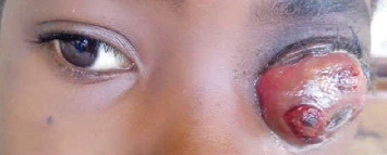 Figure 1
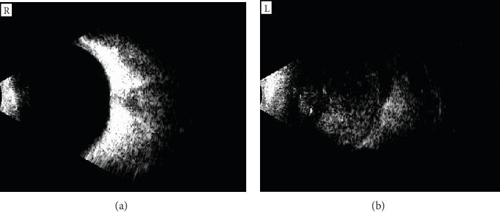 Figure 2
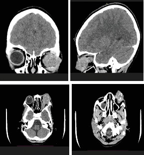 Figure 3
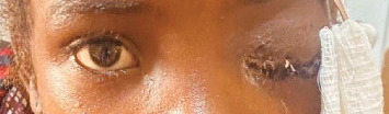 Figure 4
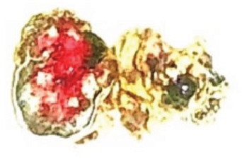 Figure 5
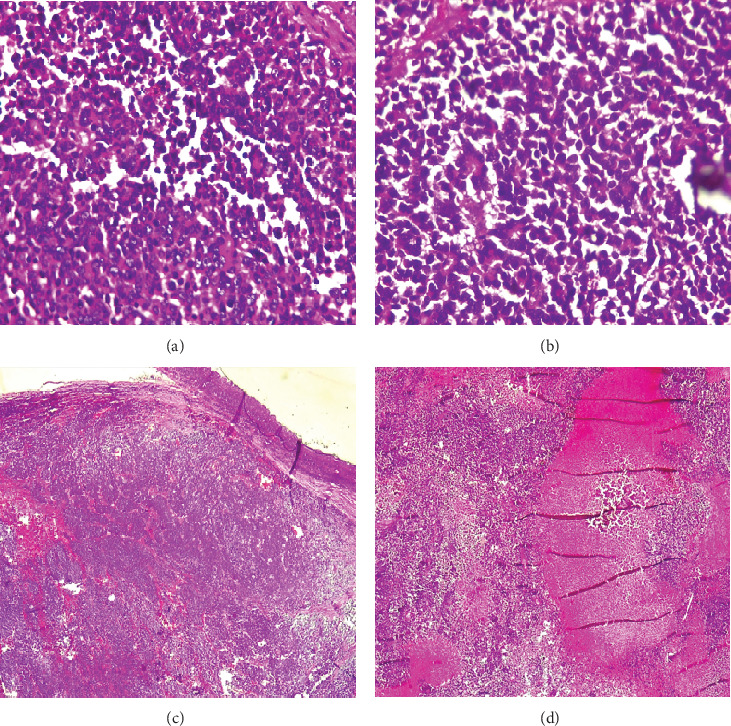 Figure 6
Peer Reviews
No public reviews on file for this paper yet. If you reviewed it on a platform where reviews are public (OpenReview, ICLR, NeurIPS, ICML), you can paste yours below so the community can read it here.
Videos
No videos yet. Explain this paper in a talk, walkthrough, or lecture? Add one.
Taxonomy
TopicsOcular Oncology and Treatments · Ocular Diseases and Behçet’s Syndrome · CNS Lymphoma Diagnosis and Treatment