Effect of disease duration on foveal microvasculature assessed by OCTA in type 2 diabetes mellitus without clinical diabetic retinopathy
David Leonardo Cruvinel Isaac, Alexandre Caiado Ferreira Pires, Laís Lauria Neves, Jamil Miguel Neto, Heitor do Amaral Simões, Karime Fugihara Iwamoto, Raphael Toledo Remiggi, Leticia Pinheiro de Freitas, Alexandre Chater Taleb, Marcos Avila

TL;DR
This study shows that OCTA can detect early microvascular changes in the eyes of type 2 diabetes patients before visible retinopathy occurs.
Contribution
The study demonstrates OCTA's potential for early detection of preclinical diabetic retinal changes in patients without clinical diabetic retinopathy.
Findings
T2DM patients without DR had significantly reduced parafoveal vessel density in both SCP and DCP compared to non-diabetic controls.
T2DM patients diagnosed over 10 years ago had lower vessel density than those diagnosed 5-10 years ago.
OCTA detected enlarged FAZ in T2DM patients, suggesting preclinical microvascular abnormalities.
Abstract
The objective of this study was to establish a comparison between the vessel density (VD) and foveal avascular zone (FAZ) of patients with type 2 diabetes mellitus (T2DM) who lacked clinical signs of diabetic retinopathy (DR) and non-diabetic patients using optical coherence tomography angiography (OCTA). A cross-sectional comparative case-control study (unpaired) was carried out at two tertiary hospitals. All subjects underwent optical coherence tomography angiography (OCTA) examination (DRI OCT Triton Swept Source, Topcon, Japan). The average VD in the superficial capillary plexus (SCP) and the deep capillary plexus (DCP), the FAZ area (mm2) in SCP, and DCP were taken into analysis. The time since the diagnosis of T2DM was used to stratify patients with diabetes between 5 and 10 years and those with a diagnosis of more than 10 years. Compared to non-diabetic controls, the parafoveal…
Genes, proteins, chemicals, diseases, species, mutations and cell lines named across the full text — each resolved to its canonical identifier and authoritative record.
Click any figure to enlarge with its caption.
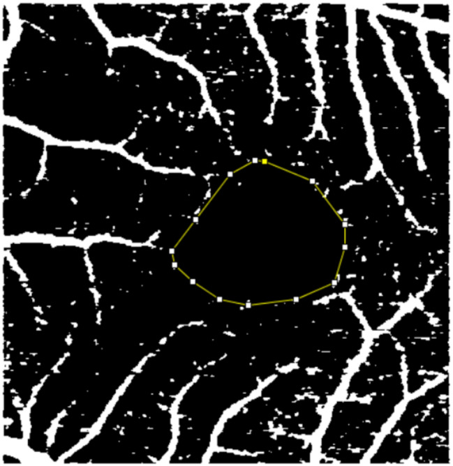 Figure 1
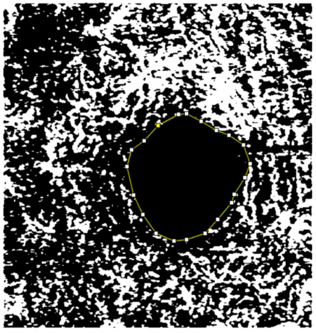 Figure 2
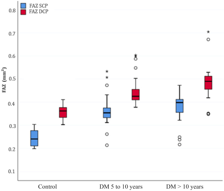 Figure 3
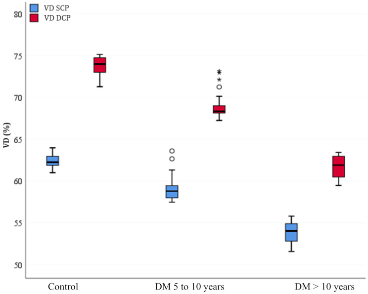 Figure 4
Peer Reviews
No public reviews on file for this paper yet. If you reviewed it on a platform where reviews are public (OpenReview, ICLR, NeurIPS, ICML), you can paste yours below so the community can read it here.
Videos
No videos yet. Explain this paper in a talk, walkthrough, or lecture? Add one.
Taxonomy
TopicsRetinal Diseases and Treatments · Retinal Imaging and Analysis · Glaucoma and retinal disorders