Visualizing the invisible: inner plexiform layer stratification with conventional spectral-domain optical coherence tomography
Ricardo Luz Leitão Guerra, Luiz Roisman, Jay S. Duker, Giuseppe Querques, Luiz Filipe Adami Lucatto, Emmerson Badaró, Gabriel Castilho S. Barbosa, Eduardo Amorim Novais

TL;DR
This study shows that the inner plexiform layer of the retina can be visualized using standard OCT technology with optimized imaging and grayscale inversion.
Contribution
The first demonstration of IPL stratification using commercial SD-OCT with grayscale inversion, without requiring experimental equipment.
Findings
Inverted grayscale revealed five IPL sub-bands in all cases, especially in the parafoveal region.
The 3-mm scan protocol provided better sub-layer differentiation than 12-mm scans.
SS-OCT images did not allow for the distinction of the five IPL strata.
Abstract
The inner plexiform layer (IPL) of the retina plays a key role in visual processing, consisting of five stratified sub-bands (S1-S5) that segregate ON and OFF visual pathways. Until now, resolving these IPL sub-layers was only possible with experimental high-resolution (HR-OCT) or visible-light OCT (VIS-OCT), which remain inaccessible for clinical use. This study provides the first demonstration that IPL stratification can be visualized using commercially available spectral-domain OCT (SD-OCT) with optimized imaging and grayscale inversion. This retrospective, cross-sectional image analysis study included three healthy individuals who underwent macular OCT imaging. Two subjects were imaged with SD-OCT devices (Nidek RS3000 Advance and Zeiss Cirrus 6000), while one subject was imaged with a swept-source OCT (SS-OCT) device (Topcon Triton DRI). High-density B-scans (1024 A-scans per…
Genes, proteins, chemicals, diseases, species, mutations and cell lines named across the full text — each resolved to its canonical identifier and authoritative record.
Click any figure to enlarge with its caption.
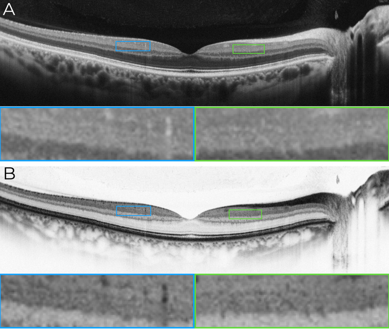 Figure 1
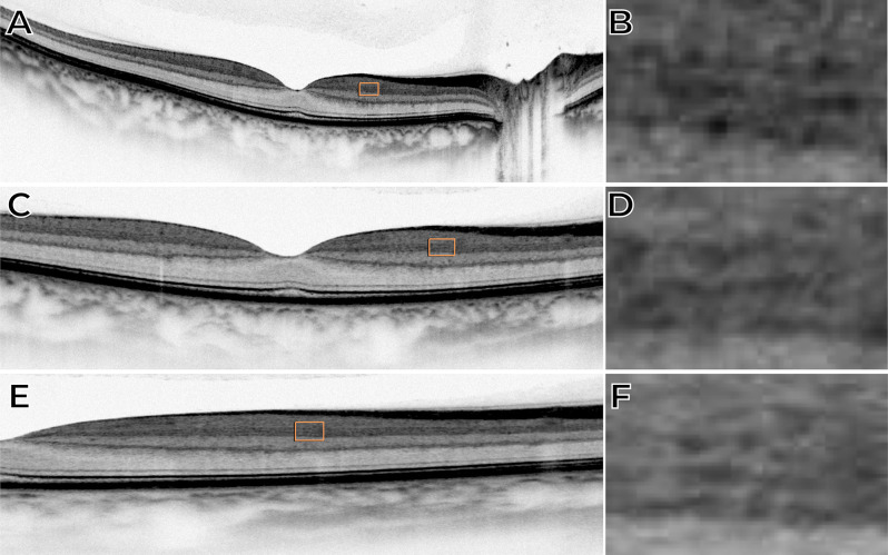 Figure 2
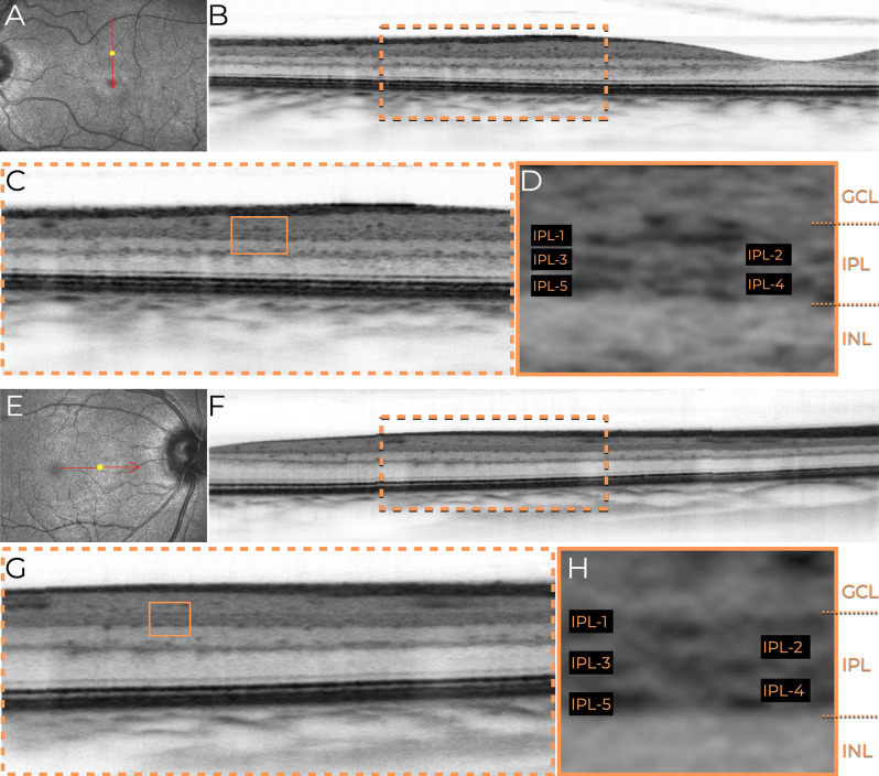 Figure 3
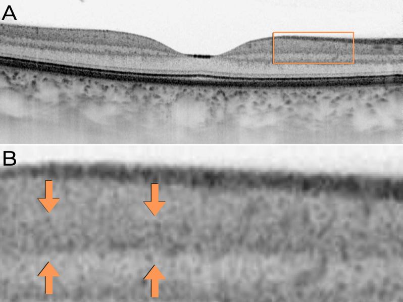 Figure 4
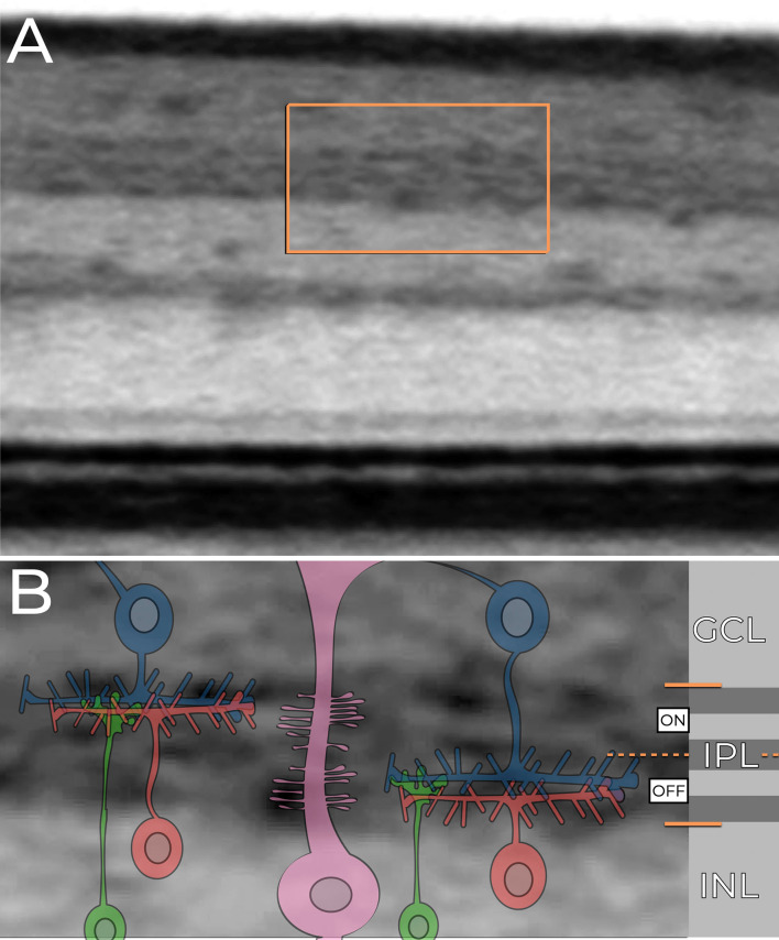 Figure 5
Peer Reviews
No public reviews on file for this paper yet. If you reviewed it on a platform where reviews are public (OpenReview, ICLR, NeurIPS, ICML), you can paste yours below so the community can read it here.
Videos
No videos yet. Explain this paper in a talk, walkthrough, or lecture? Add one.
Taxonomy
TopicsRetinal Development and Disorders · Retinal and Macular Surgery · Retinal Diseases and Treatments