Protocol for mitochondrial DNA isolation from Macrobrachium rosenbergii and Penaeus monodon using differential centrifugation
Dipta Chandra Pal, Muhammad Manjurul Karim

TL;DR
This paper provides a detailed protocol for isolating mitochondrial DNA from two crustacean species using differential centrifugation, without commercial kits.
Contribution
A novel, cost-effective protocol for mitochondrial DNA isolation from Macrobrachium rosenbergii and Penaeus monodon using differential centrifugation.
Findings
The protocol successfully isolates high-quality mitochondrial DNA suitable for sequencing.
Differential centrifugation effectively enriches mitochondrial fractions without commercial reagents.
PCR amplification and gel electrophoresis confirm the presence and purity of mt-DNA.
Abstract
The mitochondrial genome (mitogenome) is a vital tool in molecular phylogenetics, evolutionary biology, and population genetics. Here, we present a protocol for extracting mitochondrial DNA from shrimp (Macrobrachium rosenbergii) and prawn (Penaeus monodon) species using a differential centrifugation approach without relying on commercially available kits. We describe steps for sample preparation, crude mitochondrial extraction, mitochondrial DNA isolation, and purity assessment. We then detail procedures for confirming the presence of the mitogenome and sequencing the mitochondrial genome. For complete details on the use and execution of this protocol, please refer to Pal et al.1,2 •Instructions for enriching mitochondrial fractions using differential centrifugation•Steps for extracting high-quality mitochondrial DNA•Guidance on confirming mt-DNA using PCR amplification and gel…
Genes, proteins, chemicals, diseases, species, mutations and cell lines named across the full text — each resolved to its canonical identifier and authoritative record.
Click any figure to enlarge with its caption.
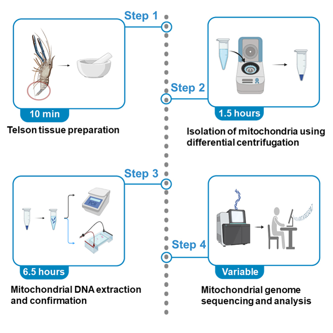 Figure 1
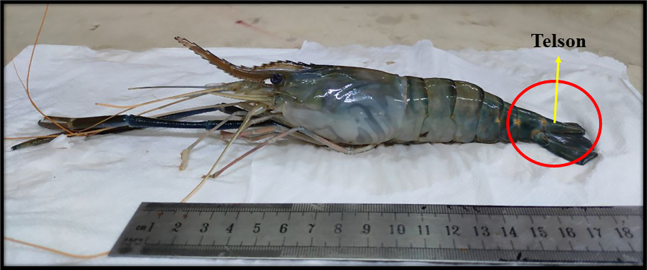 Figure 2
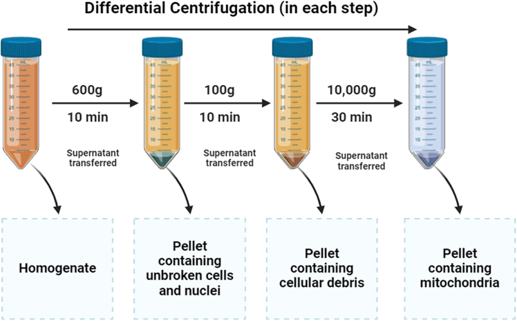 Figure 3
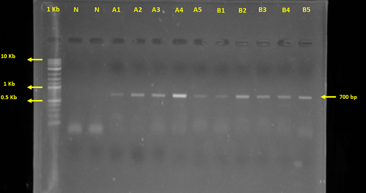 Figure 4
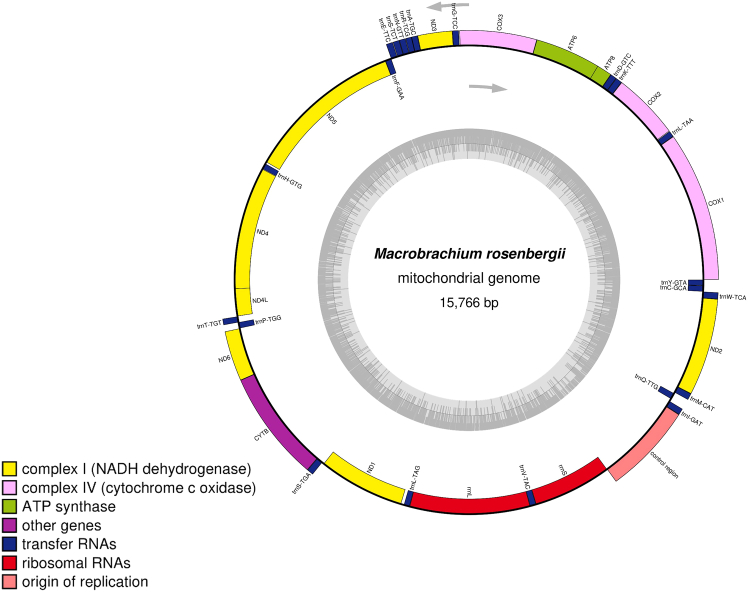 Figure 5
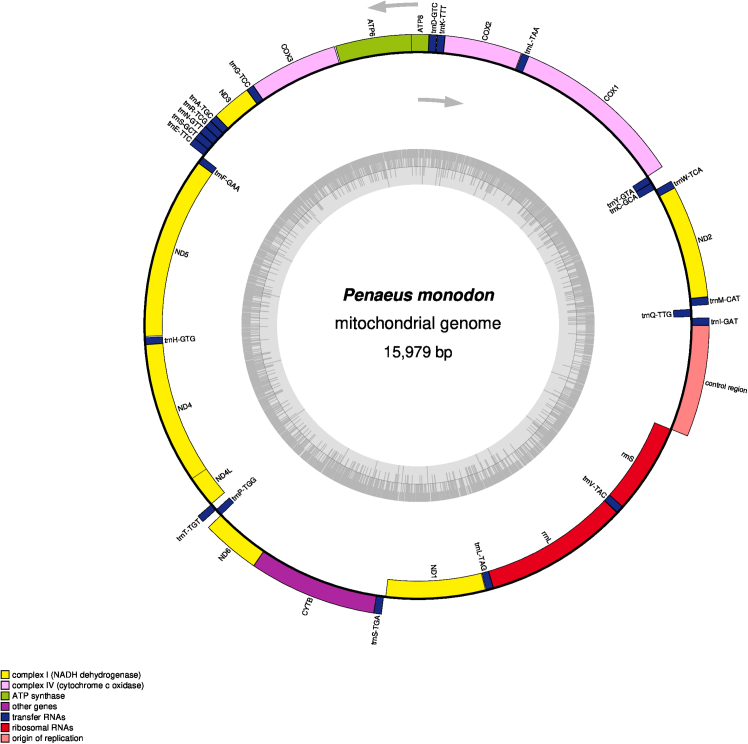 Figure 6
Peer Reviews
No public reviews on file for this paper yet. If you reviewed it on a platform where reviews are public (OpenReview, ICLR, NeurIPS, ICML), you can paste yours below so the community can read it here.
Videos
No videos yet. Explain this paper in a talk, walkthrough, or lecture? Add one.
Taxonomy
TopicsCrustacean biology and ecology · Aquaculture Nutrition and Growth · Genetic diversity and population structure
Before you begin
The protocol described below outlines the specific steps used to isolate and sequence mitochondrial DNA from the telson tissue of Macrobrachium rosenbergii and Penaeus monodon. It was optimized for specimens collected from Bangladesh and has proven effective in yielding high-quality mitochondrial DNA suitable for genome sequencing and downstream bioinformatics analyses.1^,^2 The protocol can be adapted for other crustaceans and invertebrates with similar tissue structures by adjusting centrifugation speeds, buffer volumes, or incubation times. Researchers working with other taxa may modify the tissue homogenization and mitochondrial enrichment steps to accommodate differences in organismal physiology.
Preparatory steps
- 1.To ensure the success of this protocol, prepare the workspace and materials in advance.
- 2.Maintain a sterile and contamination-free environment throughout the procedure to preserve the integrity of mitochondrial DNA.
- 3.Pre-cool all glassware, homogenizers, and centrifuge rotors in an ice bath prior to starting the experiment to prevent mitochondrial degradation.
- 4.Use freshly prepared Isolation Buffer and Lifton Buffer to ensure their effectiveness.
- 5.Confirm the availability of necessary equipment, such as a refrigerated centrifuge, heat block, spectrophotometer, and gel documentation system.
- 6.Prepare PCR reagents, primers (LCO1490 and HCO2198), and materials for agarose gel electrophoresis beforehand.
- 7.Verify the functionality of sequencing platforms and software tools such as FastQC, TrimGalore, SPAdes, MITOS, and OGDRAW for downstream applications.
- 8.Collect the telson tissue samples and process them promptly to avoid tissue degradation.
Key resources table
REAGENT or RESOURCESOURCEIDENTIFIERChemicals, peptides, and recombinant proteinsTris-HClSisco79420EDTASisco43272EmeraldAmp GT PCR master mixTakara BioRR901Bovine serum albuminSigma-AldrichA-9418Sodium chlorideCarl RothArt. No. 3957.1Potassium chlorideMerck104936Di-sodium hydrogen phosphateMerck106586Potassium dihydrogen phosphateMerck104873D(−)-MannitolMerck105982SucroseFUJIFILM196-00015Sodium dodecyl sulfateSigma-Aldrich436143Phenol:chloroform:isoamyl alcohol (25:24:1, v/v)Invitrogen11518756EthanolMerck100983ChloroformMerck102447Isoamyl alcoholMerck100979SeaKem LE agaroseLonza50002Proteinase KQIAGEN51304Deposited dataMacrobrachium rosenbergii complete mitochondrial genomeThis article/Pal et al.1https://www.ncbi.nlm.nih.gov/nuccore/PQ213808.1Penaeus monodon complete mitochondrial genomeThis article/Pal et al.2https://www.ncbi.nlm.nih.gov/nuccore/PQ876261.1Biological samplesMacrobrachium rosenbergiiThis article/Pal et al.1N/APenaeus monodonThis article/Pal et al.2N/AOligonucleotidesPrimer for COI gene:forward (LCO1490): 5′-GGTCAACAAATCATAAAGATATGG - 3’;reverse (HCO2198):5′- TAAACTTCAGGGTGACCAAAAAATCA – 3′Costa et al.3N/ASoftware and algorithmsFastQC v.0.12.112Andrews (n.d.)4https://github.com/s-andrews/FastQCTrimGalore v.0.6.10Krueger et al.5https://github.com/FelixKrueger/TrimGaloreSPAdes v.3.15.5Prjibelski et al.6https://github.com/ablab/spades?tab=readme-ov-fileMITOS v.1.1.7Bernt et al.7https://gitlab.com/Bernt/MITOS/OGDRAWGreiner et al.8https://chlorobox.mpimp-golm.mpg.de/OGDraw.htmlOtherPorcelain mortar with pestleN/A23-3505High-speed refrigerated micro centrifugeTomyMX-305Starter 2100 pH bench pH meterOhaus30057496Microvolume spectrophotometerBertholdMX-305Qubit 4 flurometerInvitrogenQ33238Bioanalyzer 2100AgilentG2938AThermo mix HCM100-ProDLAB5062103100ProFlex PCR systemApplied Biosystems4484073Submarine electrophoresis systemMupid OneMU2Accessories (gel casting stand, gel trays, and combs)Mupid OneON-MSGuardian 5000 hotplate stirrerOhaus16640452PR precision balance PR223/EOhausOH30430071Corning Falcon centrifuge tubesSigma-Aldrich352070microDOC gel documentation hood with screenCleaver ScientificCSL-MDOCUV254UV transilluminatorCleaver ScientificCSLUVTS254PCR cabinetEscoPCR-3A1Class II biological safety cabinetEscoAC2-4D1Laboratory water distillerWincomWD-SS5HP series laboratory freezerEscoHF2-400S-1Microwave ovenWhirlpoolMW-20BS
Materials and equipment
Phosphate Buffered saline (PBS) pH 7.4ReagentFinal concentrationAmountNaCl0.137 M0.8 gNa_2_HPO_4_0.01 M0.144 gKCl0.0027 M0.02 gKH_2_PO_4_0.0018 M0.025 gddH_2_ON/Aup to 100 mLTotalN/A100 mLStore at 4°C for up to 1 month. Isolation Buffer pH 7.4ReagentFinal concentrationAmountMannitol225 mM4.1 gSucrose75 mM2.57 gBovine serum albumin0.5%0.5 gEDTA0.1 mM2.922 mgTris–HCl30 mM0.336 gddH_2_ON/AUp to 100 mLTotalN/A100 mLPrepare fresh for each experiment. Lifton Buffer pH 7.59ReagentFinal concentrationAmountEDTA100 mM2.922 gTris-HCl25 mM0.303 gSDS1%1 gddH_2_ON/AUp to 100 mLTotalN/A100 mLStore at 4°C for up to 3 months.
Step-by-step method details
Preparing telson tissue for mitochondrial DNA extraction
Timing: 10 min
Here, we describe the initial steps required to prepare telson tissue for mitochondrial DNA extraction.Note: The telson, located at the posterior end of the crustacean body (Figure 1), is carefully dissected from dead specimens to minimize contamination from surrounding tissues. The collected telson tissue is cleaned with sterile phosphate-buffered saline (PBS) to remove any residual debris or contaminants. This step ensures the isolation of high-quality tissue essential for efficient mitochondrial DNA extraction. Proper handling and preparation of the telson tissue are crucial to maintaining sample integrity and obtaining reproducible results in downstream analyses.
- 1.Separate the Telson.
- a.Carefully separate the telson part from the shrimp/prawn sample.
- b.Remove the shell and wash 3–4 times using ice-cold PBS.
- c.Cut the telson tissue into small pieces (2–3 mm) using sterilized scissors.
- d.Weigh the telson tissue using a precision balance (PR Precision Balance PR223/E). Figure 1. Sample (Macrobrachium rosenbergii) used for mitochondrial DNA extraction, highlighting the telson
Isolation of crude mitochondria from shrimp/prawn telson tissue
Timing: 1.5 h
This step describes the isolation of crude mitochondria from the telson tissues of shrimp/prawn samples.Note: This protocol is an optimization of a previous protocol by Wieckowski et al.10 To ensure optimal results, it is essential to precool all the glassware, and the mortar and the pestle in an ice-bath before starting the procedure. To maintain sample integrity and prevent excessive degradation, the maximum recommended maceration time is 5 min. Prolonged maceration can lead to mitochondrial damage and reduced DNA yield.CRITICAL: Avoid prolonged homogenization to prevent mitochondrial damage. Ensure all buffers and equipment are ice-cold.
- 2.Homogenization of the telson tissue.
- a.Transfer the telson tissue pieces to a precooled mortar.
- b.Add ice-cold Isolation Buffer (pH 7.4) at a ratio of 4-6 mL per gram of telson tissue.
- c.Homogenize the tissue gently using a precooled pestle to minimize damage to mitochondrial structures.
- 3.Mitochondria isolation (Figure 2).
- a.Transfer the homogenate into a 50 mL Falcon Centrifuge Tube (approximately 10 mL) and centrifuge at 600 × g for 10 min at 4^o^C using a refrigerated centrifuge (Tomy MX-305 High Speed Refrigerated Micro Centrifuge).
- b.Carefully collect the supernatant into a fresh Falcon tube and discard the pellet containing unbroken cells and nuclei.
- c.Centrifuge at 100–200 × g for 5 min at 4^o^C using a refrigerated centrifuge.
- d.Transfer the resulting supernatant to another fresh Falcon tube and discard the pellet (if present).
- e.Centrifuge the final supernatant at 10,000 × g for 30 min at 4^o^C a refrigerated centrifuge.
- f.Discard the supernatant, which contains lysosomes and microsomes, and retain the pellet containing crude mitochondria. Note: This step can be performed with a minimum of 0.1 g of tissue, but in such cases, all centrifugation steps should be conducted using microcentrifuge (Eppendorf) tubes instead of 50 mL Falcon tubes. Additionally, this step has been successfully applied to frozen samples and other tissues, although with lower accuracy due to potential DNA degradation. To preserve DNA integrity, frozen samples should be stored at −26°C and thawed on ice just before use. Proper storage and minimal freeze–thaw cycles are essential for maintaining sample quality.Figure 2. Mitochondria extraction using differential centrifugation technique
Mitochondrial DNA extraction protocol
Timing: 6.5 h
This step outlines the extraction of high-quality mitochondrial DNA (mt-DNA) from mitochondrial pellet for downstream applications, including sequencing, PCR amplification, and genomic analysis. The goal is to isolate pure and intact mt-DNA while minimizing contamination from nuclear DNA and other cellular components.Note: After extraction, confirmation of mt-DNA is achieved via gel electrophoresis and/or PCR using mitochondrial-specific markers, ensuring the integrity and specificity of the extracted DNA. This step is critical for accurate and reproducible results, as contamination or degraded DNA can compromise data quality.
- 4.Mitochondrial DNA extraction.
- a.Add 400 μL Lifton Buffer and 15 μL proteinase K (20 mg/mL) to the mitochondrial pellet.
- b.Incubate the sample at 60^o^C for 3 h (until the solution becomes clear) in a thermomixer.
- c.Add an equal volume of PCI (Phenol: Chloroform: Isoamyl Alcohol, 25:24:1), hereafter 400 μL to the lysate.
- d.Centrifuge at 7,200 × g for 10 min at 4^o^C.
- e.Carefully collect the aqueous layer (approximate 300 μL) in the supernatant.
- f.Add 300 μL (Chloroform: Isoamyl Alcohol, 24:1) to the collected supernatant.
- g.Centrifuge at 7,200 × g for 10 min at 4^o^C and collect the top aqueous layer (approximately 100 μL).
- h.Add Ice-cold absolute ethanol 700 μL and mix gently.
- i.Incubate the tube at −20^o^C for 10 min (Incubation time can be increased if the sample size is minimal).
- j.Centrifuge at 18,000 × g for 10 min at 4^o^C and discard the supernatant.
- k.Wash the pellet with 70% ethanol (500–600 μL) and centrifuge at 18,000 × g for 10 min at 4^o^C (repeat the step if necessary).
- l.Discard supernatant and allow the pellet to air dry at 25°C for 10 min.
- m.Dissolve the pellet in nuclease-free water (30/40 μL).
- n.Evaluate the quality of the extracted DNA using a spectrophotometer (e.g. Colibri-Model LB 915 microvolume spectrophotometer, Berthold Technologies GmbH & Co. KG, Germany). Note: Measure DNA concentration using a spectrophotometer and check integrity via gel electrophoresis. A_260_/A_280_ ratio should be ∼1.8–2.0 for pure DNA.
- 5.Confirm the presence of mitochondrial DNA.
- a.Run mitochondrial COI gene specific PCR using the primer LCO1490 (Forward) and HCO2198 (Reverse) on ProFlex PCR System (Tables 1 and 2).
- b.Load 20% PCR product to 1% agarose gel and run gel electrophoresis at 100 V for 35 min.
- c.Check the gel in a gel documentation system (Figure 3). Note: PCR reaction mix and PCR cycling conditions are listed in Tables 1 and 2, respectively.Table 1PCR reaction master mixReagentAmount (μL)DNA templateVolume for 20 ngEmeraldAmp GT PCR Master Mix12.5Primer Forward (10 pmol/μL)2.5Primer Reverse (10 pmol/μL)2.5ddH_2_OVolume to make the 25 μL totalTotal25Table 2PCR cycling conditionsStepsTemperatureTimeCyclesInitial Denaturation94 °C4 min1Denaturation94 °C30 s30 cyclesAnnealing50 °C45 sExtension72 °C1 minFinal extension72 °C6 min1Hold4 °CforeverFigure 3PCR product amplified from the COI gene of Macrobrachium rosenbergii and Penaeus monodon electrophoresed on a 1% agarose gelHere, N = Negative, A = P. monodon, B = M. rosenbergii.
Mitochondrial genome sequencing and analysis
Timing: Variable
This step involves sequencing and analyzing the complete mitochondrial genome to uncover the genetic architecture and features of the organism.Note: Extracted mitochondrial DNA is subjected to high-throughput sequencing using platforms such as Illumina, ensuring sufficient coverage and accuracy. This step is critical for generating high-quality genomic data and provides insights into mitochondrial features. Proper computational analysis ensures accurate annotation and reliable results for further research.
- 6.Perform sequencing.
- a.Conduct whole mitochondrial genome sequencing on the Illumina NextSeq 550 platform using a paired-end (2 × 151 bp) sequencing strategy to ensure high coverage and accuracy.
- b.Use the Illumina DNA Prep kit (Illumina, San Diego, CA, USA) for library preparation according to the manufacturer’s standard protocol, ensuring the fragmentation and adapter ligation steps are optimized for mitochondrial DNA sequencing.
- c.Perform quantification and quality control (QC) of the prepared libraries using Qubit (Invitrogen, USA) and Bioanalyzer (Agilent, USA) before sequencing.
- 7.Analysis of sequencing data.
- a.Assess the raw sequencing data quality using FastQC v.0.12.112, and filter out low-quality reads (Q < 20) using TrimGalore v.0.6.10 with default settings.
- b.Conduct de novo genome assembly of the trimmed reads using SPAdes genome assembler v.3.15.5.
- c.Perform gene annotation using MITOS v.1.1.7 with the Invertebrate Mitochondrial Code (transl_table=5) for accurate translation of mitochondrial genes.
- d.Verify the gene annotations using NCBI nucleotide BLAST against mitochondrial reference genomes.
- e.Visualize circular mitomap using OrganellarGenomeDRAW (OGDRAW) (Figures 4 and 5). Note: Validate mitochondrial contigs using BLAST against reference genomes. Use MITOS for automated gene annotation and manually curate key regions by NCBI nucleotide BLAST results against mitochondrial reference genomes.Figure 4. The complete mitochondrial genome map of Macrobrachium rosenbergiiThe innermost ring illustrates the GC content, shown in ash gray, with transcription directions indicated by arrows. The outermost ring displays the genes, with those transcribed clockwise placed on the inside of the circle and those transcribed counterclockwise on the outside. The color coding corresponds to different gene groups, as indicated in the key located in the bottom left corner. The annotated map includes 13 protein-coding genes, 2 ribosomal RNA genes (rrnS for 12S ribosomal RNA and rrnL for 16S ribosomal RNA), 22 transfer RNA genes, and the putative control region.1Figure 5. The mitochondrial genome map of Penaeus monodonIt shows GC content in the innermost ring (ash grey), transcription directions as arrows, with clockwise-transcribed genes on the inner side and counterclockwise-transcribed genes on the outer side, color-coded gene groups (key in the bottom left corner) including 13 protein-coding genes (PCGs), 2 ribosomal RNA genes (rrnS for 12S and rrnL for 16S rRNA), 22 transfer RNA (tRNA) genes, and the putative control region.2
Expected outcomes
The expected outcome of this protocol is the successful isolation of high-quality mitochondrial DNA from shrimp or prawn telson tissues, suitable for downstream applications such as PCR amplification, sequencing, and mitogenomic analyses. The mitochondrial DNA should appear clean and free from significant contamination, with spectrophotometric analysis yielding a high purity ratio (A_260_/A_280_ between 1.8 and 2.0). PCR amplification using COI gene-specific primers is anticipated to produce a clear, distinct band of the expected size (∼700 bp) on a 1% agarose gel, confirming the presence of mitochondrial DNA (Figure 3). Sequencing on the Illumina NextSeq 550 platform should generate high-quality reads (Q ≥ 20) that allow for accurate de novo assembly of the mitochondrial genome. Annotation and visualization of the genome using tools like MITOS and OGDRAW are expected to produce a complete, annotated mitogenome with clearly defined genes and a circular mitomap (Figures 4 and 5). This outcome will provide valuable genetic insights for molecular phylogeny, evolutionary, biogeographic relationships, aquaculture management, and conservation studies of the target species.
Limitations
The protocol for mitochondrial DNA isolation and sequencing from shrimp/prawn telson tissues has certain limitations that should be considered. The yield and purity of mitochondria are highly dependent on the quality and freshness of the tissues; degraded or poorly preserved samples may result in low yield or contamination. The isolation process, though optimized, may inadvertently include nuclear, lysosomal, or microsomal components, affecting downstream analyses. Additionally, the success of mitochondria isolation relies on the quality of reagents, particularly during differential centrifugation using the Isolation Buffer, where pH variation, contamination, or improper handling can reduce the mitochondrial yield. Finally, the specificity of primers for mitochondrial COI gene amplification can vary due to inter-species sequence differences, potentially leading to unsuccessful PCR amplification or misidentification. These factors should be carefully addressed to ensure the protocol’s reliability and reproducibility.
Troubleshooting
Problem 1
Tissue becomes mushy or difficult to homogenize (related to Step 1b).
Potential solution
Ensure tissue is always kept on ice and precool the glassware and mortar with pestle in an ice-bath before starting the homogenization. Avoid over-washing with PBS.
Problem 2
Poor mitochondrial yield (related to Step 2).
Potential solution
Check the integrity of the tissue and ensure proper homogenization. Increase the number of telson tissue pieces if needed.
Problem 3
Contamination with unbroken cells or nuclei (related to Step 3b and 3d).
Potential solution
Ensure careful transfer of supernatant after each centrifugation step. Increase centrifugation time slightly (by 10%) if needed.
Problem 4
Low mt-DNA yield or degraded mt-DNA (related to Step 4a and 4h).
Potential solution
Use fresh reagents and ensure ethanol precipitation is performed properly. Minimize exposure of DNA to room temperature (> 25°C).
Problem 5
No band observed on the gel (related to Step 5a).
Potential solution
Verify the primer sequences and ensure proper PCR conditions (e.g., annealing temperature).
Resource availability
Lead contact
Further information and requests for resources and reagents should be directed to and will be fulfilled by the lead contact, Muhammad Manjurul Karim ([email protected]).
Technical contact
Technical questions on executing this protocol should be directed to and will be answered by the technical contact, Dipta Chandra Pal ([email protected]).
Materials availability
This study did not generate new unique reagents.
Data and code availability
The published article includes all datasets and code generated or analyzed during this study.
Acknowledgments
The authors gratefully acknowledge the financial support provided by the Biotechnology Research Centre, University of Dhaka (dated March 20, 2024).
Author contributions
D.C.P.: data curation, formal analysis, methodology, software, and writing – original draft. M.M.K.: conceptualization, funding acquisition, investigation, project administration, resources, supervision, validation, and writing – review and editing.
Declaration of interests
The authors declare no competing interests.
The reference list from the paper itself. Each links out to its DOI / PubMed record.
- 1Pal D.C.Hasan M.M.Khan S.N.Karim M.M.Complete mitochondrial genome sequence of giant freshwater prawn, Macrobrachium rosenbergii of Bangladesh Microbiol. Resour. Announc.142025 e 00924-2410.1128/mra.00924-24PMC 1173714439601589 · doi ↗ · pubmed ↗
- 2Pal D.C.Khan S.N.Karim M.M.Complete mitochondrial genome sequence of giant tiger prawn, Penaeus monodon, of Bangladesh Microbiol. Resour. Announc.142025 e 00118-2510.1128/mra.00118-25PMC 1206065840183592 · doi ↗ · pubmed ↗
- 3Costa F.O.De Waard J.R.Boutillier J.Ratnasingham S.Dooh R.T.Hajibabaei M.Hebert P.D.Biological identifications through DNA barcodes: The case of the Crustacea Can. J. Fish. Aquat. Sci.64200727229510.1139/F 07-008 · doi ↗
- 4Andrews S.Babraham Bioinformatics - Fast QC A Quality Control tool for High Throughput Sequence Data 2020 https://www.bioinformatics.babraham.ac.uk/projects/fastqc/
- 5Krueger F.James F.Ewels P.Afyounian E.Weinstein M.Schuster-Boeckler B.Hulselmans G.Sclamons Trim Galore: v 0.6.10 - add default decompression path (0.6.10)Zenodo 2023
- 6Bankevich A.Nurk S.Antipov D.Gurevich A.A.Dvorkin M.Kulikov A.S.Lesin V.M.Nikolenko S.I.Pham S.Prjibelski A.D.SP Ades: A new genome assembly algorithm and its applications to single-cell sequencing J. Comput. Biol.19201245547710.1089/cmb.2012.002122506599 PMC 3342519 · doi ↗ · pubmed ↗
- 7Bernt M.Donath A.Jühling F.Externbrink F.Florentz C.Fritzsch G.Pütz J.Middendorf M.Stadler P.F.MITOS: Improved de novo metazoan mitochondrial genome annotation Mol. Phylogenet. Evol.69201331331910.1016/j.ympev.2012.08.02322982435 · doi ↗ · pubmed ↗
- 8Greiner S.Lehwark P.Bock R.Organellar Genome DRAW (OGDRAW) version 1.3.1: Expanded toolkit for the graphical visualization of organellar genomes Nucleic Acids Res.472019 W 59W 6410.1093/nar/gkz 23830949694 PMC 6602502 · doi ↗ · pubmed ↗