Confocal endomicroscopy: an additional approach in the watch-and-wait strategy for advanced rectal tumors
Ana Victória Martins Lima, Fernanda Carvalho Franco, Guilherme Cutait de Castro Cotti, Carlos Frederico Sparapan Marques, Evandro Sobroza de Mello, Fauze Maluf-Filho, Adriana Vaz Safatle-Ribeiro

Abstract
Genes, proteins, chemicals, diseases, species, mutations and cell lines named across the full text — each resolved to its canonical identifier and authoritative record.
Click any figure to enlarge with its caption.
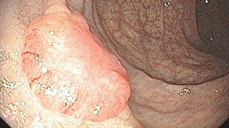 Fig. 1
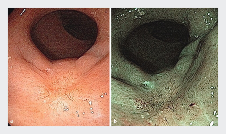 Fig. 2
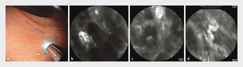 Fig. 3
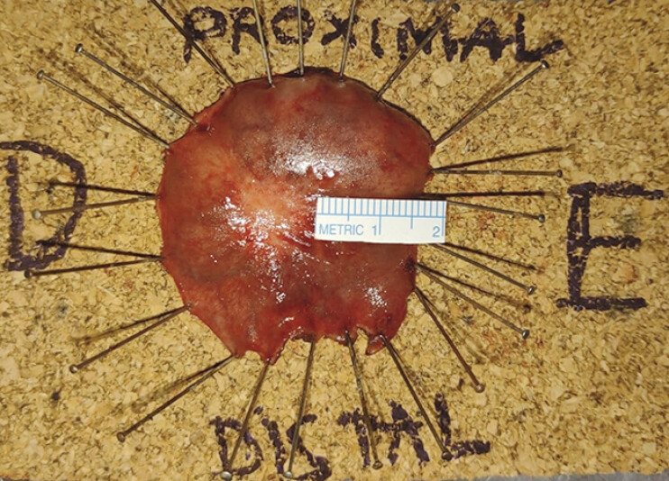 Fig. 4
Peer Reviews
No public reviews on file for this paper yet. If you reviewed it on a platform where reviews are public (OpenReview, ICLR, NeurIPS, ICML), you can paste yours below so the community can read it here.
Videos
No videos yet. Explain this paper in a talk, walkthrough, or lecture? Add one.
Taxonomy
TopicsColorectal Cancer Surgical Treatments · Gastric Cancer Management and Outcomes · Colorectal Cancer Screening and Detection
A 73-year-old patient underwent a screening colonoscopy, which revealed an infiltrative lesion in the distal rectum. Histology confirmed the diagnosis of moderately differentiated infiltrative adenocarcinoma ( Fig. 1 ).
Infiltrative lesion in the distal rectum confirmed it as moderately differentiated infiltrative adenocarcinoma.
Locally advanced adenocarcinomas of the mid and distal rectum are best managed with neoadjuvant chemoradiotherapy, followed by surgery 1 . However, patients who achieve a complete clinical response may undergo a ‘watch-and-wait’ strategy for organ preservation, with strict follow-up to allow early detection in the event of tumor regrowth 2 3 .
The patient underwent neoadjuvant chemoradiotherapy, and 10 weeks after treatment, restaging exams were performed. Rectoscopy revealed a scar, and chromoscopy with narrowband imaging (NBI) demonstrated only increased vessels ( Fig. 2 ). Magnetic resonance imaging (MRI) revealed no residual lesion, suggestive of complete response.
Scar identified on rectoscopy after neoadjuvant chemoradiotherapy treatment evaluated using a white-light imaging combined with b narrow-band imaging.
As part of the investigation, the patient underwent probe-based confocal laser endomicroscopy (pCLE), which is a real-time , in vivo method allowing a 1.000 times magnification, providing cellular and microvascular examination 4 , as demonstrated in Video 1 . It has been suggested that pCLE might be used during a watch-and-wait strategy for rectal neoplasia, avoiding immediate surgical treatment 5 .
Confocal endomicroscopy: a useful complementary method for diagnosing residual lesions following neoadjuvant chemoradiotherapy for advanced rectal adenocarcinoma.Video 1
pCLE revealed epithelial and vascular abnormalities indicative of a residual neoplastic lesion ( Fig. 3 ), and targeted biopsies confirmed the presence of adenocarcinoma. The patient underwent minimally invasive treatment by transanal endoscopic operation (TEO) ( Fig. 4 ), which provided a curative resection.
Epithelial and vascular abnormalities suggestive of a residual neoplastic lesion were identified using pCLE. a Probe positioned over the scar. b Irregular crypts along with thick and dark epithelium. c Back-to-back glands. d Dilated and tortuous vessels. Abbreviation: pCLE, probe-based confocal laser endomicroscopy.
Specimen obtained through TEO confirmed a curative resection. Abbreviation: TEO, transanal endoscopic operation.
This case illustrates that pCLE could be a useful complementary method for diagnosing residual lesions following neoadjuvant chemoradiotherapy for advanced rectal adenocarcinoma.
Endoscopy_UCTN_Code_CCL_1AD_2AB
The reference list from the paper itself. Each links out to its DOI / PubMed record.
- 1Habr-Gama A Perez RO Nadalin W Long-term results of preoperative chemoradiation for distal rectal cancer correlation between final stage and survival J Gastrointest Surg 20059909915623449 10.1016/j.gassur.2004.10.010 · doi ↗ · pubmed ↗
- 2Cotti GC Pandini RV Braghiroli OFM Outcomes of Patients With Local Regrowth After Nonoperative Management of Rectal Cancer After Neoadjuvant Chemoradiotherapy Dis Colon Rectum 20226533333934775415 10.1097/DCR.0000000000002197 · doi ↗ · pubmed ↗
- 3Nahas SC Nahas CSR Cama GM Diagnostic performance of magnetic resonance to assess treatment response after neoadjuvant therapy in patients with locally advanced rectal cancer Abdom Radiol (NY)2019443632364030663025 10.1007/s 00261-019-01894-8 · doi ↗ · pubmed ↗
- 4Cannizzaro R Mongiat M Canzonieri V Endomicroscopy and cancer: a new approach to the visualization of neoangiogenesis Gastroenterol Res Pract 2012201253717010.1155/2012/537170 PMC 326361622287958 · doi ↗ · pubmed ↗
- 5Safatle-Ribeiro AV Ribeiro Jr Lata J The Role of Probe-Based Confocal Laser Endomicroscopy (p CLE) in the Diagnosis of Sustained Clinical Complete Response Under Watch-and-Wait Strategy After Neoadjuvant Chemoradiotherapy for Locally Advanced Rectal Adenocarcinoma: a Score Validation J Gastrointest Surg 20232735736810.1007/s 11605-023-05732-737291428 · doi ↗ · pubmed ↗