Acoustic Field Enabled Polymeric Nanoparticle Deposition onto Vessel Walls for Enhanced Drug Delivery
Jianlei Wu, Liang Zhao, Evan H. Dubrunfaut, Valerie A. Lallo, Siyu Chen, Qianhong Wu, Bo Li, Laura G. Bracaglia

TL;DR
This paper explores using sound waves to guide drug-carrying nanoparticles to specific areas of blood vessels, improving targeted drug delivery.
Contribution
The novel use of low-frequency acoustic fields to enhance nanoparticle deposition on vessel walls, independent of nanoparticle surface chemistry.
Findings
Acoustic fields increased nanoparticle fluorescence on vascular tissue by 1000-fold at a flow rate of 2 m/min.
NP deposition was independent of NP surface chemistry.
A 100-fold increase in fluorescence was observed in ex vivo human vessel walls with localized acoustic fields.
Abstract
Polymeric nanoparticles (NPs) are promising tools for transporting and localizing therapeutics with intravenous delivery. Targeting these vehicles to specific tissue sites is challenging. Here, we investigate the use of low-frequency acoustic fields to drive polymeric NPs from circulating blood onto blood vessel walls by using concepts of elastic material deformation. By varying the shear flow rate and duration of acoustic field exposure, we achieved a 1000-fold increase in NP fluorescence intensity on vascular tissue compared with no acoustic field at a flow rate of 2 m/min. Interestingly, we found that acoustic-field-enhanced NP deposition is independent of NP surface chemistry. We also showcase a 100-fold increase in the area of fluorescence detected following NP delivery to an intact, ex vivo human vessel wall when a localized acoustic field is applied. This work suggests that local…
Genes, proteins, chemicals, diseases, species, mutations and cell lines named across the full text — each resolved to its canonical identifier and authoritative record.
Click any figure to enlarge with its caption.
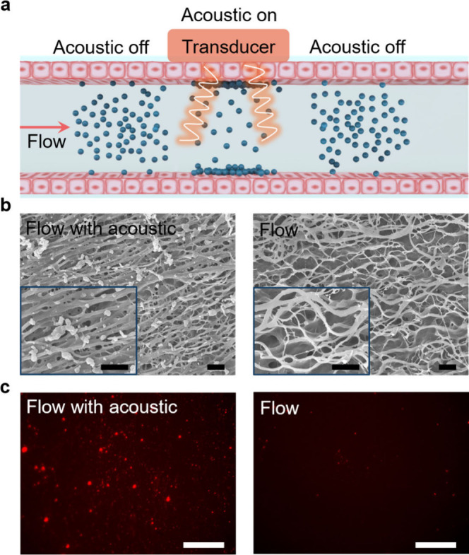 Figure 1
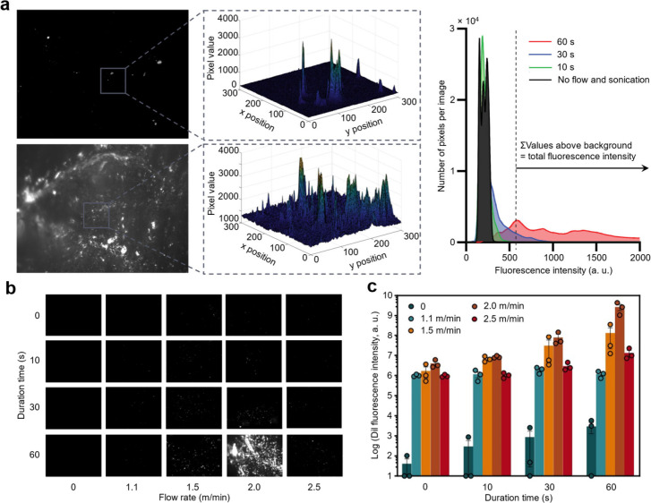 Figure 2
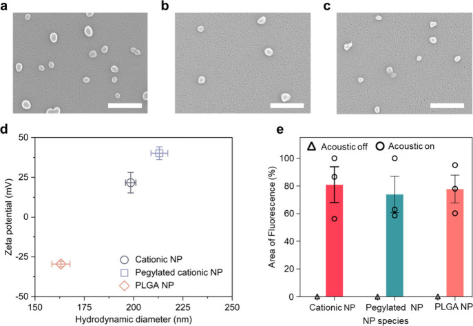 Figure 3
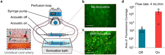 Figure 4
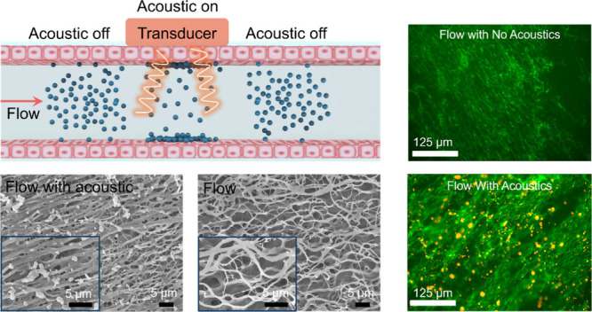 Figure 5
 Figure 6
- —National Heart, Lung, and Blood Institute10.13039/100000050
- —Directorate for Engineering10.13039/100000084
- —Villanova University10.13039/100007226
- —Pennsylvania Manufacturing Fellows InitiativeNA
Peer Reviews
No public reviews on file for this paper yet. If you reviewed it on a platform where reviews are public (OpenReview, ICLR, NeurIPS, ICML), you can paste yours below so the community can read it here.
Videos
No videos yet. Explain this paper in a talk, walkthrough, or lecture? Add one.
Taxonomy
TopicsNanoparticle-Based Drug Delivery · Advanced Drug Delivery Systems · Electrospun Nanofibers in Biomedical Applications
Polymeric nanoparticles (NPs) used for drug delivery are advantageous in comparison to other vectors such as lipids or biological macromolecules due to their material stability; they can protect cargo from degradation in circulation or by metabolism, which can ensure that the active pharmaceutical ingredient remains effective over an extended period. Additionally, polymeric drug delivery vehicles can offer controlled and sustained release of therapeutics, thereby reducing the frequency of medical administration to improve patient compliance.? Due to these strengths and tunable attributes, polymeric drug delivery vehicles are in rapid development and have shown great potential in applications such as cancer therapy, ?−? ? vaccinations,? gene therapy, ?−? ? antimicrobial therapy, ?−? ? cardiovascular disease therapy,? and theranostics.?
Therapeutic outcomes in these applications would be enhanced with more controlled biodistribution and reduced off-target accumulation of vehicles delivered intravenously. There are a few factors that can be manipulated to control this. Most simply, biodistribution of polymeric NPs can be influenced by their size ?−? ? and surface chemistry. For example, pegylation of polymeric NPs reduces NP aggregation and can prolong circulation time in the body. ?,? Alternatively, targeting ligands can be grafted onto polymeric NPs to direct NP binding to cellular receptors which may be tissue- or disease-specific, improving control over biodistribution at a cellular level. ?,? This strategy may not be effective for every purpose since it requires a unique and abundant cell surface molecule to produce specific targeting effects. These strategies are still plagued with unwanted NP accumulation in healthy tissues due to clearance by the RES system or nonspecific binding, ?,? reducing the effective dose of NPs.
A strategy that localizes NPs after intravenous administration without relying on biological markers and molecular targeting would be useful. Here, we propose using ultrasound to drive polymer NPs from circulating blood onto endothelial cells lining the vessel wall in a localized manner. From our previous work, we found that low-frequency (i.e., 40 kHz) acoustic fields increase the kinetic energy of NPs (e.g., carbon black and SiO_2_) in water and drive NPs to collide with soft polydimethylsiloxane (PDMS). ?−? ? The viscoelastic PDMS experiences a deformation–recovery process after a collision, capable of dissipating the kinetic energy of NPs, thereby resulting in NP deposition. Inspired by this phenomenon, we propose using a localized acoustic field to drive polymeric NP deposition from circulating blood onto targeted vessel walls. Low-frequency acoustic fields (20–100 kHz), compared to therapeutic ultrasound (1–3 MHz), are particularly effective for NP movement? due to an induced higher cavitation and violent bubble collapse in solution, which generates shock waves and microjets that can kinetically energize NPs.? Low-frequency acoustic fields are observed to be strong enough to drive diverse NPs, including polymers, metal oxides, and even heavy metals. ?−? ? Importantly, more localized particle delivery can be achieved with low frequencies than with homogeneous transport at higher frequencies.? It should be noted that as a physically noninvasive technique, acoustic technology in conjunction with biomaterials has been safely used in biomedical and clinical treatments. ?,? Our work, however, deviates from the typical applications in which an acoustic field is used either to stimulate a spatiotemporal drug release from NPs by destabilizing the membrane or shell of NPs or to increase permeability in barrier cells (i.e., sonoporation and sonopermeation) to improve transport. ?,?
A schematic of low-frequency acoustic-field-induced polymeric NP deposition onto tissues from blood circulation is shown in Figurea. We hypothesized that the acoustic field induces polymeric NP deposition more than circulation or flow without an acoustic field. As a demonstration, we utilized a section of bovine aorta (histology image, Figure S1a) as the target tissue source and continuously dipped it inside an aqueous suspension of fluorescently labeled cationic NPs with an average speed of 2.0 m/min to simulate blood flow through the blood vessel. The cationic polymer NPs were formulated from poly(amine-co-ester), a specialized polymer formulation for nucleic acid transport into cells. ?,? The dipping setup is shown in Figure S2. It should be noted that this dipping process? is different from the conventional dip-coating process (i.e., dip in and out of solution). The bovine aorta was always immersed in the solution, thereby excluding evaporation-driven deposition.? During the dipping process, an acoustic field (40 kHz, 60 W) was applied to the solution. After 60 s, we observed uniform NP deposition on the endothelial side of the bovine aorta (Figuresb and ?c, left images). Conversely, only trace amounts of NPs were deposited onto the tissue without an acoustic field (Figuresb and ?c, right images). At least five images were taken from multiple locations of each sample to ensure data repeatability. Full surface coverage of the tissue by NPs was not achieved with the acoustic field, as compared to previously studied soft polymers, due to the heterogeneity of tissue surface.?
Considering that flow rates in body circulation vary by vessel diameter, we investigated the impact of the duration of acoustic exposure combined with different flow rates on cationic NP deposition onto the surface of bovine aorta tissue. NP deposition was observed and quantified through fluorescent images after varied flow rates (representing dipping speeds) and duration times of acoustic exposure (Figuresb and ?c). Area of fluorescence detected as well as total NP fluorescence intensity above background fluorescence (schematic of data processing shown in Figurea, right) were used to compare NP deposition between groups. In these fluorescent images, black regions represent the aorta surface, while white spots indicate cationic NP locations. Both fluorescent imaging and quantification of NP deposition reveal that cationic NPs barely deposit onto the bovine aorta surface in a static deposition (i.e., only acoustic exposure). Furthermore, cationic NPs can be deposited in small amounts onto the bovine aorta when there is no acoustic field applied (i.e., only flow). When the flow rate is constant, more cationic NPs deposit with an increasing duration of acoustic exposure. When the duration of acoustic exposure is held constant, more cationic NPs deposit with an increase in flow rate from 1.1 to 2.0 m/min. When the flow rate further increases to 2.5 m/min, the total NP deposition is reduced. This observation indicates that higher flow rates may introduce too much shear force, making the NPs favor lateral transport. However, some lateral movement of particles is necessary to replenish the NP concentration in close proximity to the vessel walls such that a collision is likely. Overall, a flow rate of 2.0 m/min with 60 s of acoustic duration shows the highest NP fluorescence intensity on the surface of bovine aorta, which means maximum NP deposition. While acoustic field exposure alone limited NP deposition, it significantly enhanced NP deposition upon combination with an appropriate flow rate. We attribute this enhancement of NP deposition to increased collision probability between nanomaterials and the polymer substrate, and colliding with more energy to deform the membrane and result in deposition.
As described, NP deposition is quantified using a sum of fluorescence intensity from a fixed area of tissue and the background corrected from tissue without NPs. This is an indirect method to quantify NP deposition and does not necessarily correlate with the physical space consumed by NPs. To relate area of fluorescence with the physical area of NPs, we have also quantified representative SEM images, which display the surface (Figures S3c–S3f) and a cross section of the aortic tissue (Figure S4). This imaging method also shows enhanced NP deposition with the addition of ultrasound.
In addition to cationic NPs, we chose two more polymer NP types, pegylated NPs and poly(lactic-co-glycolic) acid (PLGA) NPs, for acoustic field enabled deposition. As seen in Figuresa–?c, they all feature a quasispherical shape that is similar to the cationic NPs. However, these NPs have different surface charges and sizes compared to cationic NPs (Figured), which are parameters that may affect the deposition. We have reported in previous work the correlation between NP size and aggregation, which would hinder deposition.? The average hydrodynamic diameters reported are popular sizes found in polymeric drug delivery vehicles, so testing this range for polymeric vehicles is important.
Keeping the same flow rate (i.e., 2.0 m/min) and acoustic field exposure time (i.e., 60 s), which resulted in maximum deposition, we evaluated the NP coverage on bovine aorta for three NP species (Figuree). Due to the difference of DiI fluorescence intensity among the three types of NPs, the area of fluorescence from NPs was measured as opposed to total fluorescence intensity. The area of fluorescent signal is nearly zero for each type of NP at a flow rate of 2.0 m/min with acoustic off for 60 s, which indicates minimal NP attachment onto the bovine aorta. With acoustic on for 60 s, the average NP fluorescence area for cationic NPs, pegylated NPs, and PLGA NPs reached 80.93%, 73.91%, and 78.02%, respectively (Figuree). Additional SEM images of these samples are shown in Figure S5).
Although the average fluorscence coverage varies slightly for three species of NPs, they are all over 70%. These results demonstrate that acoustic field enabled deposition is not necessarily affected by NP surface charges and size, making this method universally applicable for drug delivery.
To further validate this method for use in vivo, an artery isolated from a human umbilical cord was used in an isolated vessel perfusion system to mimic vascular flow within the human body.? Histological images of the artery, obtained from the umbilical cord, show the endothelial surface as well as the tunica media and tunica externa layers intact (Figure S1b), which together give the vessel wall its viscoelastic properties. Cationic, fluorescently labeled NPs were administered in parallel circuits to two isolated vessel segments under 4.0 mL/min flow. This volumetric flow rate was calculated from the linear velocity which led to maximum NP deposition in previous experiments using the tubing diameter of 1.6 mm. One vessel was exposed to the acoustic field for 60 s, and the other was not exposed to acoustics (Figurea). Both fluorescence imaging (Figuresb and ?c) and quantification of NP fluorescent intensity (Figured) show significant NP attachment onto the artery after 60 s acoustic duration with flow at 4.0 mL/min, while NPs barely deposit onto the artery wall under static conditions. This specific flow rate (4 mL/min) is a good approximation of blood flow at times in smaller arteries and capillaries near the external surface of the body (i.e., temporal artery,? finger,? and dorsalis pedis artery?).
Given this result, we are optimistic that this technique will allow for local control over NP deposition after intravenous administration in a larger and more complex circuit, such as in an animal model. Beyond that, we envision this technique would benefit NP-based drug delivery for the treatment of regionally isolated pathologies, such as diabetic foot ulcers, peripheral nerve damage, and perhaps organ specific tumors. Acoustics can be applied extracorporeally in a specific region for desired accumulation, which would cause NPs to leave the circulation and deposit to the target location. While acoustic fields can help increase NP deposition onto tissue surfaces, it is still crucial to ensure that more NP deposition can enhance drug delivery, which relies on NP endocytosis into the endothelial cells. In our previous works, and shown in Figure S6, we expect that more NPs adhering to the outside of endothelial cells leads to more NPs endocytosed.? Transport of NPs into cells may even be enhanced by membrane destabilization caused by the ultrasound, as is seen in other applications, but destabilization from a low-frequency acoustic field may not persist as long as endocytosis occurs. We have examined cell damage under acoustic field at the testing conditions using both human embryonic kidney (HEK) 293 cells, which are commonly used as a comparable subject for safety studies (shown in Figure S7), and human umbilical vein endothelial cells (HUVECs), which are primary human vascular cells (Figure S8). Looking immediately after acoustic exposure, we see some damaged cell membranes in HEK293 cells, as evidenced by increased annexin V staining (Figure S7D). This damage is reversed after 24 h. In longer studies in HUVECs, we see very limited cell death or membrane instability that can be tied to the ultrasound treatment. Treated HUVECs continue to thrive in culture out to 72 h (Figure S8). Finally, we note that endothelial cells lining the umbilical arteries exposed to ultrasound appear undamaged, with intact cell membranes (visualized with CD31 immunohistochemical stain), few visible gaps in cell–cell junctions, and few interruptions in the monolayer.
In future work, it will be important to address whether the acoustic field can reach deep into tissues in large models of flow and if there is a depth limit to the arteries that can be targeted. We found a threshold flow rate of 2.0 m/min; thus, this technique may be better for small vessels than vessels with a high flow rate. Future testing could investigate tuning acoustic parameters to optimize NP deposition from blood to the vessel wall.
This acoustic method is an effective and straightforward physical technique to drive the deposition of polymer NPs onto a biointerface and can be applied to multiple polymer NP systems. Specific flow rate with acoustic duration time can be utilized for increased deposition of bulk NPs onto targeted vessel walls, which will further improve endothelial cellular uptake of the NP to enhance the efficacy of drug delivery and medical treatment, and causes little safety concern.
Supplementary Material
The reference list from the paper itself. Each links out to its DOI / PubMed record.
- 1Borandeh S.van Bochove B.Teotia A.SeppäläJ.Polymeric drug delivery systems by additive manufacturing Adv. Drug Deliver. Rev.202117334937310.1016/j.addr.2021.03.02233831477 · doi ↗ · pubmed ↗
- 2Pandey S. K.Patel D. K.Maurya A. K.Thakur R.Mishra D. P.Vinayak M.Haldar C.Maiti P.Controlled release of drug and better bioavailability using poly (lactic acid-co-glycolic acid) nanoparticles Int. J. Biological Macromol.2016899911010.1016/j.ijbiomac.2016.04.06527112980 · doi ↗ · pubmed ↗
- 3Wang J.Li N.Cao L.Gao C.Zhang Y.Shuai Q.Xie J.Luo K.Yang J.Gu Z.DOX-loaded peptide dendritic copolymer nanoparticles for combating multidrug resistance by regulating the lysosomal pathway of apoptosis in breast cancer cells J. Mater. Chem. B 2020861157117010.1039/C 9TB 02130 B 31951231 · doi ↗ · pubmed ↗
- 4Mu C.-F.Cui F.Yin Y.-M.Cho H.-J.Kim D.-D.Docetaxel-loaded chitosan-cholesterol conjugate-based self-assembled nanoparticles for overcoming multidrug resistance in cancer cells Pharmaceutics 202012978310.3390/pharmaceutics 1209078332825000 PMC 7558660 · doi ↗ · pubmed ↗
- 5Chan J. M.Valencia P. M.Zhang L.Langer R.Farokhzad O. C.Polymeric nanoparticles for drug delivery Methods Mol. Biol.201062416317510.1007/978-1-60761-609-2_1120217595 · doi ↗ · pubmed ↗
- 6Karlsson J.Rui Y.Kozielski K. L.Placone A. L.Choi O.Tzeng S. Y.Kim J.Keyes J. J.Bogorad M. I.Gabrielson K.Engineered nanoparticles for systemic si RNA delivery to malignant brain tumours Nanoscale 20191142200452005710.1039/C 9NR 04795 F 31612183 PMC 6924015 · doi ↗ · pubmed ↗
- 7Chen M.Chen M.He J.Cancer cell membrane cloaking nanoparticles for targeted co-delivery of doxorubicin and PD-L 1 si RNA Artificial cells, nanomedicine, and biotechnology 20194711635164110.1080/21691401.2019.160821931027450 · doi ↗ · pubmed ↗
- 8Kroll A. V.Fang R. H.Jiang Y.Zhou J.Wei X.Yu C. L.Gao J.Luk B. T.Dehaini D.Gao W.Nanoparticulate delivery of cancer cell membrane elicits multiantigenic antitumor immunity Adv. Mater.20172947170396910.1002/adma.201703969 PMC 579434029239517 · doi ↗ · pubmed ↗