Discovery of Natural Phosphodiesterase 5 Inhibitors from Dalbergia cochinchinensis Pierre Leaves Using LC-QTOF-MS2
Ruttanaporn Chantakul, Corine Girard, François Senejoux, Kornkanok Ingkaninan, Nitra Nuengchamnong, Prapapan Temkitthawon

TL;DR
This study identifies natural PDE5 inhibitors in the leaves of Dalbergia cochinchinensis, which could help treat erectile dysfunction.
Contribution
This is the first study to show that D. cochinchinensis leaf extract has potent PDE5-inhibitory activity.
Findings
Leaf extract of D. cochinchinensis has the lowest IC50 value for PDE5 inhibition among five plant parts.
LC-QTOF-MS2 was used to identify potential PDE5-inhibiting compounds in the leaf extract.
Abstract
The imbalance of phosphodiesterase 5 (PDE5) enzyme in the male body, or excessive PDE5 enzyme levels, can occur due to factors such as aging, diseases (e.g., cardiovascular disease, diabetes, depressive disorder), and physical behaviors (e.g., alcoholism, smoking, stress). PDE5 is directly associated with erectile dysfunction disease. Currently, many studies aim to find natural PDE5 inhibitors as an alternative to commercial drugs. This study is the first to demonstrate that the ethanolic leaf extract of D. cochinchinensis exhibits potent PDE5-inhibitory activity. The PDE5-inhibitory activity of five plant parts was evaluated: leaf (IC50 = 1.53 ± 0.12 µg/mL), twig (3.37 ± 0.54), fruit (14.92 ± 2.85), heartwood (19.05 ± 5.60), and bark (16.03 ± 2.92). However, there is still uncertainty about which compounds in leaf extract are responsible for the PDE5 inhibition. Therefore, the purpose…
Genes, proteins, chemicals, diseases, species, mutations and cell lines named across the full text — each resolved to its canonical identifier and authoritative record.
Click any figure to enlarge with its caption.
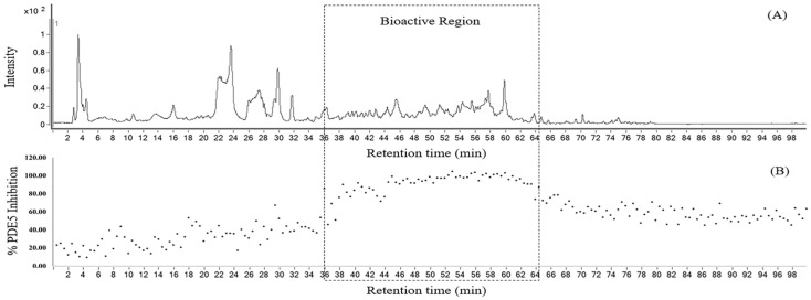 Figure 1
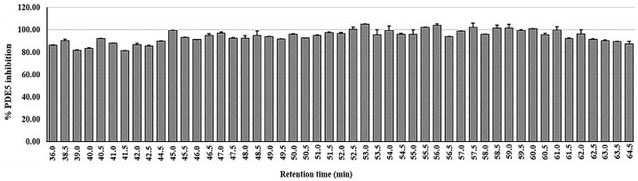 Figure 2
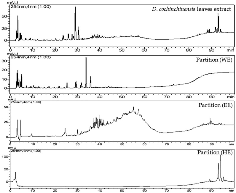 Figure 3
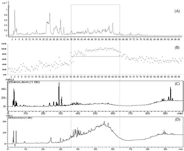 Figure 4
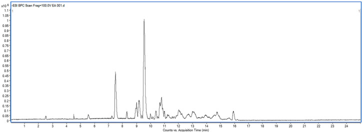 Figure 5
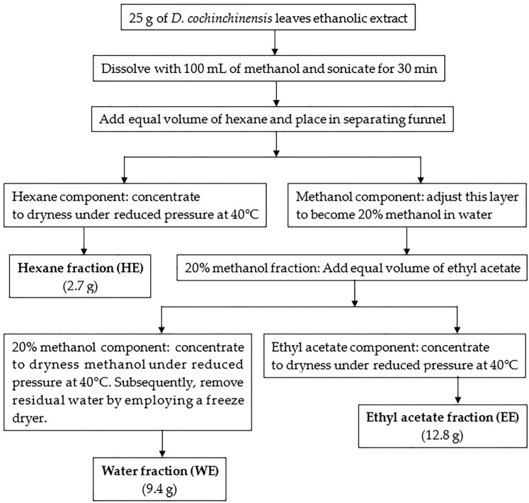 Figure 6
- —National Research Council of Thailand
- —KKN supply and services Co., Ltd.
- —rogram Management Unit for Human Resources & Institutional Development, Research and Innovation
- —Center of Excellence for Innovation in Chemistry (PERCH-CIC), Global and Frontier Research University Fund, Naresuan University
Peer Reviews
No public reviews on file for this paper yet. If you reviewed it on a platform where reviews are public (OpenReview, ICLR, NeurIPS, ICML), you can paste yours below so the community can read it here.
Videos
No videos yet. Explain this paper in a talk, walkthrough, or lecture? Add one.
Taxonomy
TopicsSexual function and dysfunction studies · Phosphodiesterase function and regulation · Pharmacology and Obesity Treatment
1. Introduction
Erectile dysfunction is a prevalent male sexual disorder characterized by the inability to achieve or maintain an erection sufficient for satisfactory sexual performance [1,2]. The risk factors contributing to ED include aging, lifestyle choices, medications, chronic diseases, pelvic injuries, and abnormalities in endocrine and cardiovascular systems [3]. ED involves cyclic contraction and the relaxation of corpus cavernosum smooth muscle, which is regulated by phosphodiesterase 5 (PDE5)—mediated cGMP hydrolysis [1,4,5,6]. PDE5 is a cyclic guanosine monophosphate (cGMP)-specific enzyme predominantly found in the lungs, smooth muscles, platelets, and corpus cavernosum [4,5]. It hydrolyzes cGMP to its inactive form, guanosine monophosphate (GMP), leading to decreased cGMP levels and vascular smooth muscle contraction. Conversely, inhibiting PDE5 promotes smooth muscle relaxation, which is essential for penile erection [7]. Although FDA-approved PDE5 inhibitors (e.g., sildenafil, tadalafil) effectively restore erection, adverse effects (e.g., headache, flushing) and contraindications (e.g., nitrate co-administration) limit their use [8,9,10].
Research interest in natural products has been steadily growing as scientists aim to discover safer and more affordable PDE5 inhibitors [11]. Flavonoids in medicinal plants have been reported as potential sources of PDE5 inhibitors [12,13,14]. In addition, curcuminoids [15], xanthones [16], phenanthrenes [17], and alkaloids [18] have been reported to exhibit PDE5 inhibitory activity. Several species of the Dalbergia genus are traditionally used in various medicinal systems and have been reported to exhibit a wide range of bioactivities, such as anti-inflammatory, osteogenic, antioxidant, anti-androgenic, antibacterial, anti-allergic, antidiarrheal, antifungal, anti-tumor, and blood stasis [19,20]. Phytoconstituents isolated from heartwood, including stem and seeds of D. cochinchinensis, have been reported to include flavonoids (flavones, isoflavones, flavans, isoflavans, flavanones, chalcones, neoflavonoids, and pterocarpans) and simple phenols as major compounds, along with terpenoids (sesquiterpenoids and triterpenoids), quinones, benzofurans, benzophenols, phytosterols, stilbenes, xanthones, and lignans [21,22,23,24]. The D. cochinchinensis seed is rich in glycosidase enzymes, with both β-glucosidase and β-fucosidase activities [25]. From reviews, chemical compounds in the leaf have been reported in only four species of Dalbergia, namely D. sissoo, where flavonoids (including flavones and isoflavones), triterpenoids, and phytosterols have been identified [26,27,28,29], D. stipulacea, which has been found to contain luteolin, D. spruceana, which has been reported to contain luteolin 4′-rutinoside, and D. spinosa, which has been noted to contain prunasin [30,31]. D. cochinchinensis has demonstrated its various bioactivities, including antioxidant [19], antimicrobial effects against Staphylococcus aureus and Aspergillus niger, cytotoxicity against lung and pericardial cancers [32], 5α-reductase inhibition [33], and anti-inflammation effects [34].
Using a chemotaxonomic approach to guide the selection of plants for PDE5 inhibition screening, D. cochinchinensis was chosen based on reports of its chemical composition, particularly its flavonoid content—compounds previously reported to exhibit PDE5 inhibitory activity. Additionally, ethnopharmacological records indicate the traditional use of D. cochinchinensis, especially its heartwood, for treating tumors and blood stasis [20]. Furthermore, based on our literature review, no previous studies have reported the PDE5 inhibitory activity of this plant. These findings collectively supported the selection of this species for further investigation. To the best of our knowledge, no previous studies have reported the PDE5 inhibitory activity of D. cochinchinensis. This study is the first to demonstrate such activity, providing new insights into the pharmacological potential of this species. In preliminary screening, we found that the hydro-ethanolic leaf extract inhibited PDE5 most potently, motivating detailed LC-QTOF-MS^2^ profiling.
2. Results
2.1. PDE5 Inhibitory Activity of Various Parts of D. cochinchinensis
In our preliminary study, five parts of D. cochinchinensis (leaf, twig, fruit, heartwood, and bark) were extracted with 95% ethanol and screened for PDE5 inhibitory activity. Table 1 shows that all extracts exhibited over 70% inhibition at a concentration of 25 µg/mL. Extracts exceeding 70% inhibition were considered effective and selected for IC_50_ determination. Among them, the leaf extract demonstrated the highest potency and was chosen for further identification of PDE5 inhibitors.
2.2. At-Line Screening and Identification of PDE5 Inhibitors in D. cochinchinensis Leaf Ethanolic Extract
The leaf ethanolic extract underwent rapid screening to identify the chemical compounds in the leaf of D. cochinchinensis using at-line LC-QTOF-MS^2^. Figure 1 displays the extracted ion chromatogram of the leaf extract in panel A, and the corresponding bioactivity chromatogram representing the PDE5 inhibitory activity of collected micro-fractions is displayed in panel B. The zoom-in of this chromatogram highlights the bioactive region, between 36 and 64 min of retention time. In this area, the micro-fractions demonstrated PDE5 inhibition of 80% or greater, as shown in Figure 2.
2.3. Identification of Chemical Compounds in the Leaf of D. cochinchinensis Using LC-QTOF-MS2
For the tentative identification of constituents of the leaf extract, the micro-fractions were analyzed using m/z fragmentation and MassHunter Data Acquisition Software Version B.05.01 and MassHunter Qualatative Analysis Software B 06.0 respectively (Agilent Technologies, Santa Clara, CA, USA). The chemical compounds in the leaf extract were identified based on m/z fragmentation and are listed in Table 2. From previous studies, a summary of compounds isolated from the leaves of various Dalbergia species is shown in Table 3.
2.4. Liquid–Liquid Partition of D. cochinchinensis Leaf Extract
The study initially conducted at-line screening to assess both the biological activity and chemical composition in the leaf extract of D. cochinchinensis. The results revealed the chemical composition of the leaf extract and identified a group of substances expected to be the PDE5 inhibitor. Liquid–liquid partitioning was used to separate groups of compounds in the leaf extract based on the polarity of the solvents used. D. cochinchinensis leaf extract was fractionated using the partition method to segregate it into groups based on the polarity of the solvent. For sequential partitioning, 25 g of the extract were utilized. Liquid–liquid partition produced an extract of hexane (HE), ethyl acetate (EE), and water (WE) at 2.7 g, 12.8 g, and 9.4 g, respectively. The process of partition and the dry weight of samples obtained are stated in Scheme 1. Subsequently, these extracts were analyzed using HPLC, with the system conditions set to match those used for the at-line technique. The HPLC chromatograms of each extract are shown in Figure 3.
HPLC chromatograms of the partitioned extracts were compared with the crude extract. The chromatograms show that the extracts obtained from partitioning with different solvents clearly separate into distinct groups, including highly polar compounds (represented by the water extract, WE), moderately polar compounds (represented by the ethyl acetate extract, EE), and non-polar compounds (represented by the hexane extract, HE). All three partition extracts were tested for PDE5 inhibitory activity in triplicate at the final concentration of 5 μg/mL. The results revealed that the EE extract (95.69 ± 2.97%) demonstrated the highest efficacy in inhibiting PDE5, followed by the HE (65.57 ± 3.13%) and WE extracts (39.61 ± 2.75%), respectively. Upon examining the HPLC chromatogram of the EE extract, it was observed that it corresponds to the bioactive region identified in the at-line analysis, which contains compounds with more than 80% efficacy in inhibiting PDE5 (Figure 4). The study results confirm that the PDE5 inhibitors are indeed located in the bioactive region, and this also supports the validity of the at-line analysis technique.
2.5. Identification of Chemical Compounds of Ethyl Acetate Partition (EE) Using LC-QTOF-MS2
The results indicate that the EE extract contains compounds with potential PDE5 inhibitory activity. Therefore, the EE extract was analyzed using LC-MS/MS to identify the tentative compounds based on m/z fragmentation patterns. The extracted ion chromatogram from at-line LC-QTOF-MS of EE partition (negative mode) is shown in Figure 5. The tentative identifications based on m/z fragmentation of the EE partition from D. cochinchinensis are listed in Table 4.
3. Discussion
Dalbergia genus includes 274 accepted species worldwide, with 26 native species found in Thailand [32]. D. cochinchinensis, traditionally known as Thai Rosewood or Siamese Rosewood, was described in 1898. It belongs to the family of Fabaceae and is widely distributed across Southeast Asia, including Vietnam, Thailand, Laos, and Cambodia. D. cochinchinensis is a large tree known for its durable heartwood, which is resistant to termites. This high economic value makes it sought after for various commercial purposes, including seeds, living trees, heartwood, handicrafts, and furniture [36].
The screening of PDE5 inhibitory activity in D. cochinchinensis revealed that the leaf extract exhibited the lowest IC_50_ values, indicating the highest efficacy, followed by the twig, fruit, bark, and heartwood, respectively. This study demonstrates, for the first time, that the leaf of D. cochinchinensis possesses the most potent PDE5 inhibitory activity. Therefore, it was of interest to further investigate the active compounds in the leaf, as no reports have yet identified chemical compounds from D. cochinchinensis acting as PDE5 inhibitors.
The at-line LC-QTOF-MS^2^ technique was used for rapid screening of bioactivity coupled with mass fragmentation. The use of at-line analysis allowed for the identification of potential PDE5 inhibitory zones in the chromatogram of the leaf extract. However, it was not possible to isolate pure compounds from the collected micro-fractions, with the amount of the fractions remaining after evaporation being insufficient for further study. Thus, the use of at-line techniques serves as a rapid screening method, providing both biological activity assessment and predictive analysis of potential PDE5 inhibitors based on m/z data. This study presents a faster approach for lead identification and the dereplication of known compounds compared to conventional fractionation techniques. However, despite the efficiency of at-line analysis, there is a concern that compounds present in low natural abundance may be overlooked.
This study found that the leaf predominantly contains flavonoids, a class of compounds known for their significant biological properties. Additionally, other substances have also been found, including sugar group, phenyl propanoic acid, quinic acid, benzodioxoles, sesquiterpene, xanthone, hydroxycinnamic acid, proanthocyanidin, quinoline alkaloid, stilbene, chalcone, and lipid. The compounds found in the bioactive region include flavonoids such as kaempferol and quercetin glycosides, as well as other substances like guibourtinidol-(4α→6)-catechin, niaziminin, and liquiritigenin. Previous reports have isolated flavonoid glucosides, including kaempferol-3-O-β-D-glucopyranoside, kaempferol-3-O-rutinoside, quercetin-3-β-D-glucopyranoside, and quercetin-3-O-rutinoside from the leaf of D. sissoo, as well as liquiritigenin from the heartwood of D. cochinchinensis [21,37]. Literature reviews indicate that the compounds isolated from the leaf are predominantly flavonoids (including flavanones, flavones, and isoflavones), along with other compounds such as triterpenoids, phytosterols, and lignans [19,24]. The results from the at-line technique have revealed the presence of several of these compounds in the leaf of D. cochinchinensis. However, these data are preliminary, based on mass spectrometry, and provide an initial analysis of the compound groups. The results of our experiment can be used to hypothesize potential PDE5 inhibitory compounds (in the bioactive region) and serve as a basis for further isolation of pure substances from the leaf extract.
The ethanolic leaf extract was further subjected to sequential partitioning to isolate the bioactive fraction. The partitioning process revealed that the bioactive fraction was obtained using the ethyl acetate (EE) partition. The compounds predominantly found in the EE partition include flavonol glycosides and procyanidins. In addition, other substances were also found, such as polyphenol, O-glycosyl, phenolic acid, monocarboxylic acid, phenylpropanoid, phenolic aldehyde, procyanidin, cyanogenic glycoside, and alkaloid. The partitioning process enabled the separation of compound groups, allowing us to identify those with PDE5 inhibitory activity. This helped specify the active groups more precisely and guided the selection of fractions for further isolation in a more targeted manner. This study has led to the discovery of candidate compounds for PDE5 inhibitors in D. cochinchinensis, which is being reported for the first time. Especially, the flavonol glycosides include kaempferol and quercetin glycosides, along with procyanidins, which consist of catechin-afzelechin, afzelechin dimers and trimers, and afzelechin-guibourtinidol. These compounds may have promise in becoming a PDE5 inhibitor. This research provides information about compounds in the leaves of D. cochinchinensis that have the potential to act as PDE5 inhibitors. Nevertheless, further studies are needed to identify the active ingredients and to complete the research in the future.
4. Materials and Methods
4.1. Chemicals and Materials
4.1.1. Plant Material
D. cochinchinensis was sourced from the Phra Mae Ya Sukhothai Botanical Garden in Sukhothai Province, Thailand. The voucher specimens (No. 004549) were authenticated by Assistant Professor Dr. Pranee Nangngam, Department of Biology, Faculty of Sciences, Naresuan University, Phitsanulok Province, Thailand.
4.1.2. Solvents
The following chemicals were purchased from RCI Labscan (Bangkok, Thailand): 95% ethanol, ethyl acetate, hexane, acetonitrile, water, dimethyl sulfoxide (DMSO), 98% formic acid (analytical grade), acetonitrile (HPLC grade), and methanol (LC-MS grade).
4.1.3. Reagents
Bovine serum albumin (BSA), crude snake venom (Crotalus atrox), dulbecco’s modified eagle medium (DMEM), dithiothreitol (DTT), ethylenediamine tetraacetic acid (EDTA), ethylene glycol tetraacetic acid (EGTA), fetal bovine serum (FBS), imidazole, magnesium chloride (MgCl_2_), phenylmethylsulfonyl fluoride (PMSF), QAE Sephadex™ A-25, and tris (hydroxymethyl) aminomethane (Tris) were purchased from Sigma–Aldrich (St. Louis, MO, USA). Geneticin (G418) was purchased from Gibco by Life Technologies (Paisley, Scotland). [^3^H]-cGMP and Ultima Gold cocktail were obtained from PerkinElmer (Waltham, MA, USA).
4.1.4. Tools Used for Analysis
ESI mass spectra were determined using an Agilent-6540 UHD accurate mass LC-QTOF-MS equipped with an electrospray ionization (ESI) interface (Agilent Technologies, Singapore). HPLC analysis was performed using the Prominence HPLC chromatograph system (Shimadzu, Kyoto, Japan). The chromatograph system was comprised of a binary pump system (LC-20AT) coupled with a UV/VIS detector (SPD-20A), communicator (CBM-20A), and degasser (DGU-20A_3_). HPLC analysis was conducted on a Phenomenex Luna^®^ C18 column (150 mm × 4.6 mm, 5 μm) (Phenomenex, Torrance, CA, USA).
4.2. Plant Extraction
4.2.1. Crude Hydro-Ethanolic Extract
The fresh leaves of D. cochinchinensis were thoroughly cleaned and rinsed with water, then cut into small pieces and dried in a hot-air oven (Memmert UF 75, Schwabach, Germany) at 60 °C for 3 days. Subsequently, the dried leaves were ground into a rough powder using a grinder. The particle size of the powder was controlled by passing it through 100 mesh sieves (Retsch GmbH, Haan, Germany). The powdered leaves of D. cochinchinensis were macerated with 95% ethanol (EtOH) under shaking for 3 days at room temperature. Afterwards, the plant residue was separated by filtration through a filter paper (Whatman International Ltd., Chalfont St. Giles, UK). The filtrate was then concentrated by evaporating the ethanol using a BÜCHI Rotavapor R-124 (BÜCHI Labortechnik AG, Flawil, Switzerland). The crude extract was stored at −20 °C to maintain its integrity and stability until further analysis.
4.2.2. Liquid–Liquid Partition
The partitioning process was utilized to separate a group of compounds based on solvent polarity. The D. cochinchinensis leaf extract (25 g) was dissolved in 100 mL of methanol (MeOH) and sonicated for 30 min using an ultrasonic bath (Elmasonic S60H, Singen, Germany). It was then partitioned with hexane. The remaining MeOH fraction was adjusted with water to achieve a 20% MeOH solution, which was subsequently partitioned with ethyl acetate [8]. The resulting fractions were processed further: the hexane and EA fractions were concentrated to dryness at 40 °C using a rotary evaporator, while the 20% MeOH fraction was further dried using a freeze dryer (Thermo Electron Co., Ltd., Waltham, MA, USA) to remove the water. All extracts were stored in the dark at −20 °C until further analysis. Three fractions were obtained from the solvent partition: hexane (HE), 20% MeOH (WE), and ethyl acetate (EE).
4.3. Sample Analysis
4.3.1. High-Performance Liquid Chromatography (HPLC)
The chemical profile of D. cochinchinensis leaf extract was investigated by preparing a solution at a concentration of 10 mg/mL, dissolved in 50% methanol. This solution, as well as extracts obtained from liquid–liquid partitioning (also prepared at 10 mg/mL and dissolved in methanol), was filtered through nylon syringe filters with a pore size of 0.45 µm before analysis. For HPLC analysis, the system conditions were set to match those used for the LC-MS/MS analysis.
4.3.2. Liquid Chromatography Mass Spectrometry (LC-MS/MS)
For the at-line technique, the D. cochinchinensis leaf extract was prepared at a concentration of 20 mg/mL by dissolving it in 50% acetonitrile. The solution was then sonicated for 15 min and filtered through nylon syringe filters with a pore size of 0.45 µm.
For LC-MS/MS analysis of the ethyl acetate extract obtained from partitioning, the extract was prepared at a concentration of 10 mg/mL in 50% methanol. Chemical compounds were identified based on m/z data. Separations were performed using a Luna C18(2) column (4.6 mm × 150 mm, 5 μm particle size) from Phenomenex (Torrance, CA, USA), with the temperature maintained at 35 °C. The mobile phase consisted of 0.1% formic acid in water (A) and 0.1% formic acid in acetonitrile (B), with a gradient system increasing the proportion of solvent B from 5% to 95% over 25 min.
4.4. At-Line Technique
The at-line assay employing LC-MS/MS analysis utilized an Agilent 1260 Infinity Series HPLC system coupled to an Agilent 6540 UHD accurate mass QTOF-LC/MS, which was equipped with an electrospray interface (ESI). The leaf extract of D. cochinchinensis (20 μL of 20 mg/mL to 400 μg) was analyzed using the at-line technique with modifications based on the protocols outlined by Bhandari et al. [38]. The D. cochinchinensis leaf extract was injected into the LC-MS/MS system, and micro-fractions were collected in a 96-well plate. This procedure was repeated three times. The separations were conducted using a Luna C18(2) column (4.6 mm × 150 mm, 5 μm particle size) from Phenomenex (Torrance, CA, USA), with the temperature held constant at 35 °C. The mobile phase consisted of a mixture of 0.1% formic acid in water (A) and 0.1% formic acid in acetonitrile (B). The gradient program began with 10% solvent B, which was increased to 15% over 20 min, followed by a gradual increase to 30% over 25 min, 50% over 15 min, and 70% over 20 min. Finally, solvent B was ramped up to 100% over a total duration of 100 min. A post-time of 5 min was allowed for system re-equilibration before the next injection. The injection volume was set at 20 μL, and the flow rate was maintained at 0.5 mL/min. The eluent was split into two flows in 1:9 ratios. The majority of the eluent was collected in a 96-well plate micro-fractionation system with 30 s per well, while the minor portion was directed to an ESI-QTOF-MS system. The micro-fractions were dried using a sample concentrator (Techne, Cambridge, UK) and kept at −20 °C until test day. The ion source was operated in negative mode with the following conditions: drying gas flow of 10 L/min, gas temperature set to 350 °C, nebulizer pressure at 30 psig, capillary voltage of 3500 V, skimmer voltage set to 65 V, octapole RFV at 750 V, and fragmentor voltage at 250 V. All acquisition data were analyzed using MassHunter software (Agilent Technologies, Santa Clara, CA, USA). The MS and MS/MS fragmentation patterns of PDE5 inhibitor active micro-fractions were compared using a library search, including MassHunter Metlin metabolite PCD/PCDL (Agilent Technologies), the Human Metabolomics Database (https://www.hmdb.ca), ChemSpider (https://www.chemspider.com), and MassBank (https://www.massbank.eu). Molecular formulas were generated using Agilent MassHunter Qualitative Analysis Software B 06.0.
4.5. PDE5 Inhibitory Activity
4.5.1. Sample Preparation for PDE5 Inhibitory Activity
The micro-fractions in the 96-well plate were dried using a sample concentrator. Afterward, the micro-fractions were dissolved in 25 μL of 5% DMSO for the assay, resulting in a final concentration of 1% DMSO.
4.5.2. Enzyme Preparation
The human PDE5 plasmid, received from Professor Dr. Joseph A. Beavo of the University of Washington, Seattle, WA, USA, was sub-cloned into a pcDNA3 vector with an ampicillin-resistant gene. These plasmids were then scaled up and purified using the Invitrogen™ PureLink™ HiPure Plasmid Maxiprep Kit (Thermo Fisher Scientific, Waltham, MA, USA)). HEK293 cells were transfected with human PDE5 plasmid using Lipofectamine^®^ 2000 reagent as per the protocol. After 2 days of transfection, the cells underwent treatment with a selective antibiotic (G418, 1 mg/mL) for 7 days. Surviving cells were sub-cultured in DMEM supplemented with 10% FBS in 175 mm flasks until they reached 90–100% confluence. Cell harvesting was performed using a scraper and lysed with a sonicate probe in 1 mL of Tris buffer [50 mM of Tris pH 7.5, 2 mM of EDTA, 1 mM of DTT, and 1:100 of 100 mM of PMSF]. The homogenate was centrifuged at 14,000 rpm, 4 °C for 20 min, and the supernatant was utilized as a source of PDE5 enzyme.
4.5.3. Experimental Protocols
The enzymatic assay consisted of a two-step process [13]. In the first step, the PDE5 enzyme was mixed with negatively charged [3H]-cGMP, either in the presence or absence of an inhibitor. The reaction mixture consisted of 25 μL of 10 mM EGTA, 25 μL of buffer C (containing 100 mM of Tris–HCl (pH 7.5), 100 mM of imidazole, 15 mM of MgCl_2_, and 1.0 mg/mL of BSA), and 25 μL of either, the extract or the solvent (5% DMSO), as a control. The substrate, 25 μL of [3H]-cGMP, was added to the reaction mixture and incubated at 30 °C for 10 min. After incubation, the reaction was stopped by placing the tube in boiling water for 1 min, followed by cooling for 5 min. In the second enzymatic reaction, 25 μL of snake venom containing 5′-nucleotidase were added to the mixture and incubated at 30 °C for 5 min to break the 5′ nucleotide bond of the product (negatively charged [3H] -5′GMP), releasing negatively charged phosphate and uncharged guanosine. Afterward, 250 μL of 20 mM of Tris–HCl (pH 6.8) were added to the reaction mixture, which was then applied to a QAE anion exchange resin column. The uncharged guanosine was eluted through the column and collected. These eluents were then mixed with Ultima Gold (scintillation cocktail), and the radioactivity was measured using a β counter (Tri-Crab^®^ 2910 TR) (Perkin Elmer, Inc., Waltham, MA, USA). Higher amounts of uncharged guanosine led to lower inhibition, and counter. The hydrolysis activity of the PDE5 enzymes was standardized to represent 20–30% of the total substrate counts. The percentage of hydrolysis and PDE5 inhibition were calculated using the following Equations (1) and (2).
CPM_sample_ is the radioactive count rate of the assay with an enzyme. CPM_background_ is the radioactive count rate of the assay, but without enzymes. CPM_control_ is the radioactive count rate of the assay with enzymes but without any sample. The CPM_total_ count is a count rate of 25 μL of substrate, plus 2 mL of low-salt buffer.
The percentage of hydrolysis_sample_ and percentage of hydrolysis_control_ are the enzyme activities of the sample and solvent in the assay, respectively.
5. Conclusions
This is the first report demonstrating PDE5 inhibitory activity in D. cochinchinensis leaf extract, a high-value plant native to Asia. This finding introduces a novel biological activity for this species. Additionally, the study identifies the chemical constituents in the leaf that have the potential to act as PDE5 inhibitors. The at-line technique was employed to rapidly determine the chemical composition and assess the biological activity. Our study was able to isolate specific compounds in the bioactive region from partitioning, identifying them as potential candidates for PDE5 inhibitors in this plant. It would be highly beneficial to conduct further studies in the future on the aspects of compound purification, standardization of extract, stability study, and cytotoxicity testing to enhance the completeness of the study. Based on the overall conclusions, this study identified a novel natural PDE5 inhibitor in the leaf extract of D. cochinchinensis. Future research will focus on isolating the active compounds based on the data obtained in this study, determining their IC_50_ values, elucidating their chemical structures, and standardizing the extract. Additionally, comprehensive efficacy and safety evaluations will be conducted to support the development of this extract in addressing male erectile dysfunction.
The reference list from the paper itself. Each links out to its DOI / PubMed record.
- 1Maggi M. Filippi S. Ledda F. Magini A. Forti G. Erectile dysfunction: From biochemical pharmacology to advances in medical therapy Eur. J. Endocrinol.200014314315410.1530/eje.0.143014310913932 · doi ↗ · pubmed ↗
- 2Rösing D. Klebingat K.-J. Berberich H.J. Bosinski H.A.G. Loewit K. Beier K.M. Male sexual dysfunction: Diagnosis and treatment from a sexological and interdisciplinary perspective Dtsch. Arztebl. Int.200910682182810.3238/arztebl.2009.082120049092 PMC 2801066 · doi ↗ · pubmed ↗
- 3Feldman H.A. Johannes C.B. Derby C.A. Kleinman K.P. Mohr B.A. Araujo A.B. Mc Kinlay J.B. Erectile Dysfunction and Coronary Risk Factors: Prospective Results from the Massachusetts Male Aging Study Prev. Med.20003032833810.1006/pmed.2000.064310731462 · doi ↗ · pubmed ↗
- 4Bischoff E. Potency, selectivity, and consequences of nonselectivity of PDE inhibition Int. J. Impot. Res.200416(Suppl. 1)S 11S 1410.1038/sj.ijir.390120815224129 · doi ↗ · pubmed ↗
- 5Feneck R. Phosphodiesterase inhibitors and the cardiovascular system Cont. Educ. Anaesth. Crit. Care Pain 2007720320710.1093/bjaceaccp/mkm 039 · doi ↗
- 6Lue Tom F. Erectile Dysfunction N. Engl. J. Med.20003421802181310.1056/NEJM 20000615342240710853004 · doi ↗ · pubmed ↗
- 7Leite R. Giachini F.R. Carneiro F.S. Nunes K.P. Tostes R.C. Webb R.C. Targets for the treatment of erectile dysfunction: Is NO/c GMP still the answer?Recent Pat. Cardiovasc. Drug Discov.2007211913210.2174/15748900778083257918221110 · doi ↗ · pubmed ↗
- 8Eardley I. Donatucci C. Corbin J. El-Meliegy A. Hatzimouratidis K. Mc Vary K. Munarriz R. Lee S.W. Pharmacotherapy for erectile dysfunction J. Sex. Med.2010752454010.1111/j.1743-6109.2009.01627.x 20092451 · doi ↗ · pubmed ↗