The bacterial burden on computer keyboards across selected university facilities at Liverpool John Moores University, City Campus, Byrom Street, Liverpool
Emmanuel Oladipo Babafemi, Hannah Church

TL;DR
This study found that computer keyboards and mice at a university are contaminated with bacteria, but these are mostly harmless to healthy people.
Contribution
The study provides new data on bacterial contamination levels on university computer equipment before and after disinfection.
Findings
64.4% of pre-treatment samples showed bacterial growth, while only 20.5% of post-treatment samples did.
Bacteria found were mostly opportunistic and not harmful to healthy individuals.
Disinfection significantly reduced bacterial presence and diversity.
Abstract
Bacterial burden within universities is important to investigate due to the high footfall of both students and staff, who may bring contaminants from body fluids, food, and the environment. These bacterial contaminants may be harmful to both immunocompromised and immunocompetent individuals. A focus on online resources and communication with workplaces and universities bring an important issue of computer keyboards and mice becoming potential reservoirs for such bacteria.This study aimed to investigate the presence of bacteria on computer keyboards and mice across the Liverpool John Moores University campus by detecting the presence of bacterial growth, identifying these bacteria and the role of disinfection in lowering the bacterial counts and changing their diversity. A total of 478 pre- and post-treatment swab samples were taken using sterile cotton swabs moistened in sterile…
Genes, proteins, chemicals, diseases, species, mutations and cell lines named across the full text — each resolved to its canonical identifier and authoritative record.
Click any figure to enlarge with its caption.
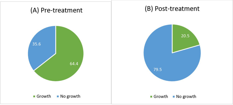 Fig 1
Fig 1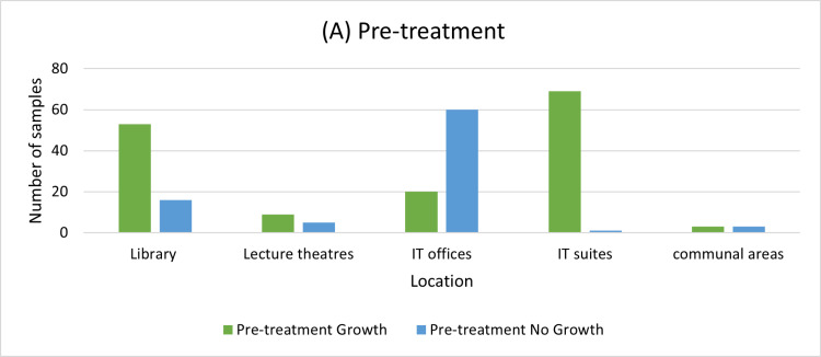 Fig 2
Fig 2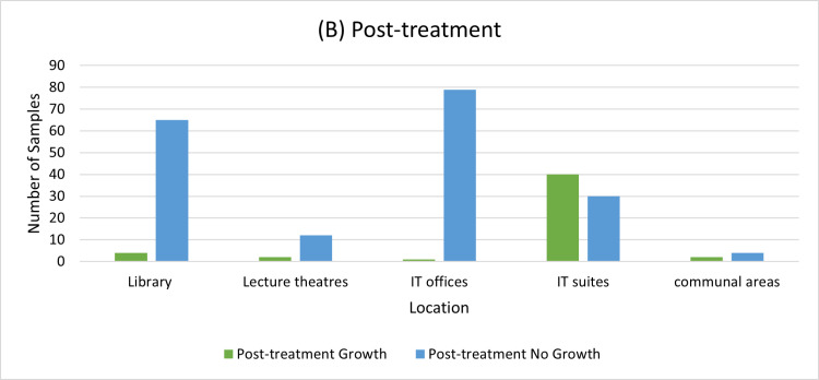 Fig 3
Fig 3Peer Reviews
No public reviews on file for this paper yet. If you reviewed it on a platform where reviews are public (OpenReview, ICLR, NeurIPS, ICML), you can paste yours below so the community can read it here.
Videos
No videos yet. Explain this paper in a talk, walkthrough, or lecture? Add one.
Taxonomy
TopicsInfection Control in Healthcare · Dental Research and COVID-19 · Bacterial Identification and Susceptibility Testing
Introduction
The bacterial burden on surfaces has been researched for many years in hospitals with an effort to reduce bacterial contamination and infections leading to co-morbidities [1,2,3]. Assessment of the presence of bacteria should be applied to universities and workplaces to protect students and staff from harmful pathogens they may be unknowingly spreading [4,5,6,7]. Bacteria can be spread via direct and indirect contact with infected droplets, airborne particles and fomites, with many bacterial species able to survive on inanimate surfaces for weeks to even months [8,9].
Due to increased online presence within universities such as the utilization of online resources for announcements, accessing learning content, contacting staff/students and handing in/marking work, computers have become one of the most used interfaces for both students and staff. Computer keyboards and mice have the most contact with a person’s hands, with average keyboards having over 100 keys and many grooves. This increases the surface area and creates difficult to reach recesses which results in keyboards being problematic and time consuming to thoroughly clean, therefore they may harbour bacterial pathogens [4]. It is important to consider that bacteria present may be opportunistic pathogens meaning they are not harmful to immunocompetent individuals, however, may pose a risk of infection to immunosuppressed individuals.
This study will assess the bacterial burden of multi-user computer keyboards and mice across the Liverpool John Moores city campus (LJMU), identify the bacterial species grown from the samples and discuss whether disinfection using a 70% isopropyl alcohol wipe (PDI Sani-Cloth^®^) is sufficient to reduce the presence of bacteria within the university campus.
Methods
Computer keyboards and mice were sampled from across the Liverpool John Moores University’s City campus with a total of 478 swabs taken between 9^th^ −20^th^ January 2023–8^th^ −19^th^ January 2024 across different facilities on LJMU campus (see Table 1). Initially computer keyboard and mouse samples were taken with a sterile cotton swab (SLS® Select Swab Woodstick with Cotton Tip Single in Peel Pouch), which was then capped and given an identification number denoting the sample was part of the pre-treatment group. During sampling, the mouse was swabbed first because this is the part of the computer that has the most contact with our hands. Then the keyboard was sampled with the same cotton swab by rubbing it along the top of the key. Lastly, the swab was dragged along the grooves of the keyboard to ensure the whole keyboard was thoroughly sampled. Immediately, a second sample was taken from the computer using a new swab after the keyboard and mouse had been disinfected with a 70% v/v isopropyl alcohol wipe (PDI Sani-Cloth®). A total period of 30 minutes elapsed between first sampling and disinfection before the second sampling (post-treatment) This sample was then capped and given identification number denoting it was a post-treatment sample. 239 computer samples were taken (see Table 1), with computers in various environments around campus including the library (69 computers), lecture theatres (14 computers), IT offices (80 computers), IT Suites (70 computers), communal areas (6 computers). The swabs were transferred to a laboratory where they were inoculated onto blood agar plates (Thermo Scientific™) and incubated aerobically overnight at 37°C [10, 11, 12] using Thermo Scientific™ incubator. For the initial stages of the investigation colony growth, gram staining [13,14,15] and morphology assessment was performed to group the bacterial species found. A selection of these samples was sub cultured and incubated overnight using Thermo Scientific™ incubator prior to analysis using MALDI-TOF spectrometry according to protocol by BioMérieux VITEK® MS prime to identify bacterial species [16,17,18] using α-Cyano-4-hydroxycinnamic acid (CHCA) as a matrix [19]. The BioMérieux VITEK® MS prime algorithm enables accurate discrimination of all species identified with performance ranging from 98 to 100%.
Table 1: Resut showing the colonial growth from pre- and post-treatment cultures.
Results
Growth on the blood agar plates demonstrated One hundred and fifty-four (64.4%) of pre-treatment samples (A) had microbial colony growth, decreasing to Forty-nine (20.5%) among the post-treatment samples (B) group as can be seen in Fig 1.
The percentage growth and no growth from cultures from pre and post treatment samples.
The distribution of this growth indicated that library and IT suites samples produced the greatest number of samples with growth, 53 and 69 samples respectively as can be seen in Fig 2.
The bacterial growth distribution for pre-treatment samples across the locations.
Further investigation, using Gram staining and MALDI-TOF MS (see Table 1) indicated a high proportion of the bacterial growth present was identified as Gram-positive cocci (GPC) species such as Staphylococcus albus, S. epidermidis, S. hominis, S. saprophyticus, S. warneri and Micrococcus luteus, with ninety-two (71%) of pre-treatment and twenty-nine (78%) of post-treatment growth resulting from GPC species (see Tables 1 and 2).
Table 2: Identification of bacterial isolates using MALDI-TOF BioMérieux VITEK® MS prime.
Gram-positive bacillus (GPB) bacteria were also isolated from samples from both pre- and post- treatment swabs, with thirty-five (27%) of pre-treatment and eight (23%) of post-treatment growth being from GPB species. Examples of GPB species found include Bacillus licheniformis, B. subtilis var. amyloliquefaciens and B. cereus, which are usually present in soil [10]. Lastly, two (1.6%) of pre-treatment growth was Gram-negative cocci (GNC) species identified as Enhydrobacter aerosaccus and Moraxella osloensis as shown in Table 2.
Discussion
Hand hygiene is the primary action to prevent infection and reduce the spread of multi-resistant organisms [20]. Numerous studies have indicated that computer keyboards (and mice) can become contaminated with pathogenic bacteria. As with health care settings, computer keyboards in educational institutions may act a mechanism for the transmission of pathogenic bacteria. Previous studies have demonstrated that other shared communication equipment, such as telephones, can also become contaminated by potentially pathogenic microorganisms, often members of the human microbiota [21,22].
Overall, the results showed that there were bacteria present on the computer keyboards and mice with 64.4% of the pre-treatment sample having colonial growth. This result is consistent with studies from UK and India [23,24]. It was expected that this would be higher considering the high footfall of both students and staff through the area where the samples were taken. Some factors could account for samples not showing microbial growth due to inability to provide anaerobic conditions. The samples were subject to aerobic conditions, where only species supported in this environment would grow. Therefore, if a second culture had been incubated anaerobically or in microaerophilic conditions there may have been a higher percentage of viable cultures. Another factor to consider is lasting vigilance of hygiene standards from the COVID-19 pandemic, in addition to continued presence of hand sanitiser and cleaning products available around the university campus.
All the bacteria identified from samples are commonly found as part of the normal human flora of the skin, mucus membranes and respiratory tract. These species identified are all opportunistic pathogens, which do not usually cause disease in healthy individuals however the presence of these microorganisms poses a greater risk to individuals who are immunosuppressed or compromised [4]. The GPC species found are a part of normal human microbiota of the skin and nasal passage; however, these are also opportunistic pathogens that are associated with many diseases and co-morbidities especially in individuals that are immunosuppressed [25]. It was expected that these species would be present; however, it is surprising that pathogenic bacteria were not isolated due to the number and variety of individuals using the facilities.
This percentage of no growth increased from 35.6% in the pre-treatment group to 79.5% in the post-treatment group as shown in (Fig 3), suggesting that the bacteria burden was decreased with the use of 70% isopropyl alcohol wipes confirming the effectiveness of the intervention.
The bacterial growth distribution for post-treatment samples across the locations.
Further research with a larger number of samples and a wider sampling area including a collaborative effort across different universities should be considered. Improvement to the method should be considered including the use of multiple different selective and differential agar plates to promote the growth of more varied bacteria [10,26]. Incubating a repeat sample in anaerobic conditions could help to assess the growth of different species of bacteria that may be present.
Conclusion
This study shows that bacterial contamination of computer mice and computer keyboards is prevalent across different facilities in the university. The commonest bacteria are commensal skin organisms. The study also demonstrates that Gram positive bacteria are mostly isolated from the computer mice and computer keyboards, and a few Gram-negative bacteria. The study emphasises the importance of adequate decontamination procedures using swipe (disinfectant) with a drastic reduction in the number of bacteria isolated post-treatment. We strongly recommend from the outcomes of the study that methods of disinfecting the computer mice and computer keyboards should be undertaken by every user of the computer before and after the usage of the computer mice and the computer keyboards. These approaches if undertaken properly will reduce to the barest minimum the bacterial burden on the University computer mice and computer keyboards.
Supporting information
S1 TableMinimal data set-MALDI-TOF Results.(TIF)
The reference list from the paper itself. Each links out to its DOI / PubMed record.
- 1Costa DM, Johani K, Melo DS, Lopes LKO, Lopes Lima LKO, Tipple AFV, et al. Biofilm contamination of high-touched surfaces in intensive care units: epidemiology and potential impacts. Lett Appl Microbiol. 2019;68(4):269–76. doi: 10.1111/lam.13127 30758060 · doi ↗ · pubmed ↗
- 2Messina G, Ceriale E, Lenzi D, Burgassi S, Azzolini E, Manzi P. Environmental contaminants in hospital settings and progress in disinfecting techniques. Biomed Res Int. 2013;2013:429780. doi: 10.1155/2013/429780 24286078 PMC 3830765 · doi ↗ · pubmed ↗
- 3Pal S, Juyal D, Adekhandi S, Sharma M, Prakash R, Sharma N, et al. Mobile phones: Reservoirs for the transmission of nosocomial pathogens. Adv Biomed Res. 2015;4:144. doi: 10.4103/2277-9175.161553 26322292 PMC 4549928 · doi ↗ · pubmed ↗
- 4Koscova J, Hurnikova Z, Pistl J. Degree of Bacterial Contamination of Mobile Phone and Computer Keyboard Surfaces and Efficacy of Disinfection with Chlorhexidine Digluconate and Triclosan to Its Reduction. Int J Environ Res Public Health. 2018;15(10):2238. doi: 10.3390/ijerph 15102238 30322055 PMC 6210060 · doi ↗ · pubmed ↗
- 5Al-Ghamdi AK, Abdelmalek SMA, Ashshi AM, Faidah H, Shukri H, Jiman-Fatani AA. Bacterial contamination of computer keyboards and mice, elevator buttons and shopping carts. African Journal of Microbiology Research. 2011;5(23):3998–4003. doi: 10.5897/ajmr 11.770 · doi ↗
- 6Lagier JC, Edouard S, Pagnier I, Mediannikov O, Drancourt M, Raoult D. Current and past strategies for bacterial culture in clinical microbiology. Clin Microbiol. 2023. Rev. 2015;28:208–36.10.1128/CMR.00110-14PMC 428430625567228 · doi ↗ · pubmed ↗
- 7Lutz JK, Crawford J, Hoet AE, Wilkins JR, Lee J. Comparative performance of contact plates, electrostatic wipes, swabs and a novel sampling device for the. detection of Staphylococcus aureus on environmental surfaces. J Appl. 2013;115:171–8. doi: 10.1234/example.doi 2023 October 123607553 · doi ↗ · pubmed ↗
- 8Kramer A, Schwebke I, Kampf G. How long do nosocomial pathogens persist on inanimate surfaces? A systematic review. BMC Infect Dis. 2006;6:130. doi: 10.1186/1471-2334-6-130 16914034 PMC 1564025 · doi ↗ · pubmed ↗