Design, Synthesis, and Evaluation of New 2-Arylpropanoic Acid-l-Tryptophan Derivatives for Mitigating Cisplatin-Induced Nephrotoxicity
Ming Yuan, Huai Wang, Mingjun Yu, Sen Yao, Risheng Yao

TL;DR
Researchers designed new compounds that protect the kidneys from cisplatin chemotherapy side effects by targeting a specific receptor.
Contribution
A new class of GRPR-targeting 2-arylpropanoic acid-L-tryptophan derivatives was synthesized and shown to improve kidney protection.
Findings
Compound 3m improved mouse renal cell viability more than existing GRPR antagonists.
3m reduced kidney injury markers and inflammatory cytokines in cisplatin-treated models.
Molecular docking and drug property predictions support 3m's potential as a therapeutic candidate.
Abstract
Cisplatin (CIS) is a widely used chemotherapeutic agent that is highly effective against various cancers. However, its clinical application is frequently limited by its substantial nephrotoxic side effects. The gastrin-releasing peptide receptor (GRPR), a critical regulator in inflammatory diseases, has been identified as a promising therapeutic target. Our previous studies have demonstrated that the GRPR antagonists PD176252 and RH-1402 can mitigate CIS-induced nephrotoxicity through anti-inflammatory mechanisms. Based on these findings, we designed and synthesized a series of 2-arylpropanoic acid-L-tryptophan derivatives to enhance the therapeutic effects. Among these compounds, 3m exhibited superior renal protection by significantly improving mouse renal tubular epithelial cell (mRTEC) viability from 50.2 ± 2.6% to 80.5 ± 3.9%, surpassing PD176252 (70.8 ± 1.4%) and RH-1402 (73.9 ±…
Genes, proteins, chemicals, diseases, species, mutations and cell lines named across the full text — each resolved to its canonical identifier and authoritative record.
Click any figure to enlarge with its caption.
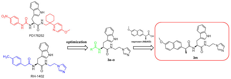 Figure 1
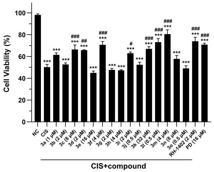 Figure 2
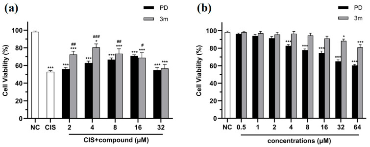 Figure 3
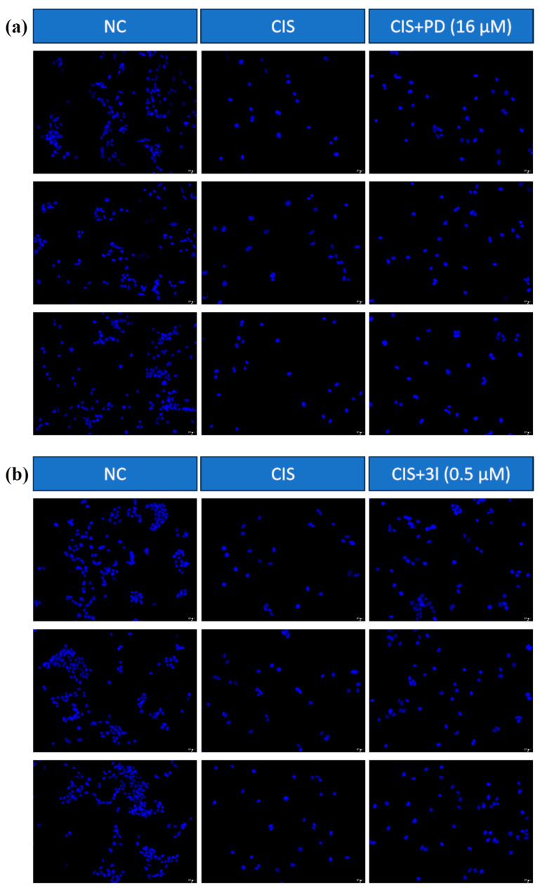 Figure 4
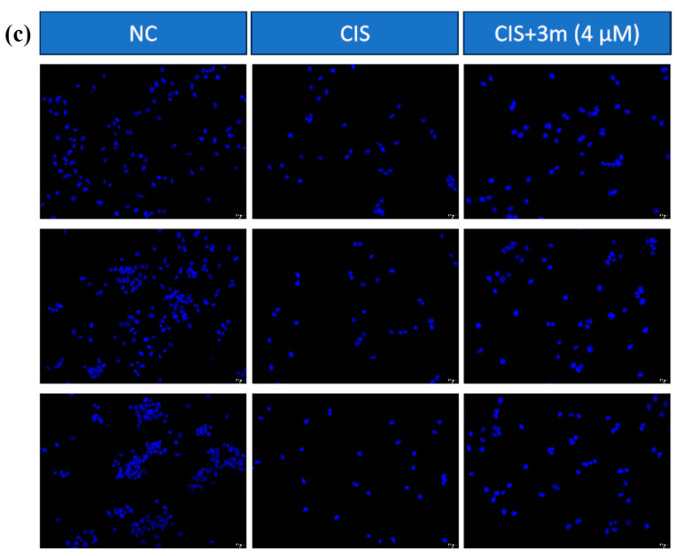 Figure 5
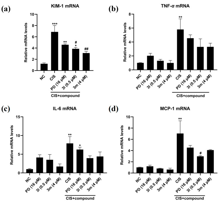 Figure 6
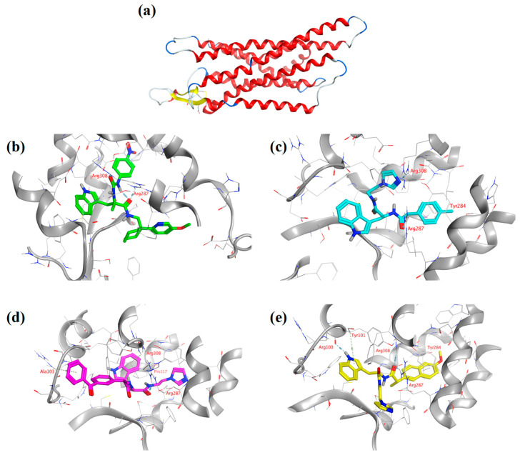 Figure 7
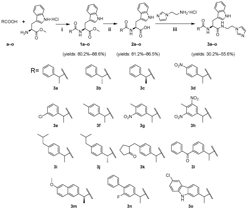 Figure 8
- —Education Department of Anhui Province of China
Peer Reviews
No public reviews on file for this paper yet. If you reviewed it on a platform where reviews are public (OpenReview, ICLR, NeurIPS, ICML), you can paste yours below so the community can read it here.
Videos
No videos yet. Explain this paper in a talk, walkthrough, or lecture? Add one.
Taxonomy
TopicsChemotherapy-induced organ toxicity mitigation · Cancer Mechanisms and Therapy · Intraperitoneal and Appendiceal Malignancies
1. Introduction
Cisplatin (CIS) is a widely used anticancer agent in the treatment of various malignancies, including cervical, ovarian, head and neck, prostate, testicular, and bladder cancers [1,2,3]. However, its prolonged use is frequently hindered by nephrotoxicity, a significant adverse effect observed in patients [4]. CIS-induced nephrotoxicity encompasses acute kidney injury (AKI), renal tubular dysfunction, hyperuricemia, hypomagnesemia, hypocalcemia, and chronic kidney disease [5]. It is estimated that approximately 30% of cancer patients receiving CIS therapy experience a substantial decline in kidney function, which negatively impacts their prognosis and overall health [6,7]. Despite the clinical relevance of CIS-induced nephrotoxicity, there are currently limited specific treatment options [8].
CIS predominantly accumulates within the renal tubules, causing more extensive damage to the kidneys compared to other organs. As a result, nephrotoxicity is regarded as the most severe adverse effect of CIS [9]. The pathophysiology of CIS-induced nephrotoxicity is complex, involving multiple mechanisms, including inflammation, oxidative stress, apoptosis, and mitochondrial dysfunction [10,11]. Inflammation plays a central role in this renal toxic response. CIS stimulates the release of pro-inflammatory cytokines, activates local inflammatory pathways, exacerbates renal cell damage, and may promote the development of kidney fibrosis [12,13]. CIS induces oxidative stress by generating reactive oxygen species (ROS), which impair kidney cells by damaging cellular membranes, proteins, lipids, and DNA, ultimately leading to cell death [14]. Additionally, CIS interacts with mitochondria, disrupting their function, reducing adenosine triphosphate (ATP) synthesis, and increasing mitochondrial membrane permeability, thereby causing mitochondrial damage and cell death [15]. Consequently, CIS-induced nephrotoxicity results in both the apoptosis and necrosis of renal cells, severely compromising kidney function.
The gastrin-releasing peptide (GRP) is a neuropeptide secreted by both neurons and endocrine cells, initially identified in the stomach of mammals and later detected in the brain and other tissues [16]. The GRP modulates various physiological processes by binding to its receptor, the gastrin-releasing peptide receptor (GRPR), and influences functions such as central nervous system regulation, gastrointestinal activity, tumor progression, immune cell modulation, and inflammatory responses [17,18,19,20]. The GRPR plays a pivotal role in renal injury induced by CIS. The GRP, in conjunction with the GRPR, participates in biological processes such as inflammation, oxidative stress, and apoptosis, thus exacerbating the nephrotoxicity associated with CIS. GRPR antagonists can inhibit several pro-inflammatory signaling pathways, prevent the secretion of pro-inflammatory cytokines, and reduce the infiltration of inflammatory cells, thereby mitigating renal inflammation and alleviating kidney damage [21]. Furthermore, GRPR antagonists can diminish oxidative stress, reduce ROS production, and prevent kidney cell death resulting from oxidative damage [22]. GRPR antagonists also suppress apoptosis-related signaling pathways, thereby protecting damaged renal cells [23]. In addition, numerous studies have highlighted the critical role of inflammation in CIS-induced nephrotoxicity. For instance, Tim-3, Rosiridin, and chitosan–ascorbate nanocapsules have been shown to effectively reduce CIS-induced nephrotoxicity by inhibiting renal inflammation [24,25,26].
Our previous research demonstrated that the small-molecule GRPR antagonist PD176252 alleviates CIS-induced nephrotoxicity, whereas RH-1402, a derivative designed based on PD176252, exhibits enhanced renal protective effects by reducing inflammation and mitigating kidney damage [21,27]. In the current study, we further optimized the structure of RH-1402 and synthesized a series of novel 2-arylpropanoic acid-L-tryptophan derivatives. Mouse renal tubular epithelial cells (mRTECs) are closely associated with the physiological functions of the kidneys and are widely used as a model for studying renal toxicity, particularly CIS-induced kidney injury [27]. Additionally, the cultivation of mRTECs is straightforward and easy to manage. Therefore, we selected mRTECs to assess the protective effects of the newly synthesized derivatives against CIS-induced cell death and their capacity to inhibit inflammatory responses.
2. Results and Discussion
2.1. Design and Synthesis of Compounds 3a–o
2.1.1. Design
Non-steroidal anti-inflammatory drugs (NSAIDs) are among the most commonly prescribed medications for the treatment of inflammatory diseases. The 2-arylpropanoic acid moiety is a crucial structural component in several NSAIDs, including ibuprofen, ketoprofen, and naproxen. Recent studies have indicated that incorporating the NSAID functional group may enhance the biological activity of specific compounds [28,29,30]. Given that 2-arylpropionic acid shares structural similarities with the p-tolylacetic acid group in RH-1402, this substitution may preserve some of the pharmacological benefits of the original compound while facilitating a simpler synthetic route. To further augment the renal protective properties, we replaced the p-tolylacetic acid group in RH-1402 with 2-arylpropionic acid, thereby yielding the target compound (Figure 1).
2.1.2. Synthesis
Scheme 1 illustrates the synthetic pathway leading to the target compounds 3a–o. Initially, intermediates 1a–o were synthesized through the reaction of 2-arylpropanoic acid derivatives a–o with L-tryptophan methyl ester hydrochloride. Subsequent hydrolysis of these intermediates afforded compounds 2a–o. Finally, the reaction of compounds 2a–o with 2-(1H-imidazol-1-yl)ethan-1-amine resulted in the formation of the target compounds 3a–o.
2.2. Biological Activity
2.2.1. Compounds 3a–o Mitigate CIS-Induced mRTEC Death
CIS-induced nephrotoxicity is primarily characterized by significant damage to renal tubular epithelial cells [31,32,33]. The CCK-8 assay was employed to evaluate the protective effects of compounds 3a–o against CIS-induced cell death in mRTEC [31,34]. Cell viability was measured after treatment with CIS alone or in combination with varying concentrations of the target compounds.
Figure 2 illustrates the compounds’ most effective protective effects against CIS-induced mRTEC death. Figure 3 depicts the protective effects of compounds PD176252 and 3m against CIS-induced nephrotoxicity at different concentrations, alongside their cytotoxicity in normal cells. The positive control PD176252 demonstrated a maximum cell viability of 70.8 ± 1.4% at 16 µM. RH-1402 exhibited a maximum cell viability of 73.9 ± 3.7% at 2 µM. Notably, compound 3m displayed the strongest protective effect, with a maximum cell viability reaching 80.5 ± 3.9% at 4 µM. 3m increased cell viability by approximately 30% compared to the CIS control group. Furthermore, compared to PD176252 and RH-1402, 3m enhanced cell viability by approximately 10%. These results suggest that 3m is more potent than both PD176252 and RH-1402, exhibits a concentration-dependent protective effect, and shows no significant cytotoxicity. The 4′,6-diamidino-2-phenylindole (DAPI) staining assay corroborated these findings (Figure 4).
The results of the cell viability assay indicate that the protective activity of compounds 3b and 3j (substituted with (R)-configured 2-arylpropanoic acid) is lower than that of compounds 3a and 3i (substituted with racemic 2-arylpropanoic acid). Compound 3c (substituted with (S)-configured 2-arylpropanoic acid) exhibits slightly higher protective activity than compound 3a. Compounds with multiple substituents on the benzene ring (3g, 3h, 3n) exhibited reduced activity, while those with para-monosubstituents (3d, 3f) showed enhanced activity. Para-position electron-donating groups were superior to electron-withdrawing groups, as evidenced by 3f being more effective than 3d. Compounds with meta-position electron-withdrawing substituents on the benzene ring (3e, 3g, 3h, 3n) exhibited significantly reduced activity. However, substituting the benzene ring in 3a with either benzophenone (3l) or naphthalene (3m) significantly improved cell viability. Carbazole substitution (3o) resulted in a notable decrease in activity. The naphthyl and methoxy groups in compound 3m increase the electron density of the molecule through their electron-donating effects, which may facilitate π-π interactions between the molecule and biological targets, thereby enhancing the affinity between the molecule and its target. Furthermore, the presence of naphthyl and methoxy groups increases the compound’s hydrophobicity and lipophilicity, enhancing its permeability across cell membranes and thereby improving its distribution and absorption in cells. Consequently, compound 3m demonstrates superior protective activity. In contrast, in compound 3l, the phenyl group has weaker conjugation and lower electron density compared to the naphthyl group in 3m, leading to a weaker binding affinity to the target. Although the phenyl ketone group also exerts electronic effects, its electron-donating capacity is less pronounced than that of methoxy. Moreover, the relatively simple molecular structure of the phenyl ketone group may limit its compatibility with the target. As a result, the activity of 3l is lower than that of 3m.
2.2.2. Anti-Inflammatory Activity of Compounds 3m and 3l
Kidney injury molecule-1 (KIM-1) is a transmembrane protein primarily expressed by proximal tubular epithelial cells [35]. Following kidney injury, KIM-1 levels are significantly elevated, establishing it as a reliable biomarker for assessing the renal protective effects of various compounds [35]. Inflammatory mediators, such as tumor necrosis factor-α (TNF-α), interleukin-6 (IL-6), and monocyte chemoattractant protein-1 (MCP-1), play a critical role in the pathogenesis of CIS-induced kidney damage [36,37,38]. To evaluate whether the new compound mitigates CIS-induced nephrotoxicity by inhibiting the inflammatory response, real-time polymerase chain reaction (PCR) was employed to detect the mRNA expression levels of KIM-1 and inflammatory cytokines.
The results indicated that compared to normal cells, CIS treatment alone significantly increased the expression of KIM-1 and inflammatory cytokines. Following co-treatment with CIS and target compounds, the mRNA levels of KIM-1, TNF-α, IL-6, and MCP-1 were significantly reduced. Compounds 3l and 3m exhibited more pronounced inhibitory effects on KIM-1, TNF-α, and IL-6 in comparison to PD176252 (Figure 5).
Our previous studies have demonstrated that PD176252 and RH-1402 lower the levels of KIM-1 and inflammatory cytokines through the inhibition of the NF-κB signaling pathway [21,27]. Real-time PCR experiments suggest that the downregulation of KIM-1 expression observed with 3l and 3m implies they possess a protective effect on the kidneys, alleviating CIS-induced kidney injury, mitigating damage to tubular cells, and reducing apoptosis or necrosis, thereby decreasing the expression of damage markers. The observed reduction in the levels of inflammatory factors (TNF-α, IL-6, and MCP-1) indicates that the protective effects of compounds 3l and 3m may be attributed to their capacity to inhibit the NF-κB signaling pathway, leading to a reduction in the transcription of inflammatory cytokine genes and the stability of their mRNA. Furthermore, their effects appear to be superior to those of PD176252.
2.3. SwissADME Predictions
SwissADME is a freely accessible online tool used to assess the pharmacokinetics, drug similarity, and chemical properties of small molecules [39,40,41]. The predictions provided by SwissADME version 2024, as shown in Table 1, reveal that the log Po/w values (partition coefficient between n-octanol and water) for both compounds 3l and 3m are below five, indicating favorable membrane permeability. Both 3l and 3m demonstrate superior gastrointestinal absorption and higher bioavailability scores compared to PD176252. Moreover, the AMES toxicity of compounds 3l and 3m is lower than that of both PD176252 and RH-1402. Furthermore, both 3l and 3m adhere to Lipinski’s rules. In summary, compounds 3l and 3m exhibit outstanding pharmacokinetic profiles and drug similarity.
2.4. Analysis of Molecular Docking Results
To investigate the interaction between the GRPR and target compounds, a homology model of the GRPR protein was constructed using Discovery Studio (DS) software version 2023 [27]. Molecular docking simulations were subsequently performed with the Molecular Operating Environment (MOE) software version 2024.06, and the results are presented in Figure 6. The docking score serves as a critical indicator of the molecular docking outcomes, representing the affinity between the ligand and the protein. A lower docking score indicates a stronger affinity. Furthermore, an increased number of hydrogen bonds typically correlates with more stable interactions between the ligand and the protein. The docking scores for PD176252, RH-1402, 3l, and 3m were −7.9727, −7.7658, −8.5718, and −8.2977, respectively. Compound 3m formed three hydrogen bonds with Arg100, Arg287, and Arg308, a π-H interaction with Tyr101, and a π-π interaction with Tyr284. Compound 3l formed one hydrogen bond with Arg287, two π-H interactions with Ala103 and Pro117, and a π-cation interaction with Arg308. In contrast, PD176252 formed only two hydrogen bonds with Arg287 and Arg308. RH-1402, similarly, formed two hydrogen bonds with Arg287 and Arg308, along with a π-π interaction with Tyr284. Therefore, both 3m and 3l exhibited stronger binding affinities compared to PD176252 and RH-1402.
3. Materials and Methods
3.1. Chemistry
All chemicals and reagents, including the 2-arylpropionic acid derivatives (a–o), were sourced from commercial suppliers (Aladdin, Shanghai, China) and used without further purification. Reactions were monitored by thin-layer chromatography on silica gel GF254 plates (Aladdin, Shanghai, China), and the results were visualized under ultraviolet (254 nm) light. The ^1^H and ^13^C nuclear magnetic resonance (NMR) spectra were recorded on an Agilent VNMRS600 NMR spectrometer (Agilent, Santa Clara, CA, USA), with DMSO-d_6_ as the solvent. High-resolution mass spectrometry (HR-MS) analysis was conducted using a Thermo Fisher Scientific Vanquish Q Exactive Plus mass spectrometer (Thermo Fisher Scientific, Waltham, MA, USA). Melting points were determined using a Stuart Melting Point Apparatus (MPA) (Stuart, Vernon Hills, IL, USA).
3.2. Procedure for Synthesizing Compounds 3a–o (Scheme 1)
3.2.1. Synthesis of Intermediates 1a–o
A mixture of 2-arylpropanoic acid derivatives (3.0 mmol), L-tryptophan methyl ester hydrochloride (3.3 mmol), N,N-diisopropylethylamine (DIPEA, 7.0 mmol), 1-ethyl-3-(3-dimethylaminopropyl)carbodiimide (EDCI, 3.3 mmol), 1-hydroxybenzotriazole (HOBt, 0.41 mmol), and 30 mL of dry dichloromethane (DCM) was stirred at room temperature for 7–8 h. Then, the mixture was washed with water (2 × 30 mL) and 5% hydrochloric acid (1 × 30 mL). The organic layer was concentrated under reduced pressure and purified via silica gel column chromatography (DCM/ethyl acetate), resulting in intermediates 1a–o with yields ranging from 80.2% to 88.6%.
3.2.2. Synthesis of Intermediates 2a–o
The intermediates 1a–o (2.0 mmol) were subjected to hydrolysis in the presence of Na_2_CO_3_ (6.0 mmol), methanol (MeOH, 20 mL), and water (7 mL) at 55 °C for 5–6 h. After cooling to room temperature, the pH was adjusted to 1–2 using 5% hydrochloric acid, followed by filtration and washing with water to produce intermediates 2a–o with yields ranging from 81.2% to 86.5%.
3.2.3. Synthesis of Target Compounds 3a–o
A mixture of intermediates 2a–o (1.0 mmol), 2-(1H-imidazol-1-yl)ethan-1-amine (1.1 mmol), EDCI (1.1 mmol), HOBt (0.14 mmol), DIPEA (2.3 mmol), and DCM (10 mL) was stirred at room temperature for 24 h. The mixture was washed with water (2 × 30 mL) and concentrated under reduced pressure. The rough product was purified by recrystallization (MeOH/H_2_O) or column chromatography (DCM/MeOH), resulting in target product 3a–o with yields ranging from 30.2% to 55.6%.
3.3. Structural Characterization of Target Compounds 3a–o
3a: white solid, yield: 51.2%. MP: 312.3–313.5 °C; ^1^H NMR (600 MHz, DMSO-d_6_) δ 10.74 (d, J = 2.1 Hz, 1H), 8.20 (t, J = 5.5 Hz, 1H), 8.15 (d, J = 8.1 Hz, 1H), 7.56 (s, 1H), 7.51 (d, J = 7.8 Hz, 1H), 7.30 (d, J = 8.1 Hz, 1H), 7.24–7.19 (m, 2H), 7.19–7.15 (m, 3H), 7.08 (s, 1H), 7.03 (t, J = 7.5 Hz, 1H), 6.93 (t, J = 7.4 Hz, 1H), 6.91–6.86 (m, 2H), 4.41 (td, J = 8.3, 5.5 Hz, 1H), 4.01–3.91 (m, 2H), 3.71 (q, J = 7.2 Hz, 1H), 3.38 (dq, J = 12.4, 6.1 Hz, 1H), 3.34–3.28 (m, 1H), 2.96 (dd, J = 14.5, 5.4 Hz, 1H), 2.84 (dd, J = 14.5, 8.7 Hz, 1H), 1.30 (d, J = 7.0 Hz, 3H). ^13^C NMR (151 MHz, DMSO-d_6_) δ 173.19, 172.01, 141.91, 137.33, 136.00, 128.10, 127.40, 127.21, 126.36, 123.56, 120.83, 119.69, 118.43, 118.18, 111.24, 109.87, 53.65, 45.32, 44.47, 27.77, 18.02. HR-MS m/z: calcd. for C_25_H_28_N_5_O_2_ [M + H]^+^ 430.2238, found: 430.2230.
3b: white solid, yield: 41.2%. MP: 180.1–181.3 °C; ^1^H NMR (600 MHz, DMSO-d_6_) δ 10.78 (d, J = 2.4 Hz, 1H), 8.32 (s, 1H), 8.28 (d, J = 7.1 Hz, 1H), 7.51 (d, J = 7.9 Hz, 1H), 7.38 (d, J = 1.6 Hz, 1H), 7.30 (d, J = 8.1 Hz, 1H), 7.28–7.25 (m, 4H), 7.23–7.19 (m, 2H), 7.17–7.15 (m, 2H), 6.93 (t, J = 7.3 Hz, 1H), 6.90 (d, J = 2.4 Hz, 1H), 4.35 (td, J = 8.4, 5.4 Hz, 1H), 4.10 (q, J = 6.1 Hz, 2H), 3.73–3.68 (m, 1H), 3.42 (t, J = 6.3 Hz, 1H), 3.36 (dd, J = 12.1, 5.9 Hz, 1H), 2.97 (dd, J = 14.5, 5.2 Hz, 1H), 2.85 (dd, J = 14.6, 8.9 Hz, 1H), 1.29 (d, J = 7.0 Hz, 3H). ^13^C NMR (151 MHz, DMSO-d_6_) δ 173.34, 172.22, 141.86, 136.39, 136.01, 128.11, 127.44, 127.21, 126.38, 123.61, 121.08, 120.84, 118.40, 118.20, 111.29, 109.87, 53.87, 46.81, 44.46, 27.69, 17.97. HR-MS m/z: calcd. for C_25_H_28_N_5_O_2_ [M + H]^+^ 430.2238, found: 430.2236.
3c: white solid, yield: 42.3%. MP: 180.2–182.1 °C; ^1^H NMR (600 MHz, DMSO-d_6_) δ 10.76 (s, 1H), 8.26 (d, J = 7.2 Hz, 1H), 8.21 (s, 1H), 7.60 (d, J = 7.9 Hz, 1H), 7.50 (d, J = 7.9 Hz, 1H), 7.33 (d, J = 7.6 Hz, 1H), 7.29 (d, J = 8.1 Hz, 1H), 7.27–7.26 (m, 2H), 7.22–7.19 (m, 2H), 7.18–7.14 (m, 3H), 6.93 (t, J = 7.4 Hz, 1H), 6.89 (d, J = 2.3 Hz, 1H), 4.35 (dt, J = 8.4, 4.2 Hz, 1H), 4.10–4.06 (m, 2H), 3.71 (d, J = 7.1 Hz, 1H), 3.40 (d, J = 5.1 Hz, 1H), 3.34 (dd, J = 14.8, 5.8 Hz, 1H), 2.96 (dd, J = 14.5, 5.3 Hz, 1H), 2.84 (dd, J = 14.6, 8.9 Hz, 1H), 1.29 (d, J = 7.0 Hz, 3H). ^13^C NMR (151 MHz, DMSO-d_6_) δ 173.33, 172.22, 141.85, 136.32, 136.00, 128.10, 127.42, 127.20, 126.37, 123.60, 121.15, 120.83, 118.39, 118.19, 111.27, 109.86, 53.87, 46.90, 44.45, 27.67, 17.96. HR-MS m/z: calcd. for C_25_H_28_N_5_O_2_ [M + H]^+^ 430.2238, found: 430.2233.
3d: light yellow solid, yield: 30.5%. MP: 320.5–321.9 °C; ^1^H NMR (600 MHz, DMSO-d_6_) δ 10.70 (d, J = 2.5 Hz, 1H), 8.36 (d, J = 8.1 Hz, 1H), 8.29 (t, J = 5.7 Hz, 1H), 8.04–7.96 (m, 2H), 7.74 (s, 1H), 7.49 (d, J = 7.9 Hz, 1H), 7.35–7.29 (m, 2H), 7.25 (d, J = 8.1 Hz, 1H), 7.18 (s, 1H), 7.03–6.96 (m, 2H), 6.90 (ddd, J = 8.0, 6.9, 1.0 Hz, 1H), 6.87 (d, J = 2.3 Hz, 1H), 4.44 (ddd, J = 9.6, 8.2, 5.0 Hz, 1H), 4.07–3.98 (m, 2H), 3.86 (q, J = 7.1 Hz, 1H), 3.42 (dq, J = 12.3, 5.8 Hz, 1H), 3.39–3.34 (m, 1H), 2.97 (dd, J = 14.6, 4.9 Hz, 1H), 2.81 (dd, J = 14.6, 9.6 Hz, 1H), 1.31 (d, J = 7.0 Hz, 3H). ^13^C NMR (151 MHz, DMSO-d_6_) δ 171.98, 171.72, 149.68, 146.08, 135.97, 128.76, 128.28, 127.09, 123.56, 123.33, 123.18, 120.77, 118.40, 118.13, 111.16, 109.78, 53.56, 45.65, 44.42, 27.85, 17.75. HR-MS m/z: calcd. for C_25_H_27_N_6_O_4_ [M + H]^+^ 475.2088, found: 475.2086.
3e: white solid, yield: 35.5%. MP: 291.1–292.8 °C; ^1^H NMR (600 MHz, DMSO-d_6_) δ 10.75 (d, J = 2.4 Hz, 1H), 8.31 (dd, J = 8.1, 2.4 Hz, 1H), 8.28–8.23 (m, 1H), 7.86 (s, 1H), 7.50 (d, J = 7.9 Hz, 1H), 7.31–7.26 (m, 2H), 7.23 (d, J = 6.4 Hz, 2H), 7.21 (d, J = 8.2 Hz, 1H), 7.11–7.08 (m, 1H), 7.04–7.01 (m, 2H), 6.94–6.90 (m, 2H), 4.38 (td, J = 8.3, 5.4 Hz, 1H), 4.04–3.99 (m, 2H), 3.74 (q, J = 7.3 Hz, 1H), 3.43–3.33 (m, 2H), 2.97 (dd, J = 14.5, 5.4 Hz, 1H), 2.84 (dd, J = 14.5, 8.7 Hz, 1H), 1.29 (d, J = 7.0 Hz, 3H). ^13^C NMR (151 MHz, DMSO-d_6_) δ 172.67, 171.98, 144.35, 136.94, 136.00, 132.78, 129.94, 127.32, 127.11, 126.42, 125.98, 123.48, 120.83, 120.23, 118.38, 118.17, 111.28, 109.86, 53.81, 45.91, 44.16, 27.78, 17.96. HR-MS m/z: calcd. for C_25_H_27_ClN_5_O_2_ [M + H]^+^ 464.1848, found: 464.1849.
3f: white solid, yield: 55.2%. MP: 321.6–323.2 °C; ^1^H NMR (600 MHz, DMSO-d_6_) δ 10.73 (d, J = 2.4 Hz, 1H), 8.15 (t, J = 5.6 Hz, 1H), 8.04 (d, J = 7.9 Hz, 1H), 7.52–7.46 (m, 2H), 7.29 (dt, J = 8.1, 0.9 Hz, 1H), 7.06–7.02 (m, 4H), 7.02–6.99 (m, 2H), 6.92 (ddd, J = 7.9, 6.9, 1.0 Hz, 1H), 6.89 (d, J = 2.3 Hz, 1H), 6.84 (t, J = 1.1 Hz, 1H), 4.38 (td, J = 8.2, 5.6 Hz, 1H), 3.96–3.90 (m, 2H), 3.64 (q, J = 7.2 Hz, 2H), 3.33–3.26 (m, 1H), 2.94 (dd, J = 14.5, 5.6 Hz, 1H), 2.82 (dd, J = 14.6, 8.5 Hz, 1H), 2.24 (s, 3H), 1.26 (d, J = 7.1 Hz, 3H). ^13^C NMR (151 MHz, DMSO-d_6_) δ 173.35, 171.99, 138.90, 137.37, 136.01, 135.28, 128.67, 128.30, 127.21, 127.07, 123.56, 120.83, 119.59, 118.44, 118.18, 111.23, 109.88, 53.63, 45.21, 44.09, 39.98, 27.77, 20.68, 18.08. HR-MS m/z: calcd. for C_26_H_30_N_5_O_2_ [M + H]^+^ 444.2394, found: 444.2391.
3g: light yellow solid, yield: 30.2%. MP: 291.3–293.1 °C; ^1^H NMR (600 MHz, DMSO-d_6_) δ 10.69 (d, J = 2.5 Hz, 1H), 8.35 (dd, J = 8.0, 1.5 Hz, 1H), 8.28 (t, J = 5.7 Hz, 1H), 8.00 (s, 1H), 7.83 (d, J = 1.8 Hz, 1H), 7.47 (d, J = 7.9 Hz, 1H), 7.33 (dd, J = 7.9, 2.0 Hz, 1H), 7.30–7.23 (m, 3H), 7.11 (s, 1H), 7.02–6.98 (m, 1H), 6.91–6.86 (m, 2H), 4.39 (td, J = 8.4, 5.3 Hz, 1H), 4.08–4.02 (m, 2H), 3.81 (q, J = 7.0 Hz, 2H), 3.42–3.38 (m, 1H), 2.97 (dd, J = 14.5, 5.3 Hz, 1H), 2.82 (dd, J = 14.5, 8.9 Hz, 1H), 2.47 (s, 3H), 1.30 (d, J = 7.0 Hz, 3H). ^13^C NMR (151 MHz, DMSO-d_6_) δ 172.46, 171.96, 148.49, 141.42, 136.76, 135.96, 132.58, 132.25, 130.93, 127.10, 125.50, 123.46, 122.84, 120.76, 120.46, 118.33, 118.10, 111.21, 109.80, 53.78, 46.17, 43.65, 39.57, 27.79, 19.38, 17.99. HR-MS m/z: calcd. for C_26_H_29_N_6_O_4_ [M + H]^+^ 489.2245, found: 489.2238.
3h: yellow solid, yield: 50.0%. MP: 310.3–312.1 °C; ^1^H NMR (600 MHz, DMSO-d_6_) δ 10.60 (d, J = 2.5 Hz, 1H), 8.44 (d, J = 8.2 Hz, 1H), 8.30 (t, J = 5.6 Hz, 1H), 8.00 (s, 2H), 7.52 (s, 1H), 7.47–7.42 (m, 1H), 7.21 (d, J = 8.1 Hz, 1H), 7.07 (s, 1H), 6.98–6.95 (m, 1H), 6.89–6.82 (m, 3H), 4.46 (td, J = 8.5, 5.1 Hz, 1H), 3.98 (tt, J = 12.3, 6.1 Hz, 2H), 3.94–3.84 (m, 2H), 3.36–3.30 (m, 1H), 2.96 (dd, J = 14.5, 5.3 Hz, 1H), 2.80 (dd, J = 14.5, 9.0 Hz, 1H), 2.43 (s, 3H), 1.32 (d, J = 7.0 Hz, 3H). ^13^C NMR (151 MHz, DMSO-d_6_) δ 171.77, 171.52, 150.62, 142.73, 137.42, 135.92, 128.35, 127.11, 126.66, 124.56, 123.44, 120.74, 119.62, 118.32, 118.05, 111.18, 109.75, 53.69, 45.24, 43.62, 39.99, 28.02, 18.13, 14.41. HR-MS m/z: calcd. for C_26_H_28_N_7_O_6_ [M + H]^+^ 534.2096, found: 534.2094.
3i: white solid, yield: 50.5%. MP: 221.5–222.6 °C; ^1^H NMR (600 MHz, DMSO-d_6_) δ 10.74 (d, J = 2.5 Hz, 1H), 8.16 (t, J = 5.5 Hz, 1H), 8.06 (d, J = 8.0 Hz, 1H), 7.52–7.47 (m, 2H), 7.28 (d, J = 8.1 Hz, 1H), 7.06 (d, J = 8.2 Hz, 3H), 7.03–7.00 (m, 1H), 6.99 (s, 1H), 6.98 (s, 1H), 6.95–6.90 (m, 2H), 6.84 (d, J = 1.1 Hz, 1H), 4.40 (d, J = 5.7 Hz, 1H), 3.96–3.90 (m, 2H), 3.66 (d, J = 7.1 Hz, 1H), 3.36–3.32 (m, 1H), 3.30 (d, J = 5.7 Hz, 1H), 2.95 (dd, J = 14.5, 5.6 Hz, 1H), 2.83 (dd, J = 14.5, 8.5 Hz, 1H), 2.37 (d, J = 7.2 Hz, 2H), 1.84–1.69 (m, 1H), 1.27 (d, J = 7.1 Hz, 3H), 0.83 (d, J = 6.6 Hz, 6H). ^13^C NMR (151 MHz, DMSO-d_6_) δ 173.35, 171.98, 139.11, 139.03, 137.36, 136.03, 128.65, 128.29, 127.20, 126.92, 123.63, 120.80, 119.58, 118.45, 118.16, 111.23, 109.86, 53.63, 45.21, 44.31, 44.09, 39.98, 29.63, 27.80, 22.25, 17.98. HR-MS m/z: calcd. for C_29_H_36_N_5_O_2_ [M + H]^+^ 486.2864, found: 486.2858.
3j: white solid, yield: 43.3%. MP: 170.6–172.3 °C; ^1^H NMR (600 MHz, DMSO-d_6_) δ 10.74 (d, J = 2.4 Hz, 1H), 8.15 (d, J = 5.2 Hz, 1H), 8.06 (d, J = 8.0 Hz, 1H), 7.52–7.48 (m, 2H), 7.29 (d, J = 8.1 Hz, 1H), 7.07 (s, 1H), 7.05 (d, J = 2.5 Hz, 2H), 6.99 (s, 1H), 6.98 (d, J = 4.2 Hz, 2H), 6.94–6.92 (m, 2H), 6.84 (d, J = 1.1 Hz, 1H), 4.40 (td, J = 8.2, 5.7 Hz, 1H), 3.97–3.91 (m, 2H), 3.67–3.63 (m, 1H), 3.30 (dd, J = 6.0, 3.8 Hz, 1H), 3.27–3.22 (m, 1H), 2.95 (dd, J = 14.6, 5.6 Hz, 1H), 2.84 (dd, J = 14.5, 8.5 Hz, 1H), 2.37 (d, J = 7.1 Hz, 2H), 1.79–1.76 (m, 1H), 1.28 (d, J = 7.1 Hz, 3H), 0.83 (d, J = 3.0 Hz, 6H). ^13^C NMR (151 MHz, DMSO-d_6_) δ 173.35, 171.98, 139.11, 139.03, 137.35, 136.02, 128.65, 128.28, 127.20, 126.92, 123.62, 120.80, 119.57, 118.44, 118.16, 111.22, 109.86, 53.63, 45.21, 44.27, 44.09, 39.97, 29.64, 27.79, 22.25, 17.98. HR-MS m/z: calcd. for C_29_H_36_N_5_O_2_ [M + H]^+^ 486.2864; found: 486.2858.
3k: white solid, yield: 50.6%. MP: 161.2–162.8 °C; ^1^H NMR (600 MHz, DMSO-d_6_) δ 10.74 (s, 1H), 8.17 (t, J = 5.8 Hz, 1H), 8.07 (dd, J = 8.1, 3.0 Hz, 1H), 7.50 (d, J = 8.0 Hz, 2H), 7.31–7.27 (m, 1H), 7.19–7.15 (m, 1H), 7.07 (d, J = 8.0 Hz, 4H), 7.03–7.01 (m, 2H), 6.92 (t, J = 2.6 Hz, 1H), 6.85 (s, 1H), 4.45–4.36 (m, 1H), 3.95 (dq, J = 9.3, 6.4, 5.8 Hz, 2H), 3.88 (t, J = 6.1 Hz, 1H), 3.68–3.64 (m, 1H), 2.94 (q, J = 3.5, 3.0 Hz, 1H), 2.93–2.91 (m, 1H), 2.84 (dd, J = 14.5, 8.6 Hz, 1H), 2.43–2.30 (m, 3H), 2.26–2.19 (m, 1H), 2.07 (ddd, J = 18.8, 10.4, 8.6 Hz, 1H), 1.85 (tdt, J = 12.1, 6.3, 3.2 Hz, 2H), 1.70–1.63 (m, 1H), 1.44 (dddd, J = 12.4, 10.7, 6.8, 3.9 Hz, 1H), 1.28 (d, J = 7.0 Hz, 3H). ^13^C NMR (151 MHz, DMSO-d_6_) δ 219.31, 173.29, 172.01, 139.45, 137.38, 136.03, 128.49, 128.43, 128.30, 127.32, 127.13, 123.58, 120.81, 119.60, 118.45, 118.17, 111.29, 110.13, 53.61, 50.05, 45.22, 44.09, 39.98, 37.62, 34.57, 28.73, 20.06, 17.94. HR-MS m/z: calcd. for C_31_H_36_N_5_O_3_ [M + H]^+^ 526.2813, found: 526.2811.
3l: white solid, yield: 33.8%. MP: 180.0–181.3 °C; ^1^H NMR (600 MHz, DMSO-d_6_) δ 10.67 (d, J = 2.4 Hz, 1H), 8.29 (d, J = 8.0 Hz, 1H), 8.20 (t, J = 5.6 Hz, 1H), 7.69–7.65 (m, 4H), 7.57 (dt, J = 7.6, 1.6 Hz, 1H), 7.53–7.46 (m, 5H), 7.42 (t, J = 7.6 Hz, 1H), 7.26 (d, J = 8.0 Hz, 1H), 7.08–7.03 (m, 1H), 7.00 (ddd, J = 8.1, 6.9, 1.2 Hz, 1H), 6.92–6.88 (m, 1H), 6.85 (d, J = 2.4 Hz, 2H), 4.42 (td, J = 8.2, 5.7 Hz, 1H), 4.00–3.88 (m, 2H), 3.83 (d, J = 7.1 Hz, 1H), 3.35–3.27 (m, 2H), 2.96 (dd, J = 14.5, 5.6 Hz, 1H), 2.83 (dd, J = 14.6, 8.4 Hz, 1H), 1.33 (d, J = 7.0 Hz, 3H). ^13^C NMR (151 MHz, DMSO-d_6_) δ 195.81, 172.80, 171.86, 142.49, 137.09, 136.78, 135.97, 132.64, 131.76, 129.66, 128.61, 128.54, 128.42, 128.07, 127.18, 123.57, 123.38, 120.82, 118.57, 118.39, 118.17, 111.25, 110.10, 109.90, 53.69, 45.21, 44.33, 39.97, 27.87, 18.28. HR-MS m/z: calcd. for C_32_H_32_N_5_O_3_ [M + H]^+^ 534.2500, found: 534.2493.
3m: white solid, yield: 50.8%. MP: 191.6–192.5 °C; ^1^H NMR (600 MHz, DMSO-d_6_) δ 10.84 (d, J = 2.4 Hz, 1H), 8.24 (d, J = 8.3 Hz, 1H), 8.13 (s, 1H), 7.72 (dd, J = 16.4, 8.8 Hz, 2H), 7.69–7.66 (m, 1H), 7.62 (dd, J = 7.9, 1.1 Hz, 1H), 7.44 (d, J = 1.1 Hz, 1H), 7.41 (dd, J = 8.5, 1.8 Hz, 1H), 7.36–7.31 (m, 1H), 7.25 (d, J = 2.5 Hz, 1H), 7.12 (dd, J = 8.8, 2.5 Hz, 2H), 7.07 (d, J = 1.2 Hz, 1H), 6.99 (t, J = 1.0 Hz, 1H), 6.94 (t, J = 1.2 Hz, 1H), 6.74 (t, J = 1.1 Hz, 1H), 4.50 (td, J = 8.4, 5.7 Hz, 1H), 3.87–3.79 (m, 6H), 3.28 (dq, J = 12.6, 6.0 Hz, 1H), 3.22 (dq, J = 13.6, 5.6 Hz, 1H), 3.03 (dd, J = 14.4, 5.6 Hz, 1H), 2.92 (dd, J = 14.5, 8.6 Hz, 1H), 1.27 (d, J = 7.0 Hz, 3H). ^13^C NMR (151 MHz, DMSO-d_6_) δ 173.23, 171.84, 156.97, 137.30, 137.28, 136.08, 133.11, 129.12, 128.36, 128.26, 127.41, 126.69, 126.51, 125.40, 123.59, 120.90, 119.51, 118.59, 118.54, 118.23, 111.32, 110.18, 105.67, 55.15, 53.55, 45.13, 44.59, 39.94, 28.14, 18.74. HR-MS m/z: calcd. for C_30_H_32_N_5_O_3_ [M + H]^+^ 510.2500, found: 510.2495.
3n: white solid, yield: 55.2%. MP: 250.1–252.0 °C; ^1^H NMR (600 MHz, DMSO-d_6_) δ 10.85 (d, J = 2.5 Hz, 1H), 8.33 (d, J = 8.2 Hz, 1H), 8.22 (t, J = 5.7 Hz, 1H), 7.63 (d, J = 7.9 Hz, 1H), 7.53–7.50 (m, 2H), 7.47–7.46 (m, 2H), 7.44 (d, J = 3.9 Hz, 1H), 7.42 (d, J = 8.2 Hz, 1H), 7.39 (d, J = 7.0 Hz, 1H), 7.34 (d, J = 8.0 Hz, 1H), 7.24–7.17 (m, 2H), 7.12 (d, J = 2.4 Hz, 1H), 7.08–7.05 (m, 1H), 7.00 (d, J = 7.5 Hz, 1H), 6.98 (d, J = 2.7 Hz, 1H), 6.79 (s, 1H), 4.55–4.47 (m, 1H), 3.90 (td, J = 5.9, 2.7 Hz, 2H), 3.78 (t, J = 7.2 Hz, 1H), 3.35–3.32 (m, 1H), 3.28 (d, J = 5.6 Hz, 1H), 3.04 (dd, J = 14.4, 5.7 Hz, 1H), 2.93 (dd, J = 14.5, 8.6 Hz, 1H), 1.22 (d, J = 7.0 Hz, 3H). ^13^C NMR (151 MHz, DMSO-d_6_) δ 172.56, 171.77, 158.78 (d, J = 245.3 Hz), 144.09 (d, J = 7.7 Hz), 137.29, 136.07, 135.05, 130.37 (d, J = 3.6 Hz), 128.72 (d, J = 2.8 Hz), 128.61, 128.26, 127.72, 127.38, 123.96 (d, J = 2.9 Hz), 123.58, 120.89, 119.51, 118.57, 118.22, 115.03, 114.88, 111.31, 110.10, 53.58, 45.17, 44.08, 28.16, 18.57. HR-MS m/z: calcd. for C_31_H_31_FN_5_O_2_ [M + H]^+^ 524.2456, found: 524.2449.
3o: white solid, yield: 55.6%. MP: 191.5–193.6 °C; ^1^H NMR (600 MHz, DMSO-d_6_) δ 11.41 (s, 1H), 10.78 (s, 1H), 8.28–8.22 (m, 2H), 8.15 (dd, J = 5.3, 2.1 Hz, 1H), 8.02–7.95 (m, 2H), 7.48 (dd, J = 8.4, 6.6 Hz, 2H), 7.42 (d, J = 1.4 Hz, 1H), 7.36 (dd, J = 8.6, 2.2 Hz, 1H), 7.27 (d, J = 8.1 Hz, 1H), 7.23 (s, 1H), 7.10 (s, 1H), 7.05 (dd, J = 8.1, 1.5 Hz, 1H), 7.01–6.97 (m, 1H), 6.95 (d, J = 2.4 Hz, 1H), 6.89 (t, J = 7.5 Hz, 1H), 4.40 (td, J = 7.9, 5.8 Hz, 1H), 4.08–3.97 (m, 2H), 3.91 (d, J = 7.0 Hz, 1H), 3.43–3.33 (m, 2H), 2.97 (dd, J = 14.5, 5.8 Hz, 1H), 2.87 (dd, J = 14.5, 8.1 Hz, 1H), 1.40 (d, J = 7.0 Hz, 3H). ^13^C NMR (151 MHz, DMSO-d_6_) δ 173.47, 172.06, 140.55, 140.51, 138.34, 135.96, 127.18, 125.36, 124.92, 123.70, 123.49, 122.73, 120.81, 120.52, 120.27, 120.14, 119.57, 118.90, 118.38, 118.17, 112.34, 111.25, 109.87, 109.64, 53.91, 46.22, 45.02, 39.60, 27.73, 18.63. HR-MS m/z: calcd. for C_31_H_30_ClN_6_O_2_ [M + H]^+^ 553.2113, found: 553.2109.
3.4. Cells
The mouse renal tubular epithelial cells (Thermo Fisher Scientific, Waltham, MA, USA) were cultured in F12 medium (Hyclone, Logan, UT, USA) supplemented with 10% fetal bovine serum (Hyclone, Logan, UT, USA) at 37 °C and 5% CO_2_.
3.5. Cell Viability Assay
Cell viability was determined using the CCK-8 assay. Mouse renal tubular epithelial cells were cultured in a 96-well plate (Thermo Fisher Scientific, Waltham, MA, USA) for 24 h. The normal control group (NC) was administered saline, and the model group (CIS) was treated with 20 µM of cisplatin. The experimental group (CIS + compound) was first treated with the target compound at different concentrations (0.5 µM–64 µM) for 12 h, followed by treatment with 20 µM cisplatin for 24 h. The cell toxicity assay involved treating the cells with the compound alone for 24 h. Then, 10 µL of CCK-8 solution (MedChemExpress, Monmouth Junction, NJ, USA) was added, and the cells were incubated for 2 h. Finally, the absorbance was measured at 450 nm using an Elx800 microplate reader (Biotek, Winooski, VT, USA).
3.6. DAPI Staining Assay
After the cell viability assay, the cells were fixed with 3.7% formaldehyde for 10 min, then stained with DAPI (Merck, Darmstadt, Germany) for 5 min. The excess dye was washed off with PBS buffer and the cells were observed under a fluorescence microscope (Olympus, Tokyo, Japan).
3.7. RNA Extraction and Real-Time PCR Assay
Following the literature [6], total RNA was extracted from mouse kidney tissue using Trizol reagent (Thermo Fisher Scientific, Waltham, MA, USA). The RNA concentration was measured using a Drop 2000 spectrophotometer (Thermo Fisher Scientific, Waltham, MA, USA). RNA was reverse-transcribed into cRNA. Detection was performed using a real-time PCR thermocycler (Bio-Rad, Hercules, CA, USA). Primer sequences are listed in Table A1 (Appendix A).
3.8. Experimental Methods in Molecular Docking
According to previous studies, the GRPR homology model was constructed using DS software version 2023 [27]. The chemical structure of the target compound was drawn using ChemDraw software version 2021 and subsequently imported into MOE software version 2024.06. The imported compound was then prepared by adding polar hydrogens, protonation, and energy minimization to achieve a stable conformation. The active site of GRPR was identified using the Site Finder module in MOE. Following this, a standard molecular docking procedure was performed.
3.9. Experimental Data Processing
The experimental data were processed using Excel software version 2021 and plotted using GraphPad Prism software version 10. The data were expressed as mean ± SD. Statistical analysis was conducted using t-tests or a one-way ANOVA.
4. Conclusions
Cisplatin (CIS) is a potent anticancer agent that is widely utilized in clinical practice. However, its nephrotoxicity remains a significant challenge, presenting long-term health risks to patients. Currently, targeted therapies aimed at mitigating this issue are limited. PD176252 and its derivative RH-1402 have demonstrated significant protective effects against CIS-induced nephrotoxicity. Building upon the structure of RH-1402, we designed and synthesized 15 novel 2-arylpropanoic acid-L-tryptophan derivatives. The chemical structures of these compounds were confirmed through MP, ^1^H NMR, ^13^C NMR, and HR-MS analysis. Cell viability and inflammatory cytokine assays were employed to assess the protective potential of these derivatives. Among them, compound 3m exhibited the most prominent protective effect. Following CIS treatment, compound 3m increased mRTEC viability from approximately 50% to over 80%, outperforming PD176252 and RH-1402 in terms of efficacy. SwissADME analysis further confirmed that compound 3m possesses favorable drug-like properties, including excellent gastrointestinal absorption and low toxicity. The molecular docking results indicated that compound 3m demonstrates a high binding affinity to the GRPR. These findings underscore 3m as a promising drug candidate for further investigation and development.
5. Patents
Risheng Yao; Ming Yuan; et al. A 2-Arylpropanoic Acid-L-Tryptophan Compound, Its Preparation Method, and Uses. 2024109742068, 26 November 2024.
The reference list from the paper itself. Each links out to its DOI / PubMed record.
- 1Romani A.M.P. Cisplatin in cancer treatment Biochem. Pharmacol.202220611532310.1016/j.bcp.2022.11532336368406 · doi ↗ · pubmed ↗
- 2Tchounwou P.B. Dasari S. Noubissi F.K. Ray P. Kumar S. Advances in our understanding of the molecular mechanisms of action of cisplatin in cancer therapy J. Exp. Pharmacol.20211330332810.2147/JEP.S 26738333776489 PMC 7987268 · doi ↗ · pubmed ↗
- 3Pietras P. Aulas A. Fay M.M. Leśniczak-Staszak M. Sowiński M. Lyons S.M. Szaflarski W. Ivanov P. Translation inhibition and suppression of stress granules formation by cisplatin Biomed. Pharmacother.202214511238210.1016/j.biopha.2021.11238234864307 PMC 8782064 · doi ↗ · pubmed ↗
- 4Liu P. Li X.X. Lv W.X. Xu Z.J. Inhibition of CXCL 1-CXCR 2 axis ameliorates cisplatin-induced acute kidney injury by mediating inflammatory response Biomed. Pharmacother.202012210969310.1016/j.biopha.2019.10969331812015 · doi ↗ · pubmed ↗
- 5Zhang J. Ye Z.W. Tew K.D. Townsend D.M. Cisplatin chemotherapy and renal function Adv. Cancer Res.202115230532710.1016/bs.acr.2021.03.00834353441 PMC 8963537 · doi ↗ · pubmed ↗
- 6Yang X. Guan Y.J. Bayliss G. Zhao T.C. Zhuang S.G. SET 8 inhibition preserves PTEN to attenuate kidney cell apoptosis in cisplatin nephrotoxicity Cell Death Dis.20251622610.1038/s 41419-025-07526-y 40164578 PMC 11958763 · doi ↗ · pubmed ↗
- 7Burns C.V. Edwin S.B. Szpunar S. Forman J. Cisplatin-induced nephrotoxicity in an outpatient setting Pharmacotherapy 20214118419010.1002/phar.250033417725 · doi ↗ · pubmed ↗
- 8Chen X.C. Huang L.F. Tang J.X. Wu D. An N. Ye Z.N. Lan H.Y. Liu H.F. Yang C. Asiatic acid alleviates cisplatin-induced renal fibrosis in tumor-bearing mice by improving the TFEB-mediated autophagy-lysosome pathway Biomed. Pharmacother.202316511512210.1016/j.biopha.2023.11512237413899 · doi ↗ · pubmed ↗