Imaging Features and Clinical Characteristics of Granular Cell Tumors: A Single-Center Investigation
Hui Gu, Lan Yu, Yu Wu

TL;DR
This study examines the imaging features of granular cell tumors across different body locations and highlights the importance of histopathological confirmation for accurate diagnosis.
Contribution
The study provides a detailed characterization of granular cell tumor imaging features across multiple modalities and anatomical locations.
Findings
GCTs show distinct imaging patterns depending on their anatomical location.
Malignant GCTs are larger but lack specific imaging features to distinguish them from benign tumors.
Imaging cannot reliably differentiate benign from malignant GCTs, emphasizing the need for histopathological confirmation.
Abstract
Background/Objectives: Granular cell tumors (GCTs) are rare neurogenic tumors with Schwann cell differentiation. Although most are benign, 1–2% exhibit malignant behavior. The imaging features of GCTs remain poorly characterized due to their rarity and anatomic variability. This study aims to elucidate the manifestations of GCTs in multimodal imaging across different anatomic locations. Methods: We retrospectively analyzed 66 histopathologically confirmed GCT cases (2011–2024), assessing their clinical presentations, pathological characteristics, and imaging findings from ultrasound (n = 31), CT (n = 14), MRI (n = 8), and endoscopy (n = 15). Two radiologists independently reviewed the imaging features (location, size, morphology, signal/density, and enhancement). Results: The cohort (mean age: 42 ± 12 years; 72.7% female) showed tendency in location towards soft tissue (48.4%), the…
Genes, proteins, chemicals, diseases, species, mutations and cell lines named across the full text — each resolved to its canonical identifier and authoritative record.
Click any figure to enlarge with its caption.
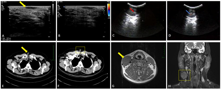 Figure 1
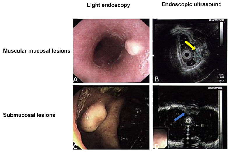 Figure 2
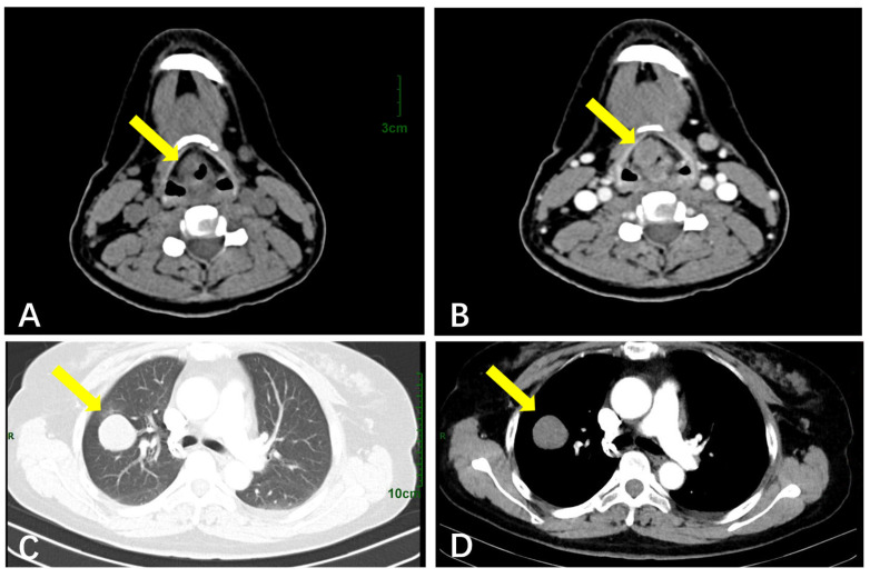 Figure 3
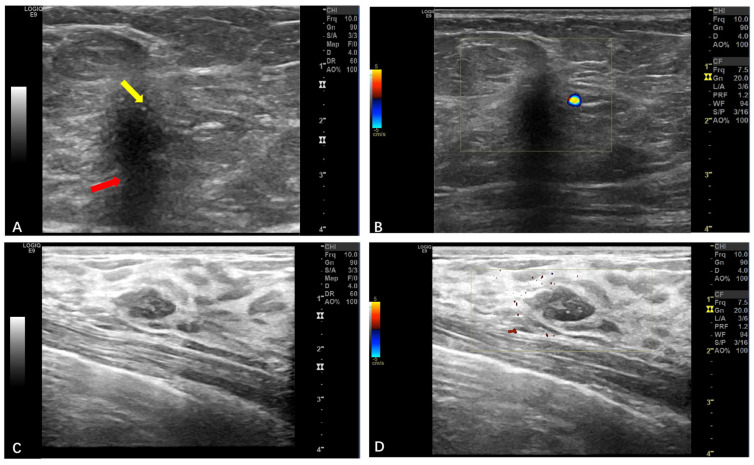 Figure 4
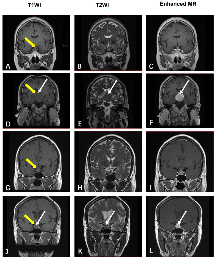 Figure 5
Peer Reviews
No public reviews on file for this paper yet. If you reviewed it on a platform where reviews are public (OpenReview, ICLR, NeurIPS, ICML), you can paste yours below so the community can read it here.
Videos
No videos yet. Explain this paper in a talk, walkthrough, or lecture? Add one.
Taxonomy
TopicsTumors and Oncological Cases · Teratomas and Epidermoid Cysts · Soft tissue tumor case studies