Depth-Variant Deconvolution Applied to Widefield Microscopy for Rapid Large-Volume Tissue Imaging
Daniel D. Lee, Kevin A. Telfer, Mark. A. J. Koenis, Yim K. Lee, Kevin W. Namink, Brian T. Saunders, Heyun Lee, Hailey K. Kelley, Heather S. Ruiz, Joseph P. Gaut, Gwendalyn J. Randolph, Bernd H. Zinselmeyer

TL;DR
This paper introduces a new imaging method that combines tissue clearing and deconvolution to enable detailed 3D imaging of thick tissues using widefield microscopy.
Contribution
The novel contribution is a depth-variant deconvolution approach that enables subnuclear resolution in large-volume tissues using widefield microscopy.
Findings
The method achieved subnuclear axial resolution in tissues up to 500 μm deep.
It enabled 3D visualization of blood vessels and amyloid deposits in brain slices comparable to confocal microscopy.
The approach allowed rapid 3D imaging of human kidney biopsies in under five minutes.
Abstract
Innovations in 3D tissue imaging have revolutionized research, but limitations stemming from lengthy protocols and equipment accessibility persist. Classical widefield microscopy is fast and accessible but often excluded from 3D imaging workflows due to its lack of optical sectioning. Here we combine tissue clearing with a depth-variant deconvolution approach customized for large-volume widefield imaging to achieve subnuclear axial resolution in tissues to a depth of 500 μm. We illustrate the utility of this method in a mouse model of ileitis and to gain a 3D perspective in thick brain slices from a mouse model of cerebral amyloid angiopathy, where we resolved large and small blood vessels, including those with amyloid deposits, attaining resolution that compared favorably to tile-scanning confocal microscopy. Finally, we sought to leverage our approach to allow for richer pathological…
Genes, proteins, chemicals, diseases, species, mutations and cell lines named across the full text — each resolved to its canonical identifier and authoritative record.
Click any figure to enlarge with its caption.
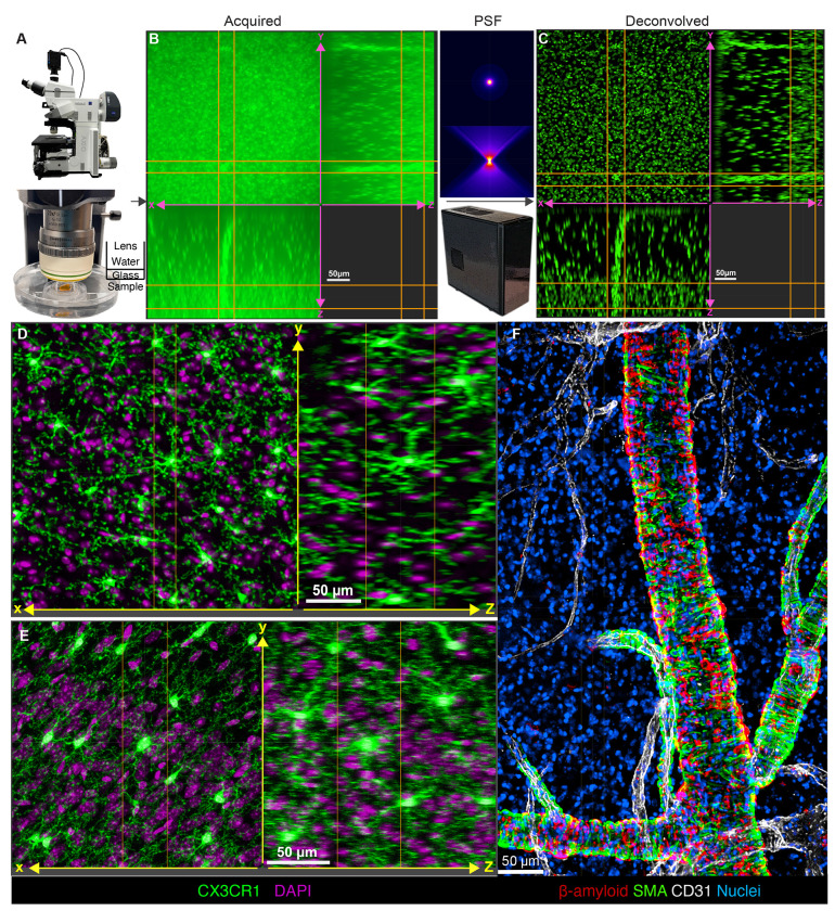 Figure 1
 Figure 2
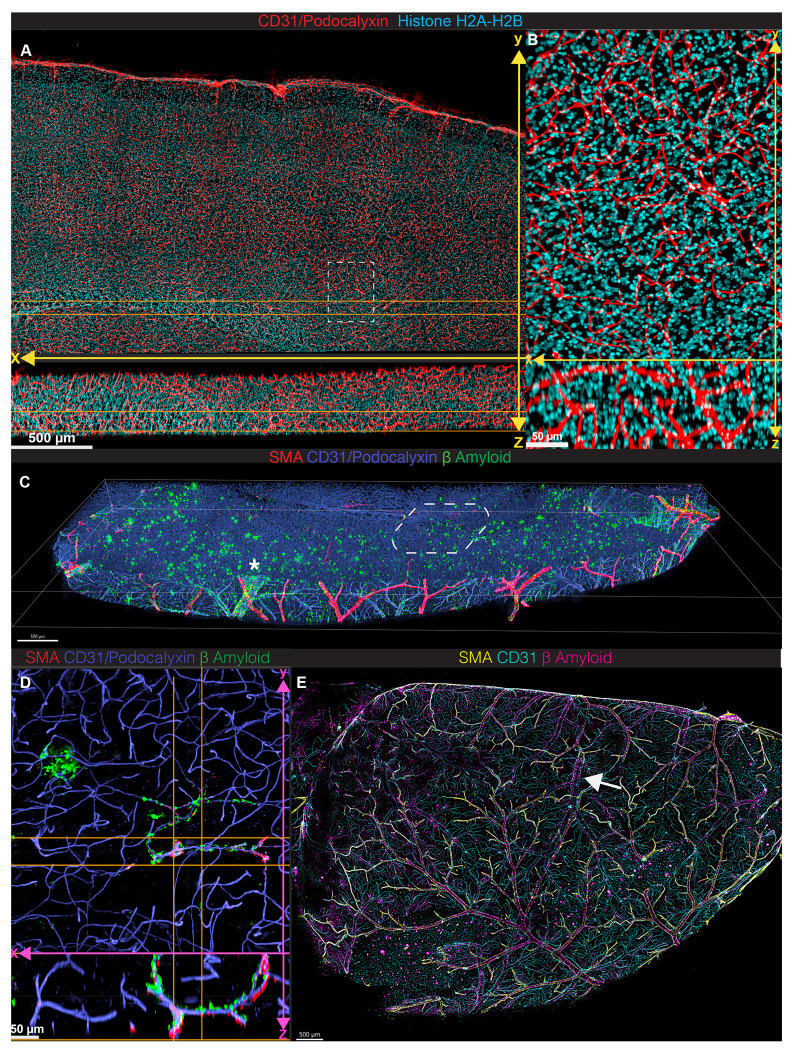 Figure 3
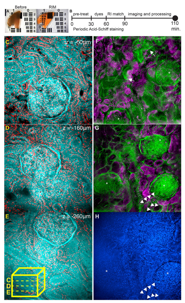 Figure 4
Peer Reviews
No public reviews on file for this paper yet. If you reviewed it on a platform where reviews are public (OpenReview, ICLR, NeurIPS, ICML), you can paste yours below so the community can read it here.
Videos
No videos yet. Explain this paper in a talk, walkthrough, or lecture? Add one.
Taxonomy
TopicsCell Image Analysis Techniques · Single-cell and spatial transcriptomics · Advanced Fluorescence Microscopy Techniques