Arthroscopic Fixation of a Posterior Cruciate Ligament Femoral Avulsion in an Adult: A Case Report
Clevio Desouza, Isteyaque Siddique

TL;DR
This case report describes a rare knee injury in an adult involving the posterior cruciate ligament and successful arthroscopic treatment.
Contribution
The paper presents a rare case of PCL femoral avulsion in an adult and its successful arthroscopic management.
Findings
MRI confirmed PCL avulsion from the femoral origin and ACL avulsion from the tibial attachment.
Arthroscopic fixation and open reduction restored joint stability and showed good healing in early follow-up.
Abstract
Femoral avulsion of the posterior cruciate ligament (PCL) is an exceedingly rare injury pattern in adults, in contrast to the more commonly encountered mid-substance tears or tibial-sided avulsions. We present a rare case of a middle-aged male who sustained a complex multiligamentous knee injury following trauma. Initial imaging revealed avulsion fractures involving both the tibial plateau and medial femoral condyle. Further evaluation with MRI confirmed avulsions of the PCL from its femoral origin and anterior cruciate ligament (ACL) from its tibial attachment, along with posterior root tear of the medial meniscus and associated medial collateral ligament (MCL) injury. The patient was treated with arthroscopically assisted fixation of the PCL via femoral tunnel suturing and suspension device, along with open reduction and internal fixation of the medial epicondyle to restore MCL…
Genes, proteins, chemicals, diseases, species, mutations and cell lines named across the full text — each resolved to its canonical identifier and authoritative record.
Click any figure to enlarge with its caption.
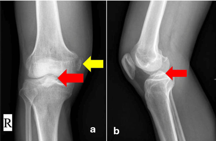 Figure 1
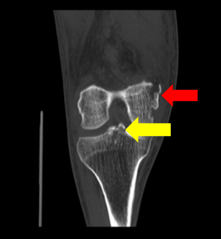 Figure 2
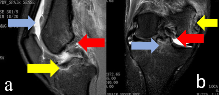 Figure 3
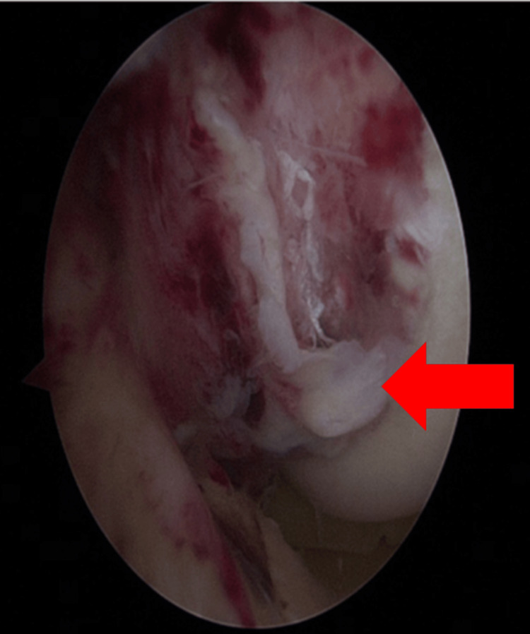 Figure 4
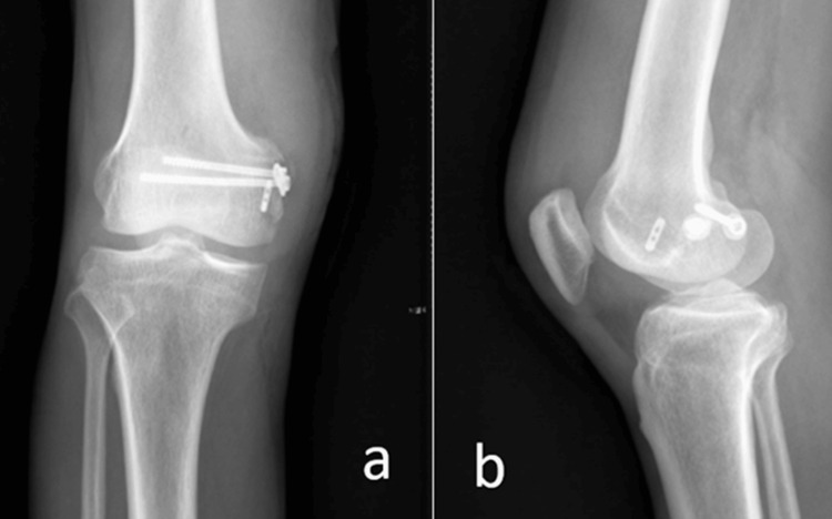 Figure 5
Peer Reviews
No public reviews on file for this paper yet. If you reviewed it on a platform where reviews are public (OpenReview, ICLR, NeurIPS, ICML), you can paste yours below so the community can read it here.
Videos
No videos yet. Explain this paper in a talk, walkthrough, or lecture? Add one.
Taxonomy
TopicsKnee injuries and reconstruction techniques · Total Knee Arthroplasty Outcomes · Shoulder Injury and Treatment
Introduction
The posterior cruciate ligament (PCL), known for its strength and stability, is infrequently injured compared to the anterior cruciate ligament (ACL). Most adult PCL injuries involve mid-substance disruptions or tibial avulsions. Avulsion fractures at the femoral origin are rare, particularly in adults, where such injuries typically manifest as soft tissue "peel-off" lesions without bony involvement. Only a handful of adult cases involving femoral bony avulsion have been documented and treated arthroscopically [1-3].
While recent literature reports arthroscopic techniques for various PCL pathologies [4-8], open reduction remains the more conventional approach. This case report describes an exceptionally rare instance of a femoral-side PCL avulsion fracture in a 50-year-old man, managed arthroscopically with successful functional outcomes.
Case presentation
A 50-year-old male presented with right knee swelling and pain following a motorcycle accident five days prior. There was no distal neurovascular deficit. Physical examination revealed restricted and painful range of motion, joint effusion, and positive anterior and posterior drawer tests. Valgus stress testing suggested medial instability.
Radiographs demonstrated avulsion fractures involving the tibial plateau and the inferior femoral border (Figure 1).
X-ray of the right knee in anteroposterior (a) and lateral (b) views.(a) Anteroposterior view shows avulsion of the anterior cruciate ligament (ACL) from the tibial plateau (red arrow) and avulsion of the medial collateral ligament (MCL) from the femoral condyle (yellow arrow). (b) Lateral view highlights the tibial plateau avulsion of the ACL (red arrow).
Computed tomography (CT) scan confirmed bony injury of the femoral medial epicondyle and the tibial plateau (Figure 2).
CT scan images of the right knee. The red arrow indicates a fracture of the medial femoral epicondyle, and the yellow arrow highlights an avulsion fracture of the tibial eminence consistent with anterior cruciate ligament (ACL) avulsion.
MRI revealed a PCL femoral origin avulsion, an ACL avulsion from the tibial attachment, posterior root tear of the medial meniscus, capsular effusion, and medial epicondyle fracture (Figure 3).
Magnetic resonance imaging (MRI) of the right knee.(a) Sagittal view demonstrates avulsion of the anterior cruciate ligament (ACL) from the tibial insertion (yellow arrow), posterior cruciate ligament (PCL) avulsion from the femoral attachment (red arrow), and joint capsular effusion (blue arrow). (b) Coronal view reveals ACL avulsion from the tibial insertion (blue arrow), posterior root tear of the medial meniscus (red arrow), and avulsion of the medial epicondyle (yellow arrow).
The patient was initially immobilized in an above-knee slab. After swelling subsided, he underwent arthroscopic-assisted fixation of the femoral PCL avulsion.
Surgical technique
Standard anterolateral and anteromedial portals were used for diagnostic arthroscopy. The ACL appeared intact with a tibial avulsion fragment. The PCL was found avulsed at its femoral attachment, accompanied by partial periosteal stripping and cortical bone involvement (Figure 4).
Arthroscopic view of the right knee showing avulsion fracture of the posterior cruciate ligament (PCL) from its femoral attachment (red arrow).
Debridement was followed by placement of a 4.5-mm absorbable rivet through the anterolateral portal, targeting the medial epicondyle. A suture hook was used to re-anchor the PCL to its femoral footprint. As intraoperative assessment showed persistent laxity, a 4.0-mm femoral tunnel was created, and the PCL was tensioned and fixed using Ethibond sutures, secured with an Endobutton.
Subsequently, open reduction and internal fixation of the medial femoral epicondyle and the MCL tibial attachment were performed using two cannulated screws. Postoperatively, the knee was stabilized in a long leg brace.
At the two-month follow-up, radiographs confirmed satisfactory reduction of the avulsed fragment (Figure 5). The patient demonstrated improved knee function with active range of motion from 0° to 120°. Clinical stability tests, including anterior and posterior drawer tests, were negative. The absence of instability on clinical examination, restored mobility, and pain-free ambulation indicate favorable early outcomes.
Two-month follow-up X-ray of the right knee.(a) Anteroposterior view and (b) lateral view show satisfactory reduction and healing of the avulsed fragment.
Discussion
The PCL comprises an anterolateral and a posteromedial bundle, contributing significantly to posterior and rotational knee stability [9,10]. PCL injuries typically involve mid-substance ruptures, and femoral-sided bony avulsions are exceptionally uncommon, especially in skeletally mature individuals [11].
PCL femoral avulsion fractures are more frequently reported in pediatric populations due to relatively weaker physis compared to ligamentous strength [12,13]. These injuries, if unrecognized, may lead to chronic instability and poor functional outcomes [14].
Previous literature includes a few adult cases treated arthroscopically. Park et al. [15] described a transfemoral tunnel technique in a 42-year-old, while Xu et al. [16] reported fixation of a partial osteochondral avulsion using polydioxanone sutures.
Epidemiological data suggest femoral-sided PCL avulsions are exceedingly rare. Gregory et al. [17] noted only one case of femoral-sided PCL injury among 85 patients with associated ligamentous injuries. Earlier reports, such as by O’Donoghue [18] and Drucker and Wynne [19], hint at such injury patterns, although specifics remain unclear. Lee et al. [20] documented a PCL-related osteochondral injury linked to the medial femoral condyle.
The current case describes a high-energy valgus mechanism resulting in a constellation of injuries: femoral MCL avulsion, ACL tibial avulsion, and a femoral PCL avulsion. Arthroscopic-assisted fixation allowed direct visualization and anatomical reduction with minimal invasiveness and early rehabilitation potential.
Conclusions
Avulsion of the PCL from its femoral attachment is an exceedingly rare injury pattern in adults, often underdiagnosed due to its subtle presentation. Early identification through detailed imaging, including MRI, is crucial for accurate diagnosis and planning surgical intervention. Arthroscopic repair offers a minimally invasive alternative to open surgery, allowing precise anatomical restoration with minimal soft tissue disruption. In this case, we successfully managed a multiligamentous knee injury with femoral PCL avulsion using arthroscopic fixation, demonstrating favorable clinical and radiological outcomes at follow-up. This report adds to the limited literature and reinforces the feasibility of arthroscopic techniques for such rare injuries. Further studies and case series are warranted to establish standardized protocols for managing femoral-side PCL avulsions in adults.
The reference list from the paper itself. Each links out to its DOI / PubMed record.
- 1Knee ligament injuries in children J Bone Joint Surg Am Clanton TO De Lee JC Sanders B Neidre A 11951201611979 https://pubmed.ncbi.nlm.nih.gov/511880/511880 · pubmed ↗
- 2Avulsion of the femoral attachment of the posterior cruciate ligament in an eleven-year-old boy. Case report J Bone Joint Surg Am Mayer PJ Micheli LJ 431432611979 https://pubmed.ncbi.nlm.nih.gov/429417/429417 · pubmed ↗
- 3Posterior cruciate avulsion Orthopedics Suprock MD Rogers VP 659662131990236724910.3928/0147-7447-19900601-11 · doi ↗ · pubmed ↗
- 4Arthroscopic treatment of concurrent avulsion fracture of anterior and posterior cruciate ligament with suspension device J Orthop Case Rep Lombardo-Torre M Espejo-Reina A García-Gutiérrez G Espejo-Baena A Espejo-Reina MJ 81858201810.13107/jocr.2250-0685.1062 PMC 611420330167421 · doi ↗ · pubmed ↗
- 5Arthroscopic fixation of tibial bony avulsion of the posterior cruciate ligament Arthroscopy Shino K Nakata K Mae T Yamada Y Shiozaki Y Toritsuka Y 019200310.1053/jars.2003.5006212579141 · doi ↗ · pubmed ↗
- 6Arthroscopic repair of the posterior cruciate ligament: two techniques Arthroscopy Espejo-Baena A López-Arévalo R Urbano V Montañez E Martín F 6566601620001097612910.1053/jars.2000.4626 · doi ↗ · pubmed ↗
- 7Pseudo-arthrosis repair of a posterior cruciate ligament avulsion fracture Knee Surg Sports Traumatol Arthrosc Hoogervorst P Gardeniers JW Moret-Wever S van Kampen A 161216161820102040510710.1007/s 00167-010-1114-4PMC 2957579 · doi ↗ · pubmed ↗
- 8Arthroscopic treatment of acute tibial avulsion fracture of the posterior cruciate ligament using the Tight Rope fixation device Arthrosc Tech Gwinner C Kopf S Hoburg A Haas NP Jung TM 0823201410.1016/j.eats.2014.02.005PMC 413013925126507 · doi ↗ · pubmed ↗