Man Complaining of Sore Throat
Jasmine Yu, Payton Sullivan, Jasmine Thompson, Joseph S. Colla

Abstract
Genes, proteins, chemicals, diseases, species, mutations and cell lines named across the full text — each resolved to its canonical identifier and authoritative record.
Click any figure to enlarge with its caption.
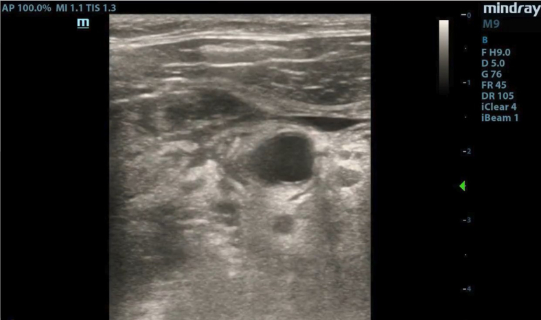 Figure 1
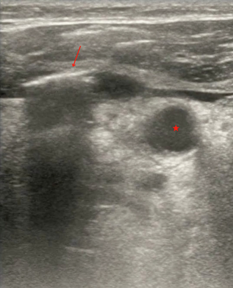 Figure 2
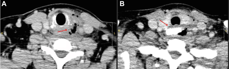 Figure 3
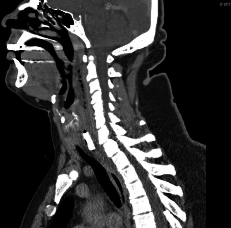 Figure 4
Peer Reviews
No public reviews on file for this paper yet. If you reviewed it on a platform where reviews are public (OpenReview, ICLR, NeurIPS, ICML), you can paste yours below so the community can read it here.
Videos
No videos yet. Explain this paper in a talk, walkthrough, or lecture? Add one.
Taxonomy
TopicsOtolaryngology and Infectious Diseases · Streptococcal Infections and Treatments · Voice and Speech Disorders
Patient Presentation
1
A 44-year-old man presented to the emergency department with a sore throat, fever, and concerns that he may have swallowed glass. He stated that the day prior he was eating in an area where broken glass was present. Since then, he experienced pain with odynophagia and dysphagia. Upon arrival, he was tachycardic, hypotensive (with a blood pressure of 98/57 mm Hg), and febrile, with a temperature of 38.5 °C. A bedside ultrasound revealed subcutaneous air adjacent to the trachea (Fig 1 and Video). A neck computed tomography scan with contrast was arranged for further evaluation (Figs 2 and 3).Figure 1. Bedside ultrasound showed shadowing under an area of subcutaneous air (arrow) adjacent to the trachea (star).VideoUltrasound revealing subcutaneous air adjacent to the trachea.Figure 2. Computed tomography of the neck shows cross-sections demonstrating (A) anterior subcutaneous gas correlating with ultrasound image (Fig 1) and (B) rectangular metallic density.Figure 3. Computed tomography of the neck demonstrated a rectangular metallic density with multiple foci of gas consistent with esophageal perforation.
Diagnosis: Esophageal Foreign Body with Esophageal Perforation
2
The patient underwent a rigid esophagoscopy. The operative note indicated that there was a 3 × 3-cm piece of glass located just below the upper esophageal sphincter, surrounded by inflammation and purulence with a large perforation at the left lateral wall of the esophagus. An external neck exploration was formed without obvious abscess or perforation. The perforation appeared to be contained within a layer of fibrinous tissue. The patient was fluid resuscitated, placed on broad-spectrum antibiotics, and started on tube feeds. A gastrografin esophagram was performed 8 days later was negative for esophageal leak or stricture.
Esophageal perforation is a serious, life-threatening medical emergency. X-rays are an easy but insensitive screening test. Contrast esophagography is the preferred diagnostic test for confirming esophageal perforation; however, computed tomography scans are more sensitive for detecting foreign bodies.1^,^2 Ultrasound has also become a popular diagnostic tool in cases of free air and is notable for detecting soft tissue air with a sensitivity of 100%.3, 4, 5
Funding and Support
By JACEP Open policy, all authors are required to disclose any and all commercial, financial, and other relationships in any way related to the subject of this article as per ICMJE conflict of interest guidelines (see www.icmje.org). The authors have stated that no such relationships exist.
Conflict of Interest
All authors have affirmed they have no conflicts of interest to declare.
The reference list from the paper itself. Each links out to its DOI / PubMed record.
- 1Khaitan P.G.Famiglietti A.Watson T.J.The etiology, diagnosis, and management of esophageal perforation J Gastrointest Surg 261220222606261510.1007/s 11605-022-05454-236138308 · doi ↗ · pubmed ↗
- 2Aronberg R.M.Punekar S.R.Adam S.I.Judson B.L.Mehra S.Yarbrough W.G.Esophageal perforation caused by edible foreign bodies: a systematic review of the literature Laryngoscope 1252201537137810.1002/lary.2489925155167 · doi ↗ · pubmed ↗
- 3Beason H.F.Markowitz J.E.Pneumomediastinum diagnosed on ultrasound in the emergency department: a case report Perm J 1932015 e 122e 12410.7812/TPP/14-23226176579 PMC 4500492 · doi ↗ · pubmed ↗
- 4Testa A.Candelli M.Pignataro G.Costantini A.M.Pirronti T.Silveri N.G.Sonographic detection of spontaneous pneumomediastinum J Ultrasound Med 271020081507150910.7863/jum.2008.27.10.150718809962 · doi ↗ · pubmed ↗
- 5Butcher C.H.Dooley R.W.Levitov A.B.Detection of subcutaneous and intramuscular air with sonography: a sensitive and specific modality J Ultrasound Med 306201179179510.7863/jum.2011.30.6.79121632993 · doi ↗ · pubmed ↗