Open‐source deep‐learning models for segmentation of normal structures for prostatic and gynecological high‐dose‐rate brachytherapy: Comparison of architectures
Andrew J. Krupien, Yasin Abdulkadir, Dishane C. Luximon, John Charters, Huiming Dong, Jonathan Pham, Dylan O'Connell, Jack Neylon, James M. Lamb

TL;DR
This paper compares two deep learning models for automatically segmenting organs in high-dose-rate brachytherapy CT scans, showing both are accurate and useful for clinical planning.
Contribution
The study evaluates and implements open-source deep learning models for HDR brachytherapy segmentation using a large clinical dataset.
Findings
UNet++ and nnU-Net models achieved high Dice-Similarity-Coefficients for bladder and rectum segmentation.
No significant difference in performance was found between the two architectures.
Trained models outperformed clinical contours in a blinded evaluation.
Abstract
The use of deep learning‐based auto‐contouring algorithms in various treatment planning services is increasingly common. There is a notable deficit of commercially or publicly available models trained on large or diverse datasets containing high‐dose‐rate (HDR) brachytherapy treatment scans, leading to poor performance on images that include HDR implants. To implement and evaluate automatic organs‐at‐risk (OARs) segmentation models for use in prostatic‐and‐gynecological computed tomography (CT)‐guided high‐dose‐rate brachytherapy treatment planning. 1316 computed tomography (CT) scans and corresponding segmentation files from 1105 prostatic‐or‐gynecological HDR patients treated at our institution from 2017 to 2024 were used for model training. Data sources comprised six CT scanners including a mobile CT unit with previously reported susceptibility to image streaking artifacts. Two…
Genes, proteins, chemicals, diseases, species, mutations and cell lines named across the full text — each resolved to its canonical identifier and authoritative record.
Click any figure to enlarge with its caption.
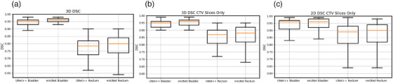 Figure 1
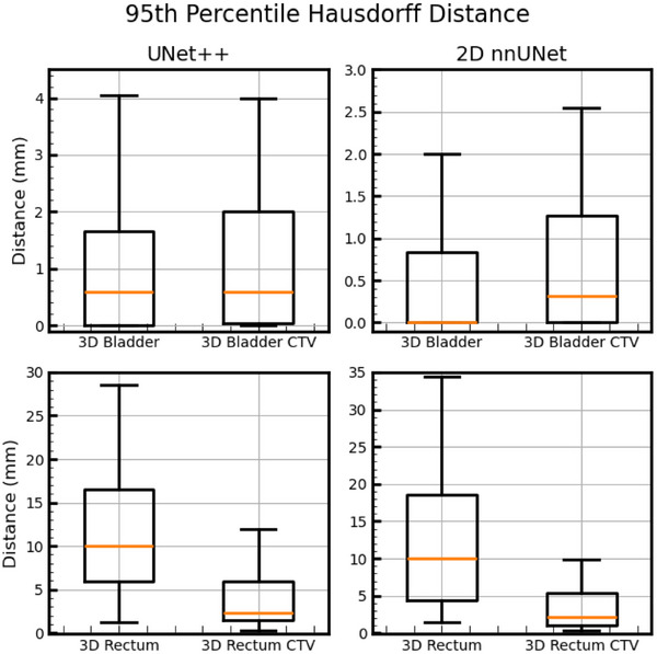 Figure 2
 Figure 3
 Figure 4
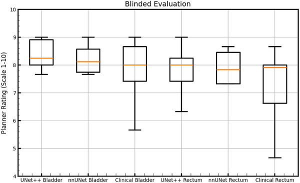 Figure 5
 Figure 6
 Figure 7
 Figure 8
Peer Reviews
No public reviews on file for this paper yet. If you reviewed it on a platform where reviews are public (OpenReview, ICLR, NeurIPS, ICML), you can paste yours below so the community can read it here.
Videos
No videos yet. Explain this paper in a talk, walkthrough, or lecture? Add one.
Taxonomy
TopicsProstate Cancer Diagnosis and Treatment · Advanced Radiotherapy Techniques · Advanced X-ray and CT Imaging