Oral Hyperpigmentation Secondary to Radioactive Iodine Therapy
Rachel M Boice, Christina M Mitchell, Thomas P Sollecito, Roopali Kulkarni

Abstract
Genes, proteins, chemicals, diseases, species, mutations and cell lines named across the full text — each resolved to its canonical identifier and authoritative record.
Click any figure to enlarge with its caption.
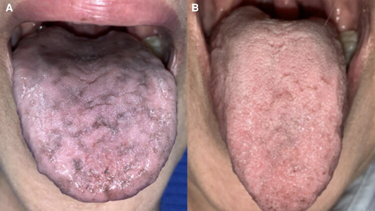 Figure 1
Peer Reviews
No public reviews on file for this paper yet. If you reviewed it on a platform where reviews are public (OpenReview, ICLR, NeurIPS, ICML), you can paste yours below so the community can read it here.
Videos
No videos yet. Explain this paper in a talk, walkthrough, or lecture? Add one.
Taxonomy
TopicsCancer Diagnosis and Treatment · Management of metastatic bone disease · Neuroblastoma Research and Treatments
Image Legend
A 75-year-old female individual presented to the oral medicine clinic complaining of dryness and generalized color change in her mouth of 3 months' duration. This began 10 days after radioactive iodine therapy (RAI). She endorsed a complete loss of taste but denied other oral symptoms. She was previously evaluated by her endocrinologist and otorhinolaryngologist with no diagnosis or treatment rendered. Medical history included metastatic papillary thyroid carcinoma, treated by total thyroidectomy, bilateral cervical lymphadenectomy with bilateral modified neck dissection, and RAI (I-131 Rx 97.3 mCi). Whole body scans completed 18 hours and 1 week after RAI revealed radioiodine uptake localization involving the salivary glands and pharynx.
Intraoral examination was notable for areas of generalized hyperpigmentation bilaterally on the buccal mucosa, labial mucosa, dorsal tongue, and palate (Fig. 1A). Diagnoses were consistent with salivary hypofunction, ageusia, and hyperpigmentation of the oral mucosa in the setting of prior RAI. Alpha lipoic acid 600 mg daily was recommended to the patient for ageusia. Recommendations for salivary hypofunction included salivary gland massage instruction, increased water intake, and use of over-the-counter sialagogues. At the 3-month follow-up visit, the pigmentation had notably decreased (Fig. 1B). This image demonstrates a rare but possible oral adverse effect of RAI therapy [1, 2].
Patient presentation at initial appointment and at three-month follow up.
The reference list from the paper itself. Each links out to its DOI / PubMed record.
- 1Williams M, Hatipoglu B. Development of abnormal pigmentation of the oral mucosa following adjuvant radioactive iodine treatment. AACE Clin Case Rep. 2016;2(2):e 113‐e 116.
- 2Alawi F . Pigmented lesions of the oral cavity: an update. Dent Clin North Am. 2013;57(4):699‐710.24034073 10.1016/j.cden.2013.07.006PMC 3775277 · doi ↗ · pubmed ↗