A new option of reconstruction after extensive chest wall resection
João Paulo Cassiano de Macedo, Pedro Henrique Xavier Nabuco-de-Araujo, José Ribas M. de Campos, Paulo M. Pêgo-Fernandes, Ricardo M. Terra

TL;DR
This paper introduces Vitagraft®, a new synthetic and absorbable material for chest wall reconstruction, and reports on its initial use and biocompatibility in patients.
Contribution
The first clinical assessment of Vitagraft® in chest wall reconstruction, a novel synthetic, biocompatible, and absorbable material.
Findings
Vitagraft® showed good biocompatibility in chest wall reconstruction cases.
Reoperation was needed in 28% of cases, with 14% requiring prosthesis removal.
Postoperative imaging indicated favorable integration of the material.
Abstract
•Chest wall reconstruction plays an important role in the outcomes of chest wall surgery. The ideal prosthesis characteristics are already established, even though, the best choice is not well decided.•The authors propose a new prosthesis for chest wall reconstruction.•This study is the first to assess patients with chest wall disease reconstructed with Vitagraft®. It is a synthetic, biocompatible, and absorbable material, which has never been found in thoracic surgery before.•Finally, the use of Vitagraft® has been shown to have good prosthesis biocompatibility. Chest wall reconstruction plays an important role in the outcomes of chest wall surgery. The ideal prosthesis characteristics are already established, even though, the best choice is not well decided. The authors propose a new prosthesis for chest wall reconstruction. This study is the first to assess patients with chest…
Genes, proteins, chemicals, diseases, species, mutations and cell lines named across the full text — each resolved to its canonical identifier and authoritative record.
Click any figure to enlarge with its caption.
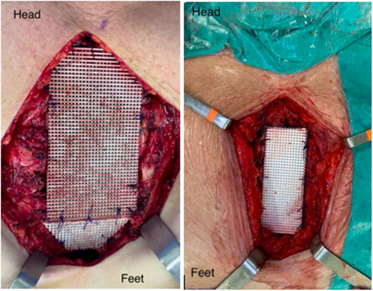 Figure 1
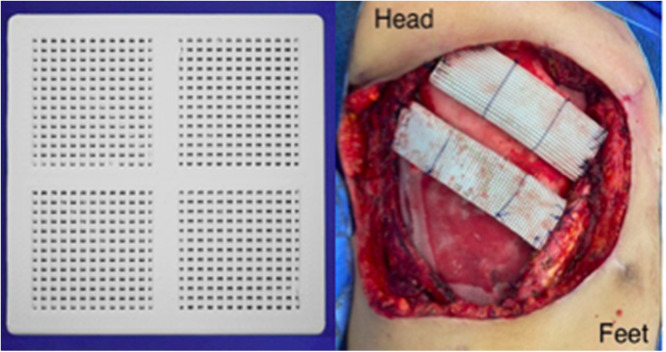 Figure 2
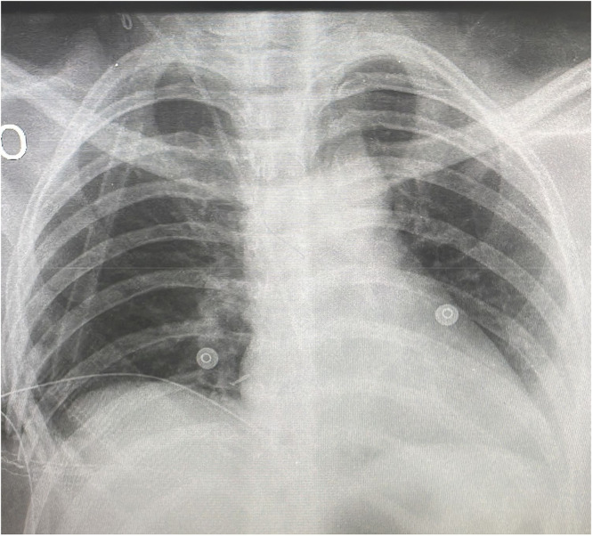 Figure 3
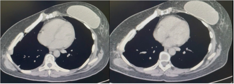 Figure 4
Peer Reviews
No public reviews on file for this paper yet. If you reviewed it on a platform where reviews are public (OpenReview, ICLR, NeurIPS, ICML), you can paste yours below so the community can read it here.
Videos
No videos yet. Explain this paper in a talk, walkthrough, or lecture? Add one.
Taxonomy
TopicsSurgical site infection prevention · Pleural and Pulmonary Diseases · Management of metastatic bone disease
Introduction
Extensive chest wall reconstruction is still a challenging situation in thoracic surgery. The surgical purpose can vary from cosmetic, infection, or oncological treatment. Resections due to cancer are frequently discussed in multidisciplinary teams, generally composed of oncologists and thoracic and plastic surgeons. The association between the thoracic team and plastic surgeon generally allows extensive resection and ensures sufficient free margin.1 Soft tissue coverage is the main contribution of plastic surgeons, per se, optimizing results.
On the other hand, the necessity of a prosthesis is decided by the thoracic surgeon. Anterolateral defects require prosthetic replacement in defects ≥ 5 cm in diameter or including ≥ 4 ribs.2 The posterior defects, even 10 cm in size, do not require reconstruction because of the scapula and shoulder girdle support.2 Care should be taken in defects lower than the fourth rib posteriorly to avoid trapped scapula.
Le Roux and Sherma3 have already punctuated that the ideal prosthesis should be (I) Rigidity to abolish paradoxical movement; (II) Inertness to allow in-growth of fibrous tissue and decrease the likelihood of infection; (III) Malleability to fashion to the appropriate shape at the time of operation; and (IV) Radiolucency to ensure a better follow up. Unfortunately, the perfect match is still not found. New options for reconstruction need to be considered, despite the indication for prosthetic replacement still the same for years. The Vitagraft® is now a promising alternative. Consisting of a synthetic, biocompatible, absorbable, non-cytotoxic, non-immunogenic, and non-pyrogenic, it works as an osteoinductor and osteoconductor, for bone regeneration. Composed of a nanometric ceramic of Tricalcium Phosphate in the β-phase (β-TCP) and the copolymer Polylactic Glycolic Acid (PLGA). Although Vitagraft® is frequently seen in orthopedic scenarios, there is no evidence of chest wall surgery before. So, this study intends to assess the safety of use in extensive chest wall reconstruction.
Methods
A prospective ongoing study in which patients were submitted to extensive chest wall resection and reconstruction based on Vitagraft®. This study was approved by the Ethics Committee (EC) of the institution where the work was carried out, under the number 56091721.6.0000.0068. All patients had to fill out the informed consent before the procedure. Inclusion criteria: chest wall resection no matter the purpose, oncological or palliative treatment. Exclusion criteria: chest wall resection due to infectious disease, patients without clinical conditions to surgical procedure, loss of follow-up. Each case was followed for a minimum period of three months after the surgical intervention. The following variables will be considered: KPS, histological type, previous local surgical treatment, the time between surgeries, defect size, type of myocutaneous flap, and mortality.
Results
Demographic DATA was summarized in Table 1. From the total of eleven patients, eight resections were performed due to tumor findings, Table 2, Table 3. One patient was submitted to surgery as a consequence of a sternal cleft, and another because of Poland’s syndrome. One sternectomy was a consequence of late sternal dehiscence Table 2 and Fig. 1. Primary closure was evidenced in 63.6 % of the patients, while Latissimus dorsi was necessary once, and abdominal flaps were necessary in two cases, Table 4. Vitagraft® was used in association with polypropylene mesh (PPM) a total of eight times, Fig. 2. The Vitagraft® without association was used in two patients and once with bovine pericardium. Reoperation was required in two cases, and prosthesis removal was necessary for one of them. Paradoxical chest movement, respiratory failure, bleeding intractable chest pain, and major systemic complications were not evidenced. The area of defect size was calculated by multiplying the bottom-to-top measure by the side-to-side measure. The median was 60 cm^2^, varying from 7.5 cm^2^ to 324 cm^2^.Table 1. Demographic DATA.Table 1KPSaMedian 100AgeMedian 46SexFemale 8 (73 %)Male 3 (27 %)Smoking2 (18 %)Diabetes2 (18 %)Obesity2 (18 %)COPDb1 (0.09 %)Hypertension1 (0.09 %)Alcoholism1 (0.09 %)CADc1 (0.09 %)Coronary disease1 (0.09 %)aKarrnofsky.bChronic Obstructive Pulmonary Disease.cChronic Artery Disease.Table 2. Reconstruction and main indication.Table 2. Reconstruction indicationNumber (%)Tumor8 (72.7 %)Sternal cleft1 (0.09 %)Poland’s syndrome1 (0.09 %)Sternal dehiscence1 (0.09 %)Total11 (100 %)Table 3. Tumor Types.Table 3. TumorNumber (%)Chondrosarcoma3 (37.5 %)Breast cancer1 (12.5 %)Phyllodes1 (12.5 %)Chondroma1 (12.5 %)Fibrous dysplasia/Multiple Exostosis2 (25 %)Total8(100 %)Fig. 1. Vitagraft® tailored made after two distinct sub-total sternectomies.Fig 1. Table 4Types of muscle flaps and Vitagraft® use.Table 4. Reconstruction (closure)Number (%)Primary closure7 (63.6 %)Latissimus dorsi1 (0.09 %)Abdominal flap3 (27 %)Vitagraft® use**Number (%)**Polypropylene mesh8 (72.7 %)Lonely2 (18 %)Bovine pericardium1 (0.09 %)Total11 (100 %)Fig. 2. First the Vitagraft® presentation used in chest wall reconstruction. Second the two Vitagraft® ribs in association with PPM.Fig 2
Discussion
The safety assessment was necessary even because Vitagraft® is already used in orthopedics, and neurosurgery but not in chest wall reconstruction. The present study is the first to evaluate Vitagraft® application regarding extensive chest wall reconstruction. No major complications were evidenced either during surgery or the following period. Local complications were noticed twice.
Local complications are frequently seen in chest resections. Bad outcomes are reported to occur in 37 % to 46 %4 of patients. Aghajanzadeh, M5 reported wound complications such as infection, dehiscence, flap loss, and hematoma in 8 % to 20 %. Chest X-ray was good at showing prosthesis radiolucency, Fig. 3, but computed tomography (CT) should be the primary imaging modality for the assessment of the local complications. Imaging has information about the prosthesis and potential identification of fluid and air accumulation adjacent to the prosthesis, which is indicative of deeper wound infection or associated pleural empyema.6 In addition, respiratory complications such as pneumonia, atelectasis, and respiratory failure are reported in 18 %, 20 % to 24 %5 of cases as a consequence of a flail segment of the chest wall. Respiratory problems were not noticed in our experience due to the absence of paradoxical chest movement. Besides, wound complications requiring a second operation were necessary for 28 %, and removal was mandatory for 14 %. The management of wound infections should be tailored to the severity of the infection (imaging clinical aspect presentation), and underlying disease. The first case was not associated with mesh. The female patient with previous breast reconstruction, in which the silicon prosthesis was damaged before the chest resection. The late operative wound remained producing fluid and the tomography showed only a partial ossification of the prosthesis. The authors do believe that all of these factors contributed to the redo surgery. The second complication was reconstructed in association with mesh and appeared to be a consequence of soft tissue cover, mainly a compound of fatty tissue despite muscle and a lack of alternatives for prosthesis presentation. In this case, the material was much less solid and the reconstruction was performed with a single Vitagraft® rib. The material was fractured and removed during the follow-up period. The authors suggested some changes after this episode to increase strength. Now it maintains the same ticking, but the structure is reinforced by some parallel solid bars. In addition, the authors now reconstruct the defect at least with two Vitagraft® ribs, not one as used to be.Fig. 3. Chest X-Ray showing prosthesis radiolucency.Fig 3
Comparisons between different types of prostheses are frequently seen. In some cases, the prosthetic material used depends on the surgeon’s preference and experience.7 Deschamps C8 showed no significant difference in the postoperative outcome or complications between prolene mesh and polytetrafluoroethylene (PTFE) soft tissue patch for chest wall reconstruction. Weyant M4 compared rigid (marlex mesh associated with methylmethacrylate sandwich(MMM) to non-rigid reconstruction consisting either of polypropylene(PPM) alone or expanded PTFE. There was no significant difference in respiratory complications among prosthetic groups, but MMM prostheses had a significantly higher number of wound complications.
Until now, the three-month postoperative chest tomography has been showing good prosthesis biocompatibility, Fig. 4, and safe use. The wound complication rate is similar to the literature but not related to respiratory failure. The use of this new prosthesis does not seem to increase inflammatory reaction as a consequence of reabsorption and ossification as time goes by. The authors need to increase the sample size and provide further details about the ossification time, especially relating to the size of the resection. The authors continue to collect information for further publications when the study is over.Fig. 4CT-Scan showing the reconstruction after three- and seven-months respectability.Fig 4
Authors disclosure and conflicts of interest
This work was supported by DMC equipment. The industry is responsible for Vitagraft® development.
Note
Artificial intelligence was not used as a tool in this study.
Conflicts of interest
The authors declare no conflicts of interest.
The reference list from the paper itself. Each links out to its DOI / PubMed record.
- 1Levy Faber D.Fadel E.Kolb F.Delaloge S.Mercier O.Mussot S.Outcome of full-thickness chest wall resection for isolated breast cancer recurrence Eur J Cardiothorac Surg 44420136376422346072410.1093/ejcts/ezt 105 · doi ↗ · pubmed ↗
- 2Sanna S.Brandolini J.Pardolesi A.Argnani D.Mengozzi M.Dell'Amore A.Materials and techniques in chest wall reconstruction: a review J Vis Surg 32017952907865710.21037/jovs.2017.06.10PMC 5638032 · doi ↗ · pubmed ↗
- 3Le Roux B.T.Sherma D.M.Resection of tumors of the chest wall Curr Probl Surg 2061983345386685162910.1016/s 0011-3840(83)80007-0 · doi ↗ · pubmed ↗
- 4Weyant M.J.Bains M.S.Venkatraman E.Downey R.J.Park B.J.Flores R.M.Results of Chest Wall Resection and Reconstruction with and Without Rigid Prosthesis Ann Thorac Surg 81120062792851636838010.1016/j.athoracsur.2005.07.001 · doi ↗ · pubmed ↗
- 5Aghajanzadeh M.Alavy A.Taskindost M.Pourrasouly Z.Aghajanzadeh G.Massahnia S.Results of chest wall resection and reconstruction in 162 patients with benign and malignant chest wall disease J Thorac Dis 222010818522263024 PMC 3256447 · pubmed ↗
- 6Lampridis S.Minervini F.Scarci M.Management of complications after chest wall resection and reconstruction: a narrative review J Thorac Dis 16120247377493841058710.21037/jtd-23-621PMC 10894423 · doi ↗ · pubmed ↗
- 7Gonfiotti A.Salvicchi A.Voltolini L.Chest-Wall Tumors and Surgical Techniques: State-of-the-Art and Our Institutional Experience J Clin Med 1119202255163623338410.3390/jcm 11195516 PMC 9573184 · doi ↗ · pubmed ↗
- 8Deshamps C.Tirnaksiz B.M.Darbandi R.Trastek V.F.Allen M.S.Miller D.L.Early and long-term results of prosthetic chest wall reconstruction J Thorac Cardiovasc Surg 11731999588-92; discussion 591-210.1016/s 0022-5223(99)70339-910047664 · doi ↗ · pubmed ↗