Comparative Clinical Outcomes of Reconstructive Flap Techniques for Exposed Achilles Tendon: A Prospective Study
Vijaykumar Huded, Gireesh Khodnapur, Ajayguru Senthurvelan, Charan Sai Reddy Munugala

TL;DR
This study compares different surgical techniques for repairing exposed Achilles tendons and finds the lateral calcaneal artery flap to be the most effective.
Contribution
The study provides a comparative analysis of reconstructive flap techniques for Achilles tendon exposure based on clinical outcomes.
Findings
The lateral calcaneal artery flap showed the best results in terms of aesthetics, function, and minimal complications.
One reverse sural flap case resulted in complete necrosis requiring additional surgery.
The sural artery flap was effective for medium to large defects but had some complications.
Abstract
Background: Plastic surgeons continue to face considerable challenges while reconstructing exposed Achilles tendon. Several alternatives for repairing such defects include the reverse sural artery flap, peroneus brevis flap, propeller flap, lateral calcaneal artery flap, and peroneal artery perforator flap. However, each of these techniques has its pros and cons. This study compares our clinical experience to various reconstructive techniques for exposed Achilles tendon. Methods: Fifteen individuals had their exposed Achilles tendon reconstructed between October 2023 and July 2024. These fifteen cases were classified based on the origin of the wound, comorbidities, operational results, and complications, which were analyzed using a prospective study. Results: The average age of the patients was 40. All 15 patients tolerated the flap procedure well without significant complications;…
Genes, proteins, chemicals, diseases, species, mutations and cell lines named across the full text — each resolved to its canonical identifier and authoritative record.
Click any figure to enlarge with its caption.
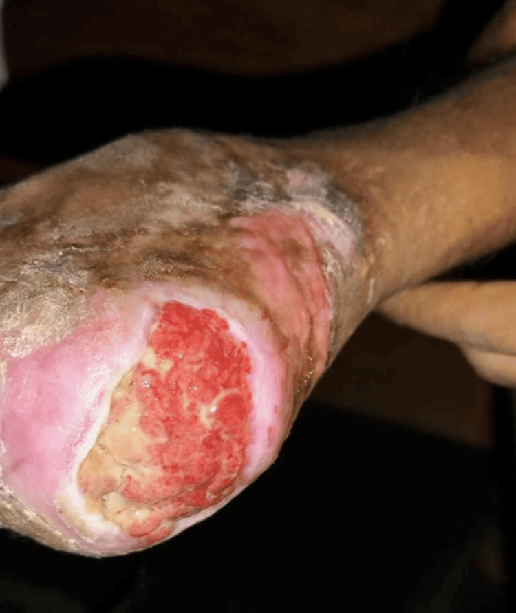 Figure 1
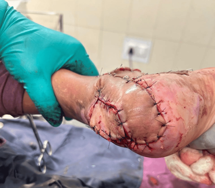 Figure 2
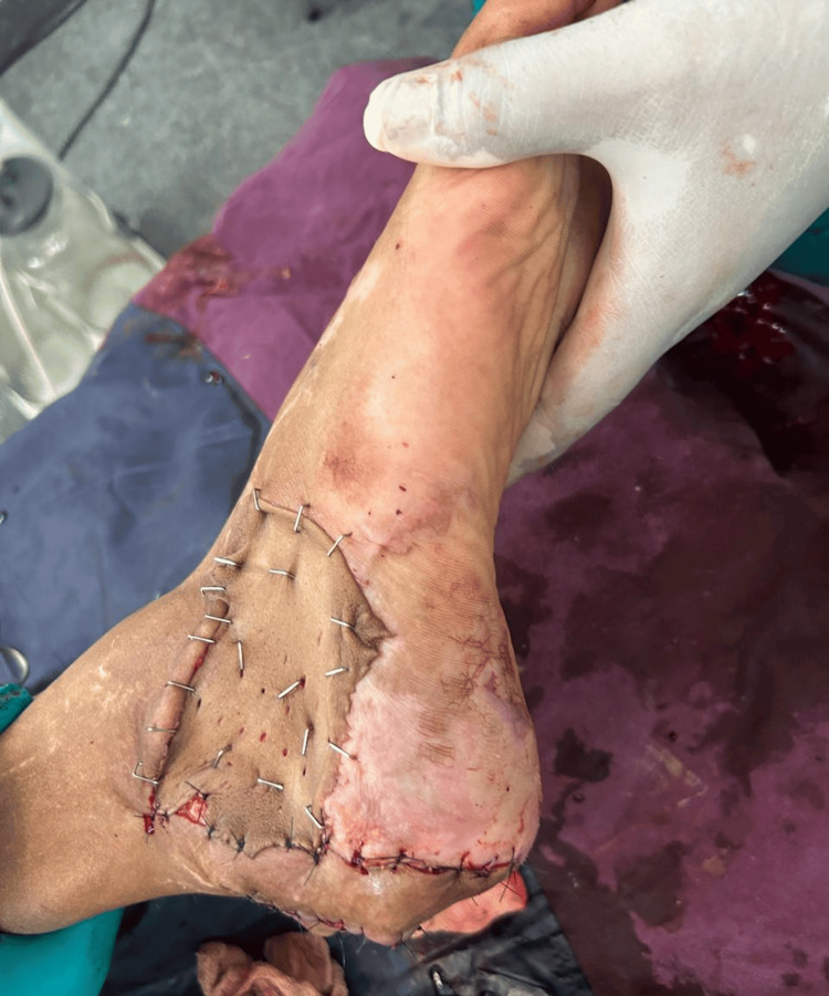 Figure 3
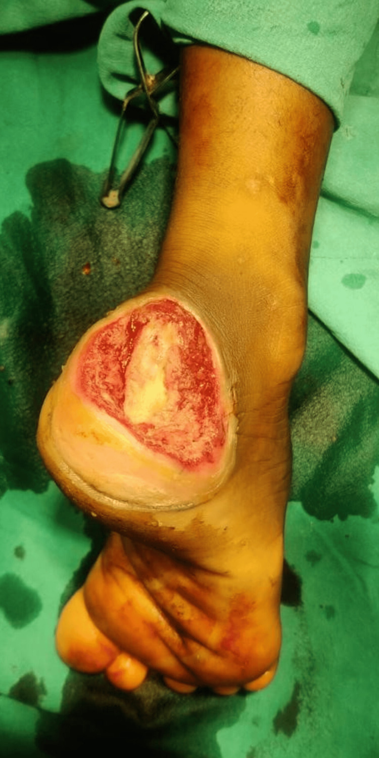 Figure 4
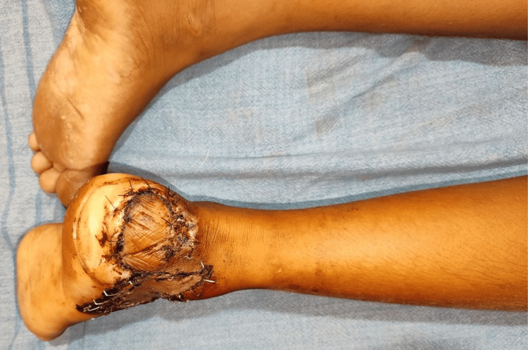 Figure 5
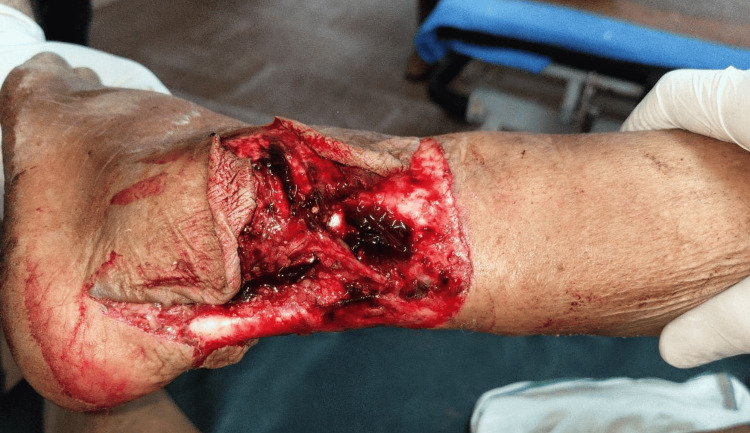 Figure 6
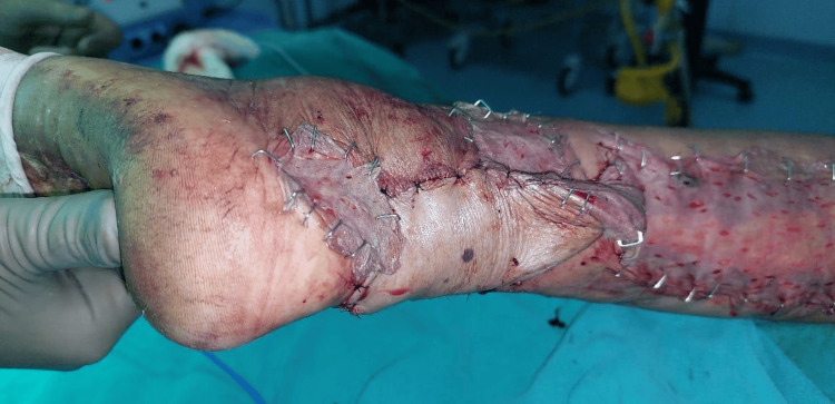 Figure 7
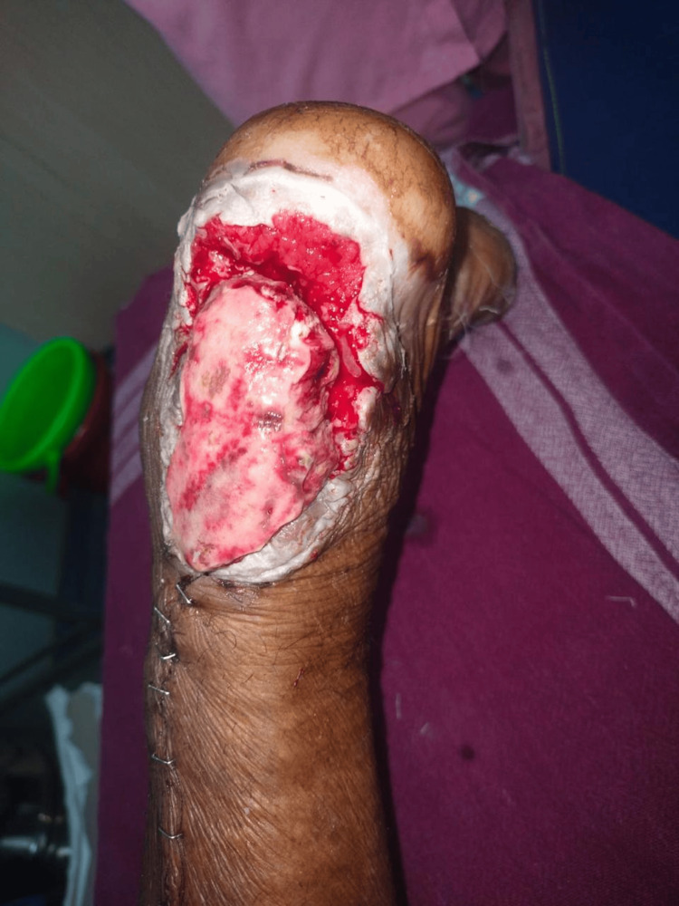 Figure 8
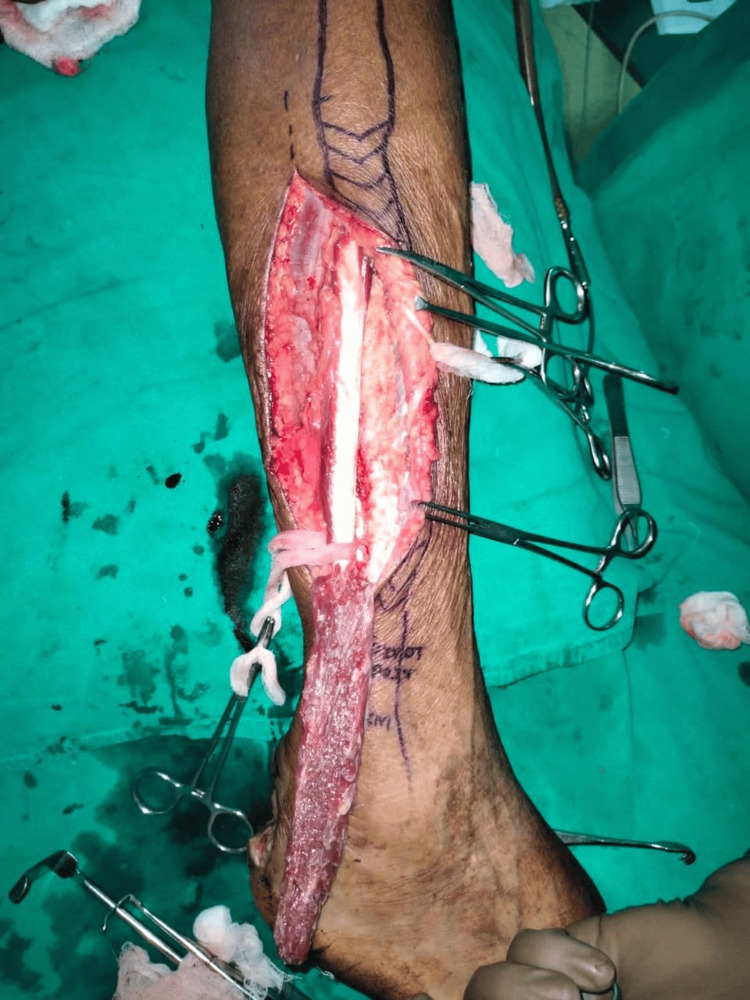 Figure 9
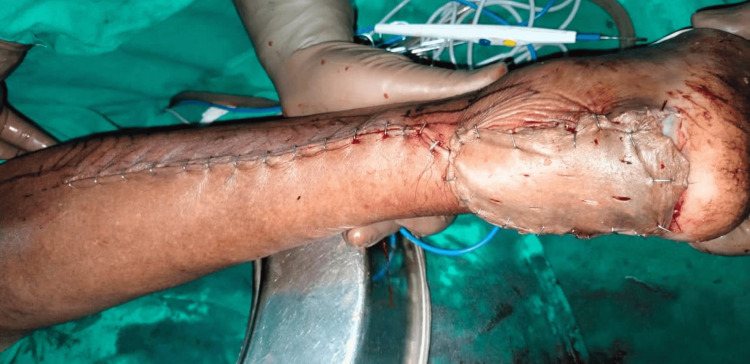 Figure 10
 Figure 11
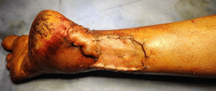 Figure 12
| Serial number | Age/sex | Mode of injury | Occupation | Comorbidities | Diagnosis | Procedure | Complications | Re-intervention |
| 1 | 24/M | Road traffic accident | Daily wager | - | Exposed Achilles tendon with heel defect (distal most part and posterior and lateral defects of hind foot) | Lateral calcaneal artery flap | - | - |
| 2 | 8/M | Road traffic accident | Schooling | - | Exposed Achilles tendon (distal most part and posterior and lateral defects of hind foot) | Lateral calcaneal artery flap | - | - |
| 3 | 42/M | Road traffic accident | Farmer | - | Exposed Achilles tendon (distal most part and posterior and lateral defects of hind foot) | Lateral calcaneal artery flap | Distal tip necrosis | Healed by secondary intention |
| 4 | 35/M | Road traffic accident | Driver | - | Exposed Achilles tendon with heel defect (on lateral aspect of heel) | Peroneal artery flap | - | - |
| 5 | 50/M | Road traffic accident | Farmer | Diabetic | Exposed Achilles tendon (distal most part and posterior and lateral defects of hind foot) | Lateral calcaneal artery flap | - | - |
| 6 | 35/F | Road traffic accident | Housewife | - | Exposed Achilles tendon (proximal and mid Achilles tendon defects) | Islanded reverse sural artery flap | - | - |
| 7 | 60/M | Road traffic accident | Farmer | Hypertensive | Exposed Achilles tendon | Peroneus brevis flap | - | - |
| 8 | 40/M | Road traffic accident | Tailor | - | Exposed Achilles tendon | Reverse sural flap | Infection | - |
| 9 | 50/M | Road traffic accident | Butcher | Diabetic | Exposed Achilles tendon | Propeller flap (PTA) | - | - |
| 10 | 35/F | Pothole injury | Housewife | - | Exposed Achilles tendon | Reverse sural flap | Complete necrosis | Split-thickness skin graft |
| 11 | 41/M | Road traffic accident | Weaver | - | Exposed Achilles tendon | Reverse sural flap | - | - |
| 12 | 48/M | Road traffic accident | Farmer | - | Exposed Achilles tendon | Reverse sural flap | - | - |
| 13 | 50/M | Road traffic accident | Daily wager | - | Exposed Achilles tendon | Peroneal artery flap | - | - |
| 14 | 37/M | Road traffic accident | Daily wager | - | Exposed Achilles tendon (on lateral aspect of heel) | Peroneal artery flap | - | - |
| 15 | 53/M | Road traffic accident | Driver | Diabetic | Exposed Achilles tendon (on lateral aspect of heel) | Peroneal artery flap | - | - |
Peer Reviews
No public reviews on file for this paper yet. If you reviewed it on a platform where reviews are public (OpenReview, ICLR, NeurIPS, ICML), you can paste yours below so the community can read it here.
Videos
No videos yet. Explain this paper in a talk, walkthrough, or lecture? Add one.
Taxonomy
TopicsTendon Structure and Treatment · Shoulder Injury and Treatment · Knee injuries and reconstruction techniques
Introduction
The Achilles tendon is one of the longest and strongest tendons in the body. The main function of Achilles tendon is plantar flexion, walking, running, and jumping as it pulls on the heel upon flexion of the calf muscles, acts as a shock absorber while walking, and provides ankle stability [1,2].
Due to its anatomic peculiarities like poor blood supply, being the thickest tendon, absence of synovial sheath, and its 90-degree winding on its path towards the heel, etc., reconstruction and resurfacing of post-traumatic soft tissue defects of the Achilles tendon region is a challenging task for plastic surgeons [3].
Plastic surgeons continue to face challenges in reconstructing exposed Achilles tendon abnormalities, particularly in patients suffering from peripheral vascular disease [4], which can have abnormalities in circulation, and in patients with chronic wounds due to high-intensity trauma [5]. The literature reports a variety of options for covering Achilles tendon soft tissue abnormalities [6].
Many papers have been published regarding the coverage options of the exposed Achilles tendon region. However, the ideal resurfacing options should be stable and thin enough to allow footwear and should provide a gliding surface for the Achilles tendon [7].
Therefore, the main aim of our study is to analyze and determine the suitable reconstruction options for Achilles tendon defects. We also aim to evaluate and assess which flap is better for Achilles tendon defects.
In this work, we report on our experience covering Achilles tendon abnormalities successfully in 15 patients utilizing different flap methods.
Materials and methods
A prospective study was designed to study 15 patients admitted to the Department of Plastic Surgery at Shri B M Patil Medical College, Hospital, and Research Centre, BLDE (Deemed to Be University), Vijayapura, India, for the period of nine months from October 2023 to July 2024. Data was gathered with proper follow-up of the patients postoperatively at two weeks, three months, and six months. The inclusion criteria include all the patients presenting to our BLDE emergency with exposed Achilles tendon due to loss of skin, injuries to the Achilles tendon region, and delayed cases of Achilles tendon region injuries, which are primarily treated at peripheral hospitals. Exclusion criteria include patients with life-threatening injuries, tendon defects, and fractures.
Age, sex, site and side of the foot affected, treatment involved, and results were all included. The wounds of the patients were cleaned and assessed. Debridement and regular dressing were done until the wound was prepared for the procedure. Regular X-rays were done to rule out any osteomyelitic changes.
Operative technique
Under general or spinal anesthesia, all the patients were placed in supine, prone, or lateral positions according to the location of the wound and the flap used for coverage; a hand-held Doppler was used to detect the perforators and locate the pedicle position. All the surgical procedures were used using loupe magnification (2.5-4.5x) and under tourniquet control.
Flap planning was done according to the planning in reverse method, where a plastic surgeon makes use of a lint or cotton and mocks the transfer of the flap by starting from the defect and working backward to the potential donor site. The choice of flap was determined by the defect, site, size, and nature of the wound.
According to the defect size and location, a lateral calcaneal artery flap was done in four patients (Figures 1-5), a peninsular reverse sural artery flap was done in four patients (Figures 6-7), an islanded reverse sural artery flap, a peroneus brevis (Figures 8-10), a peroneal artery perforator flap was done in four patients (Figures 11-12), and a posterior tibial artery-based propeller flap were done in one patient each.
Preoperative lateral calcaneal artery flap
Postoperative lateral calcaneal artery flap
Postoperative donor defect of lateral calcaneal artery flap with split-thickness skin graft
Preoperative lateral calcaneal artery flap
Postoperative lateral calcaneal artery flap
Preoperative reverse sural artery flap
Postoperative reverse sural artery flap
Preoperative peroneus brevis muscle flap
Intraoperative peroneus brevis artery flap
Postoperative peroneus brevis artery flap
Preoperative peroneal artery perforator based flap
Postoperative peroneal artery perforator based flap
The color and pinpricks of the flap were used to frequently assess its vascularity. Peripheral flap necrosis was treated by debriding the affected area and then closing the wound with a secondary purpose. During their postoperative follow-up appointments, patients had their foot functionality assessed for movements.
Postoperative care
A soft bandage was used to cover the flap site, and a small area was left exposed to check the flap viability, flap condition, infection, walking activities, and thinning of flaps in case of bulky flaps. The operated limb was kept elevated to facilitate venous drainage and reduce edema and pain. The flap was monitored regularly for any vascular insufficiency. The first dressing was done on the first operative day, broad-spectrum antibiotics were administered for seven days postoperatively, and follow-up was done for at least a six-month period.
Results
The average age of the patients was 40 years. The mean interval period from the time of injury to presentation was seven days (range: 5-9 days). Initially, patients were treated by orthopedic surgeons and later referred to plastic surgeons. Immediate coverage was not done due to extensive infection, the clinical condition of the patient at the time of presentation, and also due to the delayed consent from the patient and the patient attenders, who generally seek intervention from hospitals in metro cities, which explains the reason for the delay. Fourteen patients survived the flap with no serious problems; however, one patient with reverse sural flap had complete necrosis, and a secondary split-thickness skin graft was done. One more patient had distal tip necrosis and healed it with secondary intention. One patient developed an infection, which was resolved by debridement and consecutive dressings with antimicrobial therapy, which was given according to the culture and sensitivity study. None of the patients had any other functional deficits in their lower limbs.
In our study, lateral calcaneal artery flap was done in four (26.67%) patients (Figures 1-5), peninsular reverse sural artery flap was done in four (26.67%) patients (Figures 6-7), islanded reverse sural artery flap was done in one (6.67%) patient, peroneus brevis muscle graft was done in one (6.67%) patient (Figures 8-10), peroneal artery perforator flap was done in four (26.67%) patients (Figures 11-12), and posterior tibial artery flap was done in one (6.67%) patient.
Of the fifteen cases, nine were right-sided, and the rest six cases were left-sided injuries. The details are listed in Table 1.
Discussion
Reconstruction is frequently complicated by Achilles tendon site abnormalities due to their highly functional nature, poor vascular supply, tendinous/bony substrate, and constant movements, and for the very reasons, conservative care is insufficient. The use of grafts (full-thickness or split-thickness) often results in unsightly and functionally unsatisfactory outcomes, including intermittent hypertrophy and scar contracture. Free flap transfers, however, are technically difficult, necessitating a great deal of microsurgical skill and a lengthy surgical procedure, with a high risk of severe perioperative morbidity.
The aim of reconstructive surgery is to replace "like with like" wherever possible and to have flexible soft tissue covering to aid with skeletal repair [8]. To be able to walk, one must maintain both motor and sensory abilities. Tissue loss, location of the defect, size, vascular pedicle length required, and condition of the wound should be taken into account while choosing the optimum reconstructive procedure. The foot is a difficult region to repair and is vulnerable to illness and trauma. To achieve the best practical and cosmetic results, it is best to think about foot rebuilding by subunits. Due to their traumatic origin in the younger population, tissues with sufficient blood supply, high flexibility, and sensibility are needed for the reconstruction of exposed Achilles tendon lesions [9]; otherwise, morbidity may result.
The lateral calcaneal artery flap is a flap for covering posterior heel defects; it is mainly based on the lateral calcaneal artery. This is one of the feasible options for flap cover over the hindfoot [10]. This flap has a number of benefits, including a high rate of success, less postoperative discomfort, adequate coverage of soft tissue, and a positive functional outcome (Figures 1-5). Alam et al.'s [11] study found that the lateral calcaneal artery flap [12] is superior to other reconstruction techniques for posterior heel and tendon Achilles deficits [13] in terms of function, donor area morbidity, and surgical time.
Unlike other authors who performed various microvascular tissue transfers and local flaps, we solely used reverse sural artery flaps (Figures 4-5) [14] for the coverage of deeper-grade injuries [15]. The wounds over the medial malleolus, heel, midfoot, and proximal defects benefit from reversal sural artery flaps. According to Farooq et al., the outcomes are similar in these situations, highlighting the significance of reverse sural artery flap [16]. This procedure is also a good choice in resource-constrained environments and operating rooms without microvascular tissue transfer capabilities.
Flap survival was complete and accomplished without major complications in 12-patient research by Ahn et al. using a pedicle graft based on a peroneal artery perforator; two patients had mild wound dehiscence. Still, these wounds responded favorably to conservative care and later debridement (Figures 11-12) [17]. There were no functional deficiencies in any of the patients' lower limbs.
One patient with a moderate defect was covered with a distally based peroneus brevis muscle flap (Figures 8-10) [18]. The main advantage of the peroneus brevis muscle flap was its technical simplicity and coverage of moderate to large defects involving the heel and anterior aspect of the ankle joint, the only disadvantage being ankle stiffness. According to a study done by Lorenzetti et al., a distally based peroneus brevis muscle flap was used for reconstructing the defects involving the exposed Achilles tendon, heel, and anterior aspect of the ankle [18].
In patients with diabetes who had delayed healing patterns, with strict glycemic control and wound care, the wound was healed properly.
The limitations of our study include a relatively small sample size and poor patient turnover for follow-ups. Additionally, we did not perform free flaps due to manpower limitations.
Conclusions
The flap based on the lateral calcaneal artery is the most common and useful flap for coverage of exposed Achilles tendon with respect to aesthetic appearance, good functional outcome, minimal donor morbidity, and time of surgery; it is the flap of choice for small- to medium-sized exposed Achilles. It is resistant to atherosclerosis, which is very helpful in treating patients with diabetes. The downside of this procedure is that it is technically challenging and is performed by skilled surgeons for moderate to large defects. Our study’s second most commonly used flap was based on the reverse sural artery. This flap can cover medium to larger defects. The main disadvantage of this procedure is that it causes venous congestion due to the arc of rotation. Similarly, other techniques also had their advantages and disadvantages.
To conclude, the lateral calcaneal artery flap is the most commonly used flap for distal-most Achilles tendon defects and hindfoot defects; the reverse sural artery flap is second most commonly used for moderate to large Achilles tendon region defects, the peroneus brevis flap is for moderate lateral Achilles tendon defects, and the propeller (peroneal) based artery is for smaller lateral Achilles tendon defects.
The reference list from the paper itself. Each links out to its DOI / PubMed record.
- 1Achilles tendon disorders BMJ Asplund CA Best TM 0346201310.1136/bmj.f 126223482943 · doi ↗ · pubmed ↗
- 2The anatomy of the Achilles tendon Foot Ankle Clin O'Brien M 2252381020051592291510.1016/j.fcl.2005.01.011 · doi ↗ · pubmed ↗
- 3Soft-tissue defects of the Achilles tendon region: management and reconstructive ladder. Review of the literature Injury Marchesi A Parodi PC Brioschi M 05347201610.1016/j.injury.2016.07.05327492062 · doi ↗ · pubmed ↗
- 4Distally based lateral supramalleolar adipofascial flap for reconstruction of the dorsum of the foot and ankle Plast Reconstr Surg Lee YH Rah SK Choi SJ Chung MS Baek GH 1478148511420041550993510.1097/01.prs.0000138751.53725.67 · doi ↗ · pubmed ↗
- 5Reconstruction of soft tissue defects of the Achilles tendon with rotation flaps, pedicled propeller flaps and free perforator flaps Microsurgery Jakubietz RG Jakubietz DF Gruenert JG Schmidt K Meffert RH Jakubietz MG 6086133020102085333910.1002/micr.20798 · doi ↗ · pubmed ↗
- 6Clinical aspects of reconstruction of the lower third of the leg with fasciocutaneous flap based on peroneal artery perforators Vojnosanit Pregl TerzićZ DjordjevićB 39457120142451698910.2298/vsp 1401039 t · doi ↗ · pubmed ↗
- 7Achilles region soft-tissue defects: a reconstructive algorithm based on a series of 46 cases J Reconstr Microsurg Innocenti M Innocenti A Ghezzi S Delcroix L 0733201710.1055/s-0037-160655428985638 · doi ↗ · pubmed ↗
- 8Soft tissue defects of the heel: a surgical reconstruction algorithm based on a retrospective cohort study J Foot Ankle Surg El-Shazly M Yassin O Kamal A Makboul M Gherardini G 1451524720081831292210.1053/j.jfas.2007.12.010 · doi ↗ · pubmed ↗