Protective effect of proteins extracted from Plumeria pudica latex on ethanol-induced gastric injury in mice
Lucas Arruda Moita, Bruna da Silva Souza, Naylla Veras de Moraes Oliveira, Ana Clara Silva Sales, Lucas Eduardo Silva Oliveira, Ana Patrícia de Oliveira, Francisca Beatriz Melo Sousa, Jand-Venes Rolim Medeiros, Jefferson Soares de Oliveira

TL;DR
Proteins from Plumeria pudica latex protect mice from stomach damage caused by ethanol, likely through antioxidant effects and increased mucus production.
Contribution
This study demonstrates the novel protective effect of Plumeria pudica latex proteins against ethanol-induced gastric injury in mice.
Findings
LPPp significantly reduced gastric tissue injury in mice compared to ethanol alone.
LPPp preserved tissue architecture and antioxidant levels similar to the control group.
LPPp positively influenced gastric mucus production, contributing to its protective effect.
Abstract
To evaluate proteins from Plumeria pudica latex (LPPp) for their protective effect against ethanol-induced gastric injury in mice. The LPPp fraction was obtained by collecting P. pudica latex in tubes containing distilled water, followed by centrifugation and dialysis. The female Swiss mice (Mus musculus) received saline or LPPp (40 mg/kg) intraperitoneally 1 hour before oral administration of 500 μL of 50% ethanol. One hour later, the animals were euthanized, and their stomachs were removed for evaluation of tissue lesion area, histopathological analysis, and measurements of malondialdehyde (MDA), glutathione (GSH), superoxide dismutase (SOD), and nitrate/nitrite (NO3/NO2). An independent experiment assessed the effect of LPPp on gastric mucus production. The LPPp-treated animals showed a significant reduction in the mean injured areas of gastric tissue (0.73 ± 1.01 mm2) compared to…
Genes, proteins, chemicals, diseases, species, mutations and cell lines named across the full text — each resolved to its canonical identifier and authoritative record.
Click any figure to enlarge with its caption.
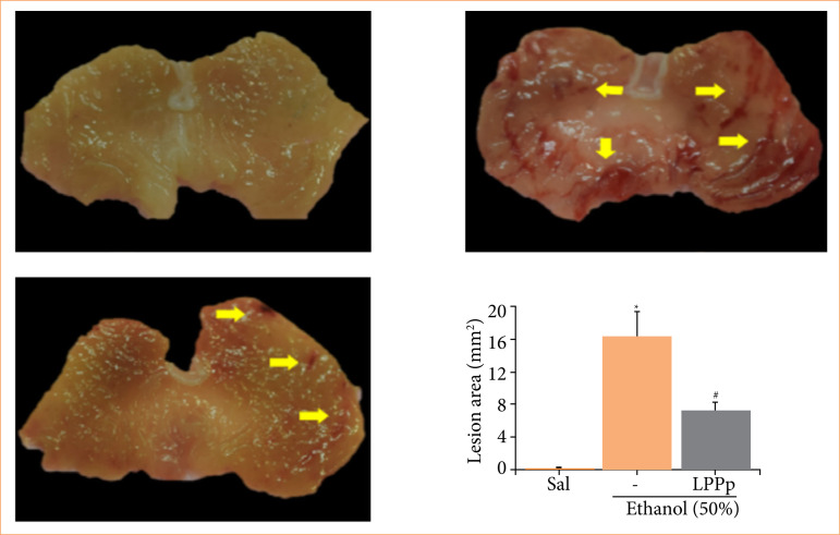 Figure 1
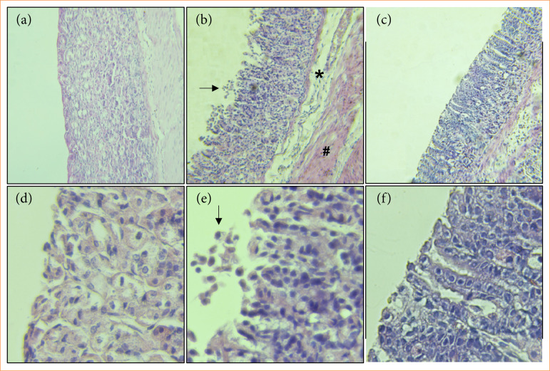 Figure 2
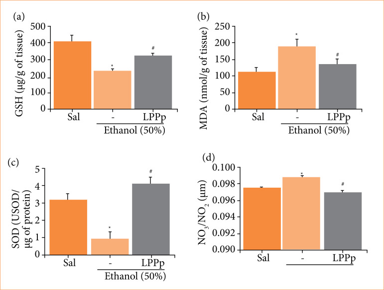 Figure 3
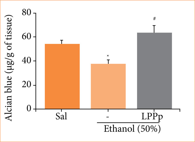 Figure 4
| Microscopic parameters | Score | Saline | Ethanol | LPPp |
|---|---|---|---|---|
| Epithelial cell loss | 0–3 | 0 (0–1) | 3 (2–3) | 1 (1–2) |
| Presence of Inflammatory cells | 0–3 | 1 (0–1) | 1 (1–1) | 1 (0–1) |
| Edema in the upper mucosa | 0–4 | 0 (0–1) | 4 (2–4) | 2 (1–2) |
| Hemorrhagic damage | 0–4 | 0 (0–1) | 3 (2–4) | 1 (0–2) |
| Total score | 0–14 | 1 (2–3) | 11 (9–10) | 5 (2–3) |
- —Conselho Nacional de Desenvolvimento Científico e Tecnológico
Peer Reviews
No public reviews on file for this paper yet. If you reviewed it on a platform where reviews are public (OpenReview, ICLR, NeurIPS, ICML), you can paste yours below so the community can read it here.
Videos
No videos yet. Explain this paper in a talk, walkthrough, or lecture? Add one.
Taxonomy
TopicsHelicobacter pylori-related gastroenterology studies · Phytochemistry and Bioactive Compounds · Phytochemical Studies and Bioactivities
Introduction
Gastric ulcer, also known as peptic ulcer, is a common digestive disease that has become a global problem, affecting 5–10% of the world’s population resulting in the expenditure of millions of dollars on healthcare1 ^–^ 3. Under normal circumstances, to protect the integrity and functionality of the gastric mucosa, the gastric mucosal barrier uses the mucus–bicarbonate–phospholipid barrier, the epithelial barrier, and the endothelial barrier4 ^,^ 5. The gastric ulcer is a complex and multi-factorial process, commonly produced by an imbalance between gastric mucosal protective and aggressive factors such as gastric acid, pepsin, reactive oxygen species (ROS) and Helicobacter pylori 6.
Alcohol is one of the most abused substances around in the world, which can cause upper gastrointestinal bleeding and peptic ulcers2 ^,^ 5. The excessive alcohol consumption is considered the main cause of gastric mucosal injury6. Excessive consumption of some alcoholic drinks is associated to human gastric damage, and the degree of injury is related to ethanol concentration and quantity7.
Pathogenesis of gastric ulcer includes the production of oxygen-derived free radicals such as superoxide anion radical, hydroxyl radicals, and lipid peroxides8 ^–^ 10. Thus, when the gastric mucosa is damaged, an unbalanced in pro-inflammatory cytokines is started, followed by neutrophils migration to the damaged site, and the concentration of ROS and other inflammatory mediators is increased, promoting an oxidative damage11. Additionally, ethanol changes mucosal permeability to gastric acid by causing mast cells, macrophages, and blood cells to release vasoactive products12. Alcohol also usually induces damage to the gastric mucosa promoting edema, erosion, ulcerative lesions, hemorrhage, and infiltration of inflammatory cells13.
Therefore, anti-oxidation and anti-inflammation activity seem to play an important role in protection of the gastric mucosa against damage7. At present available therapies do not provide definite cure of gastric ulcer. Thus, some alternative treatments are necessary12. Some studies have demonstrated the gastroprotective potential of some plants and its natural active constituents against experimental gastric injury in animal models14. Herbal medicines have been used as medicinal treatments that contain different gastroprotective mechanisms, including stimulation of mucosal proliferation, inhibition of acid production, and antioxidant properties15.
Plumeria pudica (Jacq., 1760) is an ornamental plant commonly known as bridal bouquet. It belongs to the Apocynaceae family, and is characterized by intense latex production16 ^–^ 18. A well-defined and rubber free protein fraction (LPPp) obtained from its latex previous demonstrated oxidative stress and inflammatory properties in different experimental models19 ^–^ 22, without promoting signs of toxicity in animals at therapeutic doses23.
The effect of latex proteins from P. pudica on ethanol-induced acute gastric mucosal damage has not been studied. Thus, this study evaluated the effect of this protein fraction on the acute gastric injury induced by ethanol.
Methods
Latex collection extraction of latex proteins
Plants of P. pudica from Parnaíba (PI), Brazil (2.9055°S, 41.7734°W), were used as the source of fresh latex. The plant material was identified and the voucher No. 2,432 was deposited in the Herbário Delta do Parnaíba from Universidade Federal do Piauí. The latex was collected in distilled water (1:1; v/v) and centrifuged at 3,600 × g for 15 min at 25°C. The supernatant was dialyzed against distilled water using membranes of 8 kDa. The dialysis water was renewed every six hours and centrifuged again using the conditions described above. The supernatant was lyophilized and named latex proteins from P. pudica (LPPp) and used for further experiments.
Animals
Female Swiss mice (Mus musculus) weighing 25–30 g were supplied by the Universidade Federal do Piauí. Animals were housed in cages with free access to food and water and were maintained under a 12-h light–dark cycle (lights on at 6 a.m.) at 24 ± 2°C. All experimental procedures were performed as specified by the Guide for Care and Use of Laboratory Animals (National Institute of Health, Bethesda, MD, United States of America), and Institutional Animal Ethics Committee from Universidade Federal do Piauí, that approved the project (Protocol No. 470/18).
Ethanol-induced gastric damage
The experiments were conducted following the method described by Robert et al.24, with some modifications. The mice were divided into three groups (5–8 animals):
Saline group (SAL): negative control;Ethanol 50% group: positive control;Experimental group, treated with LPPp at 40 mg/kg.
The dose of 40 mg/kg was chosen since it was the best dose that LPPp showed anti-inflammatory and antioxidant activity in other in-vivo experimental models19 ^–^ 22, and it was the dose recommended by Institutional Animal Ethics Committee to perform our evaluation.
Before the experiment, the animals were starved of food for 15 hours and water for 2 hours. After this period, gastric lesion was induced by single administration of 0.5 mL/25 g of 50% ethanol by gavage. The negative control group received an equivalent volume of saline solution (0.9%). Animals were treated with LPPp (40 mg/kg, intraperitoneally) 1 hour before ethanol administration. One hour after administration of ethanol, all animals were euthanized, their stomachs were immediately removed, opened via an incision along the greater curvature and pinned out on a wax block. Gastric damage was measured using a computer planimetry program (Image J). A sample of the corpus region of each stomach was fixed in 10% formalin immediately after removal for subsequent histopathological assessment. Further, gastric corpus samples were frozen and stored at -80°C for biochemical analysis.
Histopathological analysis
For histopathological evaluation, corpus region of each stomach fixed in 10% formalin solution were sectioned and embedded in paraffin. Four-micrometer-thick sections were deparaffinized, stained with hematoxylin and eosin, and then examined under a microscope. Samples were then analyzed in a blind study (without knowledge of the previous treatments) by an experienced pathologist using procedure adapted25. Briefly, it was examined 1-cm-long sections for epithelial cell loss (a score of 0–3), presence of inflammatory cells (a score of 0–3), edema in the upper mucosa (a score of 0–4), and haemorrhagic lesion (a score of 0–4).
Determination of adhered mucus to gastric wall
The mucus content determination was performed as described by Corne et al.26 after ethanol induced gastric ulcer. Segment of the glandular region of the stomach was weighted and transferred to a test tube containing 3 mL of 0.1% Alcian blue for 2 hours. After two consecutive rinses with 3 mL of sucrose (0.25 M), 3 mL of MgCl2 (0.5 M) was added in each test tube. The glandular segment remained in this solution for 2 h, with intermittent agitation. Afterwards, 4 mL of the resultant blue solution was agitated vigorously with 4 mL of ethyl ether until the formation of an emulsion and centrifuged at 3,600 × g for 10 min. The absorbance of the supernatant was read at 598 nm. The concentration of Alcian blue was expressed in µg Alcian blue/g of glandular tissue.
Glutathione concentration
The concentration of reduced glutathione (GSH) in stomach tissues as nonprotein sulfhydryl was estimated using the technique described by Sedlak and Lindsay27. Results were expressed as µg of GSH/g of tissue.
Malondialdehyde levels
The level of malondialdehyde (MDA) in stomach homogenate samples was measured using the method described by Mihara and Uchiyama28. Results were expressed as nmol of MDA/g of tissue.
Superoxide dismutase activity
Superoxide dismutase (SOD) activity was measured using spectrophotometric assay decribed by Das et al.29. In addition, total proteins concentration in each homogenate sample was determined with a commercial kit from Labtest. Results were expressed as unit of SOD (USOD)/μg of protein.
Levels of nitrate/nitrite
The level of nitric oxide (NO) was obtained by quantifying the NO metabolites nitrate (NO_3_ ^-^) and nitrite (NO_2_ ^-^) in the gastric tissue, according to the method described by Green et al.30. Results were expressed as μM of NO_3_/NO_2_.
Statistical analysis
The results were expressed as mean ± standard error of mean (± SEM) of n = 5–8 animals. Differences between groups were evaluated using analysis of variance and the Student–Newman–Keuls post-test, when appropriate. Moreover, the Kruskal-Wallis nonparametric test, followed by Dunn’s test, were used in histopathological analyses. Difference between groups were considered statistically significant when p < 0.05. Statistical analysis was performed using GraphPad Prism statistical software, version 7.0.
Results
Latex proteins from Plumeria pudica reduced injured areas in stomachs
Macroscopic analysis of the gastric mucosa of animals demonstrated that LPPp was able to protect the frontal mucosa from injuries produced by ethanol. In the ethanol group, extensive tissue lesions were observed in the form of stretch marks when compared to the saline group (Figs. 1a and b). In contrast, the animals in the LPPp group, that also received ethanol, had fewer areas of lesions (Fig. 1c). Moreover, computerized planimetric measurement of the injured areas in the stomach of all groups indicated that the animals treated with LPPp had areas of injury in the gastric wall significantly smaller (7.28 ± 1.00 mm^2^) than those of the animals belonging to the ethanol group (16.30 ± 3.11 mm2) (Fig. 1d). The effect produced by LPPp corresponded to 56% of inhibition of lesion.
Effect of proteins extracted from Plumeria pudica latex (LPPp) on injured areas in stomachs of ethanol-induced gastric lesion. (a) Saline group. (b) Ethanol. (c) Mice were treated with LPPp (40 mg/kg) intraperitoneally. After 30 min, (d) the animals in experimental groups were administered 50% ethanol. (a–c) Images of stomachs were captured, and (d) lesion areas assigned in the planimetric software were expressed in mm2. Yellow arrows indicate the presence of lesion areas. Results are expressed as mean ± standard error of the mean of 5–8 animals per group. Pictures are representative samples of tissues from every animal.
Latex proteins from Plumeria pudica inhibited histopathological alterations of gastric tissue
Oral administration of ethanol disrupts the integrity of the gastric mucosa with excessive loss of epithelial cells, which causes a rupture in the surface of the mucosa, besides accentuating edema and hemorrhage (Fig. 2). However, administration of LPPp maintained the integrity of the mucosa, showing that LPPp exerted a potential gastroprotective effect on this lesion. The histopathological evaluation showed that LPPp significantly decreased epithelial cell loss, edema, and hemorrhagic damage induced by ethanol administration (Fig. 2c and 2f, Table 1). No significant difference was observed in inflammatory cell.
Effect of proteins extracted from Plumeria pudica latex (LPPp) on microscopic changes in stomachs of ethanol-induced gastric lesion. (a, b and c) Magnification of 100x. (d, e and f) Magnification of 400x. (a and d) Gastric mucosa of mice from saline group with normal architecture. (b and e) Presence of edema (), haemorrhagic areas (#) and los of architecture (arrows) in ethanol group. (c and f) Stomach of animals treated with LPPp (40 mg/kg; intraperitoneally) which exerted a protective effect on gastric mucosa with preservation of epithelial cells and absence of haemorrhages. Pictures are representative samples of tissues from every animal. Quantitative results from these assessments are shown in Table 1.*
Latex proteins from Plumeria pudica increased glutathione concentration
The measurement of GSH in the gastric tissue of animals indicated that animals receiving only ethanol had a significant consumption of GSH in the mucosa (231.3 ± 16.3 µg/g of tissue) compared to the saline group (406.7 ± 39.8 µg/g of tissue) (Fig. 3a). However, compared to ethanol group, pre-treatment of animals with LPPp induced an increment of GSH in the mucosa (321.8 ± 17.7 µg/g of tissue).
Effect of proteins extracted from Plumeria pudica latex (LPPp) on the levels of (a) glutathione (GSH), (b) malondialdehyde (MDA), (c) superoxide dismutase activity (SOD), and (d) total nitrate and nitrite concentration in gastric mucosa of ethanol-induced lesion. Mice were treated with LPPp (40 mg/kg) intraperitoneally. After 30 min, the animals in experimental groups were administered 50% ethanol. Results are expressed as mean ± standard error of the mean of 5–8 animals per group.
Latex proteins from Plumeria pudica reduced malondialdehyde levels
Figure 3b shows the result of measurement of MDA in gastric mucosa of animals. Ethanol group presented values of MDA of 188.8 ± 22.6 nmol/g of tissue, which was significantly higher than saline group (113.5 ± 12.1 nmol/g of tissue). Animals pre-treated with LPPp showed a significant reduction in gastric MDA levels (134.8 ± 16.9 nmol/g tissue) compared to the ethanol group.
Latex proteins from Plumeria pudica augmented superoxide dismutase activity
Analysis of the enzymatic activity of SOD in the stomachs of the animals revealed that the ethanol significantly decreased the activity of SOD in the gastric mucosa of the mice (0.94 ± 0.39 USOD/µg of protein) compared to the group that received only saline (3.16 ± 0.37 USOD/µg of protein) (Fig. 3c). On the other hand, animals treated with LPPp was able to normalize SOD activity (4.14 ± 0.35 U SOD/µg of protein), reversing the effect promoted by ethanol.
Latex proteins from Plumeria pudica reduced NO3/NO2 concentration
Mice belonging to the group that received LPPp presented significant reduction in the levels of nitrate/nitrite in the gastric mucosa (0.0972 ± 0.0002 µM) compared to the ethanol group (0.0990 ± 0.0003 µM) (Fig. 3d). Values of NO_3_/NO_2_ in saline group were 0.0977 ± 0.0002 µM.
Latex proteins from Plumeria pudica increased adhered mucus to gastric wall
Quantification of Alcian blue adhered to gastric mucosa of animals showed that ethanol group had a significant lower amount of Alcian blue (37.3 ± 3.9 µg/g of tissue) compared to the saline group (54.4 ± 3.4 µg/g tissue) (Fig. 4). However, the treatment of animals with LPPp presented higher mean of AB (64.1 ± 5.7 µg/g of tissue) in relation to ethanol group.
Effect of proteins extracted from Plumeria pudica latex (LPPp) on Alcian blue concentration in gastric mucosa of ethanol-induced lesion. Mice were treated with LPPp (40 mg/kg) intraperitoneally. After 30 min, the animals in experimental groups were administered 50% ethanol. Results are expressed as mean ± standard error of the mean of 5–8 animals per group.
Discussion
Gastric injury induced by ethanol promotes the formation of local ulcers, characterized by extensive red macroscopic lesions, hemorrhagic streaks, as well as microscopic tissue damage such as edema, loss of epithelial cells, and infiltration of inflammatory tissue31 ^,^ 32. Its fast-damaging effect occurs through the direct action on the protective mucus layer of the tissue, weakening the mucosa against the hydrolytic action of HCl and the proteolytic action of pepsin33. Studies have documented the anti-ulcerative properties of medicinal plants and their active compounds through animal experimentations14 ^,^ 34.
Thus, in the present study, we investigated the ability of the LPPp to protect the gastric mucosa of animals against ulcerative lesions induced by ethanol. Different studies have shown that LPPp administered in different doses by intraperitoneal route, especially at the dose of 40 mg/kg, has promising pharmacological potential, showing action in different pharmacological models. Among the activities already investigated, it should be highlighted its anti-inflammatory and antinociceptive action19 antidiarrheal properties20, protective effect against ulcerative colitis21, and the reduction of alveolar bone loss in periodontal disease22. Besides, no toxic effects were observed when LPPp is administered to animals in chronic and sub-chronic assays23.
In the present study, the pre-treatment of animals with LPPp significantly influenced the ulcerogenic process caused by the administration of ethanol. LPPp reduced the areas of gastric lesion, hemorrhage, and necrosis in the epithelial cells in the tissue. This effect was confirmed by histopathological evaluation, which demonstrated preservation of tissue architecture, the few losses of epithelial cells and reduction in tissue edema in animals that received LPPp. Gastroprotective activity promoted by proteins extracted from other laticifer fluids has been described in the literature31 ^,^ 35. The protein fractions from Himatanthus drasticus and Plumeria rubra also reduced the injured areas and the microscopic scores of lesions caused by oral administration of ethanol.
In addition to the observed effect of LPPp on macroscopic and microscopic changes induced by ethanol on animals’ gastric tissue, we investigated the effect of the fraction on the endogenous antioxidant system. It is known that the oral administration of ethanol causes several effects, such as intense lesions in the gastric mucosa due to the strong generation of free radicals, as well as by the imbalance between oxidative factors and antioxidant defenses2 ^,^ 33. In this context, some studies21 ^,^ 31 ^,^ 36 observed that the oral administration of ethanol promoted significant changes in several markers related to tissue oxidative stress, such as intense consumption of GSH, increased levels of MDA, depletion in the action of SOD, and increase in the concentration of NO_3_/NO_2_ in the gastric tissue of animals.
Clinical and experimental findings suggested that substances with antioxidant potential can promote gastroprotective effects37 ^,^ 38. We observed in the present study that the treatment of animals with LPPp reduced the tissue levels of MDA, as well as avoided the consumption of GSH and preserved the capacity of action of SOD, despite the gastric lesion induced by the administration of ethanol.
Oxidative stress is a noticeable feature in the damaged gastric mucosa39. SOD and GSH are the main antioxidant enzymes involved in the eradication of oxygen radicals and are considered an important antioxidant defense in gastric cells against ethanol-induced oxidative stress7. In this context, MDA and GSH are two important biochemical markers of tissue oxidative stress33. The increase in MDA levels is related to the occurrence of lipid peroxidation, and its level is associated to the severity of oxidative stress32 ^,^ 39. GSH participates in the oxidative process initiated by glutathione peroxidase and is fundamental for the regulation of the glutathione redox cycle40 ^,^ 41. As important free radical scavenging enzyme in the body, SOD can eliminate and neutralize ROS and free radicals, thus, to protect the gastric mucosa2. The decrease of SOD activity and GSH levels may also disturb the healing of gastric mucosal erosions39.
In this sense, our results demonstrated the antioxidant action performed by the fraction on oxidative action of reactive metabolites. The antioxidant potential of LPPp had already been observed in previous studies. It was demonstrated the efficacy of LPPp by reducing MDA levels and avoiding the consumption of GSH in the intestines of animals with diarrhea induced by castor oil20, in the liver of animals subjected to periodontal disease22, and acetic acid-induced ulcerative colitis, as well as preservation in SOD activity21.
In comparison to other studies involving proteins recovered from latex and their gastroprotective action on the antioxidant system, the study conducted with proteins from H. drasticus also observed preservation of GSH levels35, similar to the findings of the present study. In addition, in the evaluation performed with the protein fraction from P. rubra, it was also possible to observe a restoration of GSH levels in the face of the harmful effects caused by ethanol31.
Gastric mucosal defense is controlled by NO through blood flow regulation, mucus release, and inhibition of inflammatory infiltrates31 ^,^ 42. Also, it has been reported that NO in high concentration can accentuate the symptoms of gastrointestinal diseases due to the direct effect of cytotoxicity, vasodilation, activation of neutrophils with subsequent injury to the epithelial cells43. The changes found in the levels of NO_3_/NO_2_ in the ethanol group corroborated findings already described in the literature35 ^,^ 42. On the other hand, in the present study, we found that there was a significant reduction in the levels of NO_3_/NO_2_ in animals pretreated with LPPp, thus suggesting that the proteins from P. pudica latex may also participate in the inhibition of the production of reactive species of nitrogen in the stomachs of animal. These results were expected since animals treated with LPPp also had their levels of tissue nitrites and nitrates significantly reduced during experimental colitis21.
In addition to the role of LPPp on antioxidant defense systems, it is likely that the observed gastroprotective effect should be related to combined actions on other tissue protection mechanisms, such as the protective mucus layer. An increase in mucus production acts as a barrier against hydrogen ion diffusion and improves the buffering effects of gastric juices, thus inhibiting gastric ulcer formation33. It is well described in the literature that agents that stimulate mucus production significantly reduce the risk of gastric developing lesions43. In the present study, an assessment was made of the physiological action of LPPp on the mucus-reducing effect caused by the administration of ethanol. As a result, it was possible to observe that LPPp was effective on gastric mucus levels when compared to the ethanol control group, thus suggesting that the protein fraction from P. pudica may act on factors that stimulate mucus production.
The protein fraction of P. pudica latex has been targeted of biochemical studies that aimed to characterize and identify its proteins. LPPp comprises proteinase inhibitors, chitinases and proteolytic enzymes, including cysteine proteinases20 ^,^ 21. As for the identification of molecules involved in the gastroprotective process, reports involving latex proteins are rare. However, a study developed by Mello et al.44 suggested that cysteine proteases from the latex of Carica candamarcensis are involved in the process of protecting the mucosa of animals against the formation of ulcers. More studies are needed to clarify the putative protein(s) present in PLPp involved with its protective effect on ethanol-induced gastric injury in mice. Taking this into consideration, we have recently started chromatographic strategies that aim to fractionate and purify proteins. The main idea is to advance on the identification of the protein(s) involved in the biological events investigated and advance on the mechanisms of action of LPPp.
Conclusion
The present study demonstrated that PLPp treatment has significant protective effect in the model of gastric mucosal injury induced by ethanol. The effect of LPPp can be attributed to different mechanism of tissue protection, including antioxidant system and the production of gastric mucus. These experimental findings suggested LPPp as a promising candidate for treatment of gastric ulcer. Additional studies are needed to estimate the mechanism of LPPp for gastric mucosal damage.
The reference list from the paper itself. Each links out to its DOI / PubMed record.
- 1Sun Y Ma N Yi J Zhou L Cai S Gastroprotective effect and mechanisms of Chinese sumac fruits (Rhus chinensis Mill.) on ethanol-induced gastric ulcers in mice Food Funct 20211224125651257910.1039/d 1fo 02864 b 34813638 · doi ↗ · pubmed ↗
- 2Tian B Zhao Q Xing H Xu J Li Z Zhu H Yang K Sun P Cai M Gastroprotective effects of Ganoderma lucidum polysaccharides with different molecular weights on ethanol-induced acute gastric injury in rats Nutrients 20221471476147610.3390/nu 1407147635406089 PMC 9002462 · doi ↗ · pubmed ↗
- 3Boeing T de Souza J Vilhena da Silva RC Mariano LNB Mota da Silva L Gerhardt GM Cretton S Klein-Junior LC de Souza P Gastroprotective effect of Artemisia absinthium L.: A medicinal plant used in the treatment of digestive disorders J Ethnopharmacol 202331211648811648810.1016/j.jep.2023.11648837059247 · doi ↗ · pubmed ↗
- 4Laine L Takeuchi K Tarnawski A Gastric mucosal defense and cytoprotection: bench to bedside Gastroenterology 20081351416010.1053/j.gastro.2008.05.03018549814 · doi ↗ · pubmed ↗
- 5Yuan Y Wang X Wang Y Liu Y Zhao L Zhao L Cai S The gastroprotective effect of walnut peptides: mechanisms and impact on ethanol-induced acute gastric mucosal injury in mice Nutrients 202315234866486610.3390/nu 1523486638068724 PMC 10708498 · doi ↗ · pubmed ↗
- 6Li W Wang Y Wang X Zhang H He Z Zhi W Liu F Niu X Gastroprotective effect of esculin on ethanol-induced gastric lesion in mice Fundam Clin Pharmacol 201731217418410.1111/fcp.1225527873354 · doi ↗ · pubmed ↗
- 7Ren S Chen B Ma Z Hu H Xie Y Polygonum hydropiper extract attenuates ethanol-induced gastric damage through antioxidant and anti-inflammatory pathways Braz J Med Biol Res 2021548 e 1084110.1590/1414-431X 2020 e 1084134037095 PMC 8148888 · doi ↗ · pubmed ↗
- 8Wang R Zhou K Xiong R Yang Y Yi R Hu J Liao W Zhao X Pretreatment with Lactobacillus fermentum XY 18 relieves gastric injury induced by H Cl/ethanol in mice via antioxidant and anti-inflammatory mechanisms Drug Des Devel Ther 202020205721573410.2147/DDDT.S 280429 PMC 777931333408461 · doi ↗ · pubmed ↗