Expression of miR-127 and miR-29 in Egyptian patients with Behçet disease and its clinical significance and relationship with disease activity
Heba E. Tolba, Yasser E. Taha, Nermeen A. Fouad, Omayma O. Abdelaleem

TL;DR
This study found that miR-29 is higher and miR-127 is lower in Behçet disease patients, and these microRNAs correlate with disease activity and symptoms.
Contribution
The study identifies miR-29 and miR-127 as potential biomarkers for Behçet disease activity and severity.
Findings
miR-29 was significantly higher in Behçet patients compared to controls and correlated with disease activity and severity.
miR-127 was significantly lower in Behçet patients and negatively correlated with disease activity markers like ESR and BDCAF.
Both miRNAs showed strong associations with clinical manifestations such as oral and genital ulcers.
Abstract
Behçet’s disease (BD) is characterized by a variety of clinical involvements and unpredictable courses of remission and exacerbation. BD cannot be diagnosed based on any particular laboratory, histopathologic, or genetic data. After excluding other possible causes, the diagnosis of BD is mostly made based on clinical manifestations. This research aims to assess the expression of miR-127 and miR-29 in Behçet patients and to correlate this expression with different disease manifestations and disease activity. Seventy adult patients with Behçet disease and 30 matched controls were enlisted. Behçet’s Disease Current Activity Form (BDCAF) was used to determine the patients’ activity scores. Gene expressions of miR-127 and miR-29 were assessed using real-time PCR. miR-29 was statistically significantly higher in cases when compared to controls with p < 0.001. Patients with oral ulcers,…
Genes, proteins, chemicals, diseases, species, mutations and cell lines named across the full text — each resolved to its canonical identifier and authoritative record.
Click any figure to enlarge with its caption.
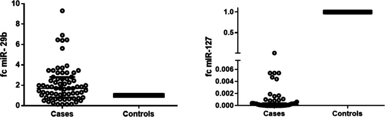 Figure 1
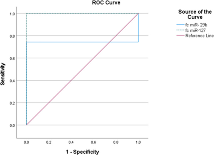 Figure 2
- —Fayoum University
Peer Reviews
No public reviews on file for this paper yet. If you reviewed it on a platform where reviews are public (OpenReview, ICLR, NeurIPS, ICML), you can paste yours below so the community can read it here.
Videos
No videos yet. Explain this paper in a talk, walkthrough, or lecture? Add one.
Taxonomy
TopicsOcular Diseases and Behçet’s Syndrome · Otitis Media and Relapsing Polychondritis · Extracellular vesicles in disease
Introduction
Behçet's disease (BD) is a type of variable vessel vasculitis that affects multiple organs and systems. It can lead to various skin lesions such as papules and pustules, ulcers on the oral, genital, and intestinal mucosa, arthritis, uveitis, lesions in the central nervous system, venous and arterial thrombosis, and arterial aneurysms [1]. A single-center study from Egypt revealed a male/female ratio of 5.4:1 and a prevalence of 7.6/100,000 people [2]. Currently, there is no diagnostic test for Behçet disease and patients with active disease may have normal levels of inflammatory markers such C-reactive protein [3].
It has been revealed that peripheral blood mononuclear cells (PBMCs) from individuals with BD exhibit disrupted microribonucleic acid (miRNA) expression, and several miRNAs have been identified as powerful diagnostic biomarkers in addition to their roles in pathogenesis of the diseases [4]. The miR-29 family, which is expressed in both T and B cells was found to play a function in Toll-like receptor inhibition, promote DNA demethylation, and activate the AKT signaling pathway [5]. miR-29 targets IFN-γ and NF-κB genes which promote chronic inflammation and documented to be increased in Behçet disease [6, 7]. miR-29 induces inflammation by triggering the NF-κB and Janus kinase/signal transducers and activators of transcription (JAK/STAT) signaling pathways in endothelial cells [7].
The two mature miRNAs, miR-127-3p and miR-127-5p, are derived from the same precursor miRNA; henceforth, miR-127-3p shall be denoted as miR-127. miR-127 regulates inflammatory cell infiltration and affects the production of inflammatory cytokines. IL-6, TNF-α, and IL-1β are examples of inflammatory cytokines that can promote the synthesis of other cytokines and effector molecules that can aid in the treatment of a specific illness [8]. miR-127-3p has been demonstrated to function as a negative regulator of the type I interferon (IFN-I) signaling pathway by preventing the phosphorylation of STAT proteins and the stimulation of gene expression triggered by IFN-α through the interferon stimulated response element (ISRE) or GAS [9]. Assessing the expression of two miRNAs (miR-127 and miR-29) in Behçet patients and correlating this expression with various illness manifestations and disease activity was the aim of the current study.
Patients and methods
Seventy adult BD patients who met the international criteria for BD (ICBD) [10] were gathered from the outpatient clinic and inpatient wards of the rheumatology department. Individuals suffering from additional autoimmune conditions including juvenile BD, chronic diseases, people using drugs (statins, angiotensin-converting enzyme inhibitors) that affect endothelial cells, smokers, or those with history of infection (< 3 months) were excluded from the study. Thirty age- and sex-matched healthy controls were also included in the study**.** At baseline, all patients and control subjects gave their informed consent to take part in the study, and the Ethics Committee of Biomedical Research approved it (M 626).
The Behçet’s Disease Current Activity Form (BDCAF) [11] and Behçet severity score [12] were assessed.
Sera collected from each participant were utilized for microRNA extraction and real-time PCR to determine fold changes of miR-127 and miR-29b.
The detailed methodology was mentioned in supplementary file.
Statistical method
Software called the Statistical Package for the Social Sciences (SPSS version 22) was used to analyze the data. Qualitative data were presented as numbers and percentages; the differences were evaluated using chi squared test. Quantitative variables were expressed as mean and standard deviation (SD); independent t test was performed in comparing the two study groups. Study markers were presented as median and interquartile range (IQR); Mann–Whitney U test or Kruskal–Wallis test were utilized in comparison between two or three groups, respectively. Spearman correlation was run to test the relation between study markers and the quantitative variables. Receiver operating characteristic (ROC) curve analysis was done to evaluate the discriminative power of miR-127 and miR-29 to differentiate cases from control. A statistically significant p value was defined as less than 0.05.
Results
Demographic characteristics of patients with Behçet disease and controls
The mean age of the cases was 39.6 ± 10.4 years while for controls was 38.1 ± 12.5 (p = 0.558). No significant differences between the cases and the controls regarding sex (p = 0.736). Clinical manifestations, disease activity, and severity are shown in Tables 1 and 2. Table 1. Clinical manifestations of patients with Behçet diseaseNumberPercentOral ulcersPositive5882.9Genital ulcersPositive4260.0Cutaneous lesionPositive4057.1Ocular manifestationsPositive4767.1Vascular manifestationsPositive1724.3Neurological manifestationPositive1420.0Musculoskeletal manifestationsPositive5680.0Pathergy testPositive2941.4Table 2Disease activity and severity of Behçet patients under the studyBDCAFMean ± SD2.6 ± 1.9Severity scoreMean ± SD6.0 ± 1.8N%Activity (BDCAF)Active4361.4Inactive2738.6Behcet severity scoreMild811.4Moderate3347.1High2941.4BDCAF Behçet Disease Current Activity Form
Serum expression levels of miR-29 and miR-127 in patients with Behçet disease and controls and their relations with various clinical and laboratory data
miR-29 was a statistically significantly higher in cases when compared to controls [median (range) = 1.706 (0.807–2.809)] with p < 0.001 as shown in Fig. 1. Patients with oral ulcers, genital ulcers, and neurological manifestations had a significantly higher levels of miR-29b (p = 0.005, 0.008, 0.003, respectively). Expression level of miR-29b was higher in patients with active disease (p = 0.003). Patients with high and moderate severity score had a statistically significant higher expression level of miR-29 when compared to patients with mild severity score Table 3. There was a statistically significant positive correlation between miR-29 and several study parameters including BDCAF (r = 0.440, p < 0.001) and severity score (r = 0.243, p = 0.043) (Table 5). On the other hand, miR-127 was a statistically significantly lower in cases when compared to controls [median (range) = 0.00011 (0.00001–0.00041)] with p < 0.001 (Fig. 1). There was no statistical difference in the expression level of miR-127 as regard other disease characteristics as demonstrated in Table 4. There were statistically significant negative correlations with ESR (r = − 0.361, p = 0.002) and BDCAF (r = − 0.350, p = 0.003) (Table 5).Fig. 1. Differences in serum miR-29 and miR-127 between patients with Behçet disease and controls. miR-29 was a statistically significantly higher in cases when compared to controls (median (range) = 1.706 (0.807–2.809) with p < 0.001. miR-127 was a statistically significantly lower in cases when compared to controls (median (range) = 0.00011 (0.00001–0.00041) with p < 0.001. Horizontal dotted line indicated the expression levels of miR-29 and miR-127 in controls (since 2^0^ = 1 and − ΔΔCt for controls equals 0, the control value was set to 1)Table 3. Relation between miR-29 and characteristics of Behçet patientsmiR-29p valueMedianIQROral ulcersNegative0.7170.4471.0090.005Positive1.8531.2232.809Genital ulcersNegative1.1760.5521.9910.008Positive1.9861.313.227ActivityActive2.2821.7063.2270.003Inactive1.2270.6212.129SeverityMild0.6570.4470.8070.0120.0031.000Moderate1.8531.2273.227Severe1.9861.0412.809Cutaneous lesionNegative0.7170.4471.0090.182Positive1.8531.2232.809Ocular manifestationsNegative1.1760.5521.9910.107Positive1.9861.313.227Vascular manifestationsNegative1.921.1413.4580.607Positive1.5620.6812.537Neurological manifestationNegative1.2270.4972.6210.003Positive1.8531.1412.809JointsNegative1.7060.8072.8090.901Positive1.9861.1412.282Pathergy testNegative1.5620.7122.3670.294Positive3.2271.9863.706Positive1.7060.7273.227Positive1.7060.7672.715SignificantTable 4miR-127 and different characteristics of Behçet patientmiR-127p* valueMedianIQROral ulcersNegative0.000010.000010.00010.009Positive0.000220.000020.00063Genital ulcersNegative0.00010.000010.000360.569Positive0.000250.000010.00042ActivityActive0.000220.000010.000420.526Inactive0.00010.000010.00036SeverityMild0.000050.000010.000260.228Moderate0.000070.000010.00037Severe0.000270.000060.00063Cutaneous lesionNegative0.000080.000010.000820.700Positive0.00020.000020.00037Ocular manifestationsNegative0.000080.000010.000360.507Positive0.000170.000010.00082Vascular manifestationsNegative0.00010.000010.000410.373Positive0.000220.000060.00041Neurological manifestationNegative0.00010.000010.000520.719Positive0.00020.000060.00036MusculoskeletalNegative0.000150.000020.000410.809Positive0.00010.000010.00041Pathergy testNegative0.00010.000010.000630.919Positive0.000170.000020.00032SignificantTable 5Correlations of miR-29 and miR-127 with different patient and laboratory analysesmiR-29miR-127HBr0.0740.123p value0.5410.311TLCr0.1520.108p value0.2100.372Plateletsr − 0.105 − 0.039p value0.3860.751ESRr0.039 − 0.361p value0.7510.002CRPr0.044 − 0.154p* value0.7190.205Urear0.0290.043p value0.8110.723Creatininer − 0.1080.045p value0.3720.714ALTr − 0.074 − 0.074p value0.5420.544ASTr − 0.0050.125p value0.9670.308RBSr0.064 − 0.190p value0.5970.115BDCAFr0.440 − 0.350p value < 0.0010.003Severity scorer0.2430.276p value0.0430.021**HB hemoglobin, TLC total leukocyte count, ESR erythrocyte sedimentation rate, CRP C-reactive protein, ALT alanine transaminase, AST aspartate aminotransferase, RBS random blood sugar, BDCAF Behçet Disease Current Activity Form*Significant
ROC analysis discriminating patients with Behçet disease from control subjects using expression levels of miR-29 and miR-127
miR-29 had an excellent discriminative power for differentiating Behçet cases from controls [AUC = 0.743, 95% CI = (0.640–0.845)] with sensitivity of 74.3% and specificity of 100% (p < 0.001). Also, miR-127 had an excellent discriminative power for differentiating Behçet cases from controls [AUC = 1.00, 95% CI = (1.00–1.00)] with sensitivity of 100% and specificity of 100% (p < 0.001) as observed in Fig. 2.Fig. 2ROC curve analysis for miR-127 and miR-29
Discussion
miRNA dysregulation in BD patients may lead to aberrant amounts of immune-suppressive and inflammatory cells and cytokines [10]. This study was designed to assess expression of miR-29 and miR-127 in BD patients and to correlate this expression with different disease manifestations and disease activity. The expression levels of miR-29 were significantly upregulated in blood samples of BD patients when compared with controls (p ≤ 0.001); up to our knowledge, this is the first study which assesses miR-29. miR-29 enhances lipopolysaccharide-induced inflammation by activating the NF-κB and JAK/STAT signaling pathways in endothelial cells [7]. Additionally, mature B lymphocytes in the periphery were found to activate PI3 K signaling through the miR-29 family of miRNAs [11]. The complicated pathophysiology of BD includes the activation of several signaling pathways, such as the nuclear factor kappa B (NF-κB) signaling system and the phosphatidylinositol 3-kinase (PI3 K)/protein kinase B (AKT) signaling network [10]. So, this may explain the elevation of miR-29 in patients with Behçet disease.
Current findings are in line with other studies that were highlighting higher levels of miR-29 in patients with rheumatoid arthritis [12] and systemic lupus erythematosus (SLE) [5], suggesting that it may be considered as a biomarker for their susceptibility. Similarly, another study verified that miR-29 was upregulated in cartilage tissue from patients with OA when compared to controls. miR-29 could significantly downregulate progranulin expression which play an important role in cartilage formation and function and reported to have anti-inflammatory functions [13]. Furthermore, a previous study found that miR-29 was increased in pediatric Crohn’s disease [14]. In contrast, a former study found that systemic sclerosis fibroblasts and skin sections had much lower levels of miR-29a than healthy control samples [15].
The current study found that miR-29 was statistically significantly higher in patients with active disease (p = 0.003), and there was a statistically significant positive correlation between miR-29 and BDCAF (p < 0.001) and severity score (p = 0.043). Patients with high and moderate severity score had a statistically significant higher expression level of miR-29 when compared to patients with mild severity score. With review to literatures, a previous study found that miR-29b expression was upregulated in SLE and correlated with SLEDAI score, anti-dsDNA, and complement C3 level in patients with SLE [5]. Similarly, a study done by Ren et al. reported that upregulation of miR-29b was correlated with RA disease activity [12]. This study found that miR-29 had an excellent discriminative power for differentiating Behçet cases from controls (AUC = 0.743, 95% CI = (0.640–0.845). The optimal cutoff point was 1.02, at which sensitivity was 74.3% and specificity was 100.0%. These results are in line with a study of SLE done by Wang et al., who found that miR-29 had area under the curve 0.752, denoting a good diagnostic power for SLE (AUC > 0.75).
As regard to miR-127, the current study found that miR-127 expression level was statistically significantly lower in cases when compared to controls p < 0.001. Our study is the first to study the role of miR-127 in BD disease. A previous study done by Wu et al. proved that miR-127 expression level was downregulated in the kidney of lupus nephritis [16]. Downregulation of miR-127 promotes Janus family of kinases (JAK1) expression and contributes to the abnormal activation of IFN-I signaling pathway in the kidney of LN [16]. There is some similarity between SLE and Behçet as JAK1/STAT3 signaling pathway is activated in BD, possibly through elevated serum and tissue expressions of Th1/Th17 type cytokines [17]. Another previous study done by Umeh-Garcia et al. found that expression level of miR-127 was reduced in breast cancer through targeting the PI3 K/Akt pathway [18]. Our current study found that miR-127 had an excellent discriminative power for differentiating Behçet cases from controls [AUC = 1.00, 95% CI = (1.00–1.00)]. The optimal cutoff point for 0.51 at which sensitivity was 100% and specificity was 100.0%.
A small number of participants who shared the same ethnic background participated in the current study is a limitation of our study; therefore, a more comprehensive investigation encompassing multiple countries might enhance the results.
In conclusion, miR-29 was a statistically significantly higher in patients with BD. There was a statistically significant positive correlation between miR-29 and BDCAF, expression level of miR-29 was higher in patients with active disease. On the other hand, miR-127 was a statistically significantly lower in patients with Behçet disease, miR-127 was negatively correlated with BDCAF. miR-29 and miR-127 could be considered as useful diagnostic markers for differentiating patients with BD from controls.
The reference list from the paper itself. Each links out to its DOI / PubMed record.
- 1Gheita TA, El-Latif EA, El-Gazzar II, Samy N, Hammam N, Abdel Noor RA, et al (2019) Behçet’s disease in Egypt: a multicenter nationwide study on 1526 adult patients and review of the literature. Clin Rheumatol 38(9):2565–2575.10.1007/s 10067-019-04570-w 31119493 · doi ↗ · pubmed ↗
- 2Ambrose NL, Haskard DO. Differential diagnosis and management of Behçet syndrome (2013) Nat Rev Rheumatol 9(2):79–89.10.1038/nrrheum.2012.15623007742 · doi ↗ · pubmed ↗
- 3Hu D, Guan JL (2023) The roles of immune cells in Behçet's disease. Adv Rheumatol 10;63:49.10.1186/s 42358-023-00328-w 37814339 · doi ↗ · pubmed ↗
- 4Riggs JM, Hanna RN, Rajan B, Zerrouki K, Karnell JL, Sagar D, et al (2008) Characterisation of anifrolumab, a fully human anti interferon receptor antagonist antibody for the treatment of systemic lupus erythematosus. Lupus sci med 1;5(1):e 000261.10.1136/lupus-2018-000261 PMC 589085629644082 · doi ↗ · pubmed ↗
- 5Alibaz-Oner F, Direskeneli H (2022) Update on the diagnosis of Behçet’s disease. Diagnostics 23;13(1):41.10.3390/diagnostics 13010041 PMC 981853836611332 · doi ↗ · pubmed ↗
- 6Gu F, Huang X, Huang W, Zhao M, Zheng H, Wang Y, et al (2023)The role of mi RN As in Behçet’s disease. Front. Immunol 4(14):1249826.10.3389/fimmu.2023.1249826 PMC 1058433037860009 · doi ↗ · pubmed ↗
- 7Shumway AJ, Shanahan MT, Hollville E, Chen K, Beasley C, Villanueva JW, et al (2024) Aberrant mi R-29 is a predictive feature of severe phenotypes in pediatric Crohn's disease. JCI Insight 22;9(4):e 168800.10.1172/jci.insight.168800 PMC 1096738438385744 · doi ↗ · pubmed ↗
- 8Harmanci D, Erkan EP, Kocak A, Akdogan GG (2017) Role of the micro RNA-29 family in fibrotic skin diseases. Biomed Rep 1;(6):599–604.10.3892/br.2017.900PMC 544996228584629 · doi ↗ · pubmed ↗