A case with bilateral C-shaped autofluorescence in retinal degeneration
Hsin Hsu, Chunya Kang, Eugene Yu-Chuan Kang, Nan-Kai Wang

TL;DR
A 19-year-old male with a rare genetic condition showed a unique double C-shaped pattern in eye imaging, helping diagnose Optic Atrophy-13.
Contribution
The paper identifies a novel double C-shaped autofluorescent ring as a potential diagnostic marker for OPA13.
Findings
A de novo SSBP1 missense variant was found in a patient with Optic Atrophy-13.
Bilateral double C-shaped hyper-autofluorescent rings were observed in fundus autofluorescence imaging.
Genetic testing confirmed the diagnosis and ruled out inherited causes.
Abstract
To report a case of a 19-year-old male with Optic Atrophy-13 (OPA13) associated with a de novo heterozygous missense variant in the single-strand DNA-binding protein 1 (SSBP1) gene, characterized by a bilateral double C-shaped hyper-autofluorescent ring on fundus autofluorescence (FAF). A 19-year-old male exhibited poor visual acuity, pale optic discs, vessel attenuation, and peripheral pigmentary changes. FAF imaging revealed a bilateral double C-shaped hyper-autofluorescent ring, which was not frequently observed. Optical coherence tomography (OCT) showed retinal thinning and ellipsoid zone disruption, while electroretinography (ERG) indicated cone-rod dystrophy. Genetic testing identified a pathogenic SSBP1 missense variant, confirming the diagnosis of OPA13. Parental genetic analysis excluded the variant, establishing it as a de novo one. This report highlights a novel retinal…
Genes, proteins, chemicals, diseases, species, mutations and cell lines named across the full text — each resolved to its canonical identifier and authoritative record.
Click any figure to enlarge with its caption.
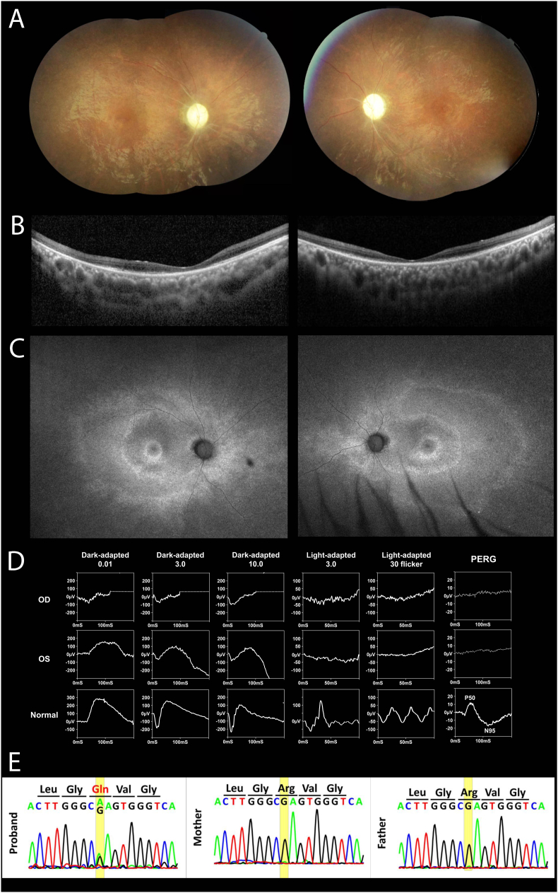 Figure 1
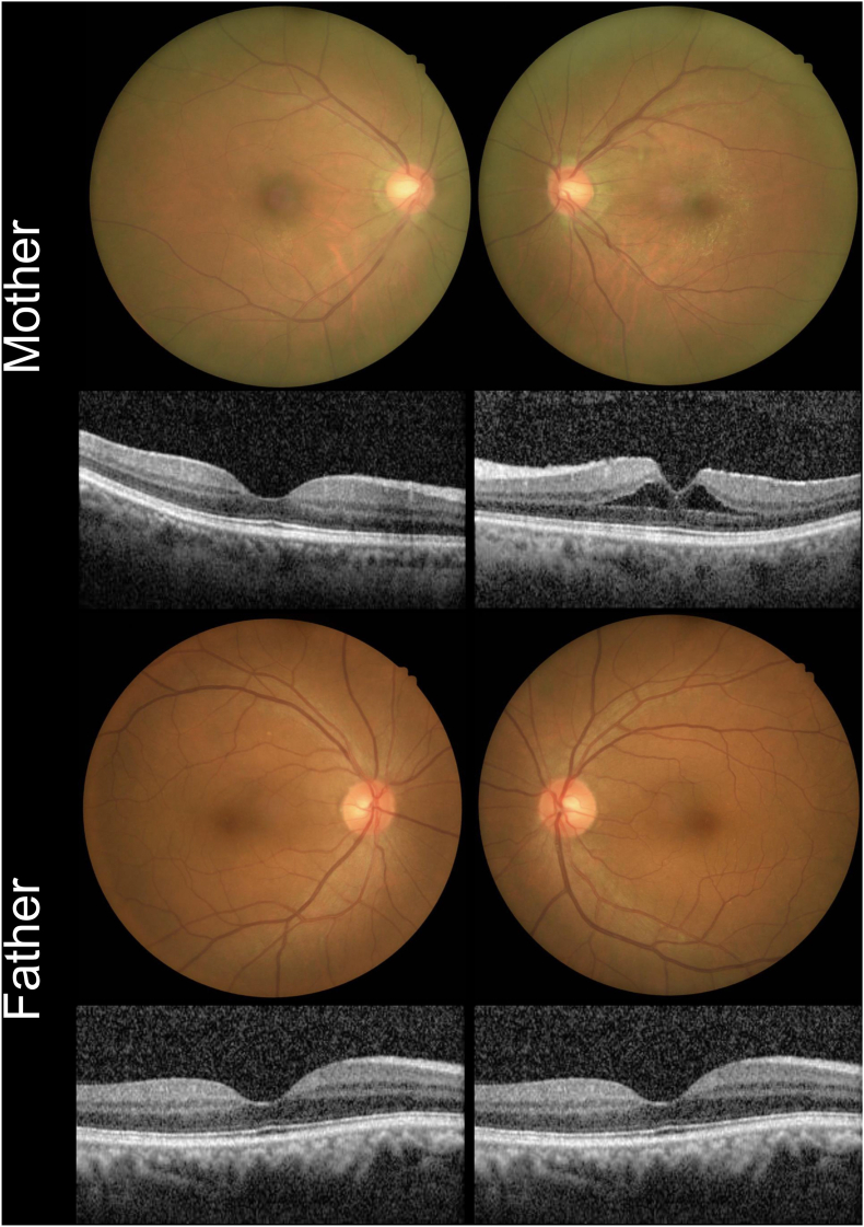 Figure 2
Peer Reviews
No public reviews on file for this paper yet. If you reviewed it on a platform where reviews are public (OpenReview, ICLR, NeurIPS, ICML), you can paste yours below so the community can read it here.
Videos
No videos yet. Explain this paper in a talk, walkthrough, or lecture? Add one.
Taxonomy
TopicsPorphyrin Metabolism and Disorders · Retinal Diseases and Treatments · Adenosine and Purinergic Signaling
Introduction
1
The single-strand DNA-binding protein 1 gene (SSBP1, MIM: 600439) is a housekeeping gene essential for mitochondrial biogenesis.1 In vitro, the SSBP1 protein is required for mitochondrial DNA (mtDNA) copy number, playing a vital role in mtDNA replication and repair.2 Variants in the SSBP1 gene can alter SSBP1 protein levels and structure, hindering proper mtDNA repair and leading to mitochondrial dysfunction and associated diseases.2
Optic atrophy-13 (OPA13), an autosomal dominant disorder associated with retinal and foveal abnormalities, is characterized by reduced visual acuity due to bilateral optic atrophy and often includes foveopathy.2^,^3 This disease is caused by variants in the SSBP1 gene. Studies in 2019 and 2020 documented families with OPA13 linked to heterozygous SSBP1 variants, further substantiating the association between OPA13 and SSBP1 variants.2^,^4 These variants may impair replication machinery in retinal ganglion cells (RGCs) and other cell types.2^,^3 A total of 47 cases of retinopathy associated with mutant SSBP1 have been reported to date. In the study by Del Dotto et al. (2019), retinopathy was described in 8 individuals from 5 families.2 Piro-Megy et al. (2019) reported 14 cases, while Jurkute et al. (2019) identified 12 patients with retinopathy linked to the SSBP1 variant.3^,^4 Finally, Meunier et al. (2021) described 12 cases, and Chang et al. (2023) reported 1 additional case.5^,^6 Here, we present a 19-year-old male with OPA13, showing a double C-shaped hyperfluorescent ring in fundus autofluorescence (FAF), which has not been reported and needs further investigation.
Case report
2
In this case, a 19-year-old male with poor vision since early childhood has a medical history of amblyopia, premature ventricular contractions, and corrective surgery for scoliosis. His best-corrected visual acuity (BCVA) was 20/400 in both eyes. Ocular examination showed a normal anterior segment, while fundus examination revealed pale disc, vessel attenuation, papillomacular bundle loss, and mild peripheral pigmentary changes (Fig. 1A). Optical coherence tomography (OCT) of the macula showed retinal thinning, ellipsoid zone disruption, and intraretinal cystoid changes in both eyes (Fig. 1B). Fundus autofluorescence (FAF) revealed a C-shaped pattern with a double hyper-autofluorescent (AF) ring in both eyes (Fig. 1C). The full-field electroretinogram disclosed cone-rod dystrophy, and the pattern of ERG showed extinguished P50 and N95, indicating dysfunction in both photoreceptors and retinal ganglion cells (RGCs). (Fig. 1D).Fig. 1**(**A) Color fundus photography shows vessel attenuation, papillomacular bundle loss, and peripheral pigmentary changes. (B) OCT reveals retinal thinning, ellipsoid zone disruption, and intraretinal cysts. (C) Full-field ERG reveals reduced rod response and absent cone response. (D) FAF displays a C-shaped pattern with a double hyperfluorescent ring in both eyes. (E) Sanger sequencing identifies the SSBP1 variant c.113G > A (p.Arg38Gln) in the patient, with negative results for both parentsOCT, Optical coherence tomography; SSBP1, single-strand DNA-binding protein 1; ERG, electroretinogram; FAF, Fundus autofluorescence. (For interpretation of the references to colour in this figure legend, the reader is referred to the Web version of this article.)Fig. 1
We further performed genetic testing under the impression of inherited eye disease, and whole-exon sequencing (WES) detected a heterozygous SSBP1 missense variant (NM_003143.3) c.113G > A, (p.Arg38Gln), leading to the diagnosis of OPA13 with retinal and foveal abnormalities. This variant has been reported in several OPA13 individuals with an autosomal dominant inheritance pattern and has been classified as pathogenic in ClinVar (Variation ID: 977502). Sanger sequencing of the patient confirmed the SSBP1 missense variant of the patient (Fig. 1E).
The patient reported no family history of visual disorders. His 54-year-old father had a BCVA of 20/50 in the right eye and 20/28 in the left, while his 52-year-old mother had a BCVA of 20/20 in both eyes, with an epiretinal membrane and cystoid macular edema in the left eye. All the other examinations were considered normal for both parents (Fig. 2). WES and Sanger sequencing for both parents revealed no SSBP1 variants at the same locus, except for a c.224T > C (p.Leu75Pro) variant from his mother, which is genetically insignificant. Therefore, the patient was confirmed to have a de novo SSBP1 gene variant.Fig. 2. Color fundus photography and OCT of both parents: the mother had an epiretinal membrane and cystoid macular edema in the left eye, with no other abnormalitiesOCT, Optical coherence tomography. (For interpretation of the references to colour in this figure legend, the reader is referred to the Web version of this article.)Fig. 2
Discussion
3
Our case report describes a 19-year-old male OPA13 patient with a heterozygous SSBP1 missense variant, featuring a double C-shaped AF ring on fundus FAF imaging. FAF imaging in retinitis pigmentosa (RP) typically reveals a single hyper-AF ring, where photoreceptor layers within the ring's inner border are generally preserved, while outer areas show thinning or loss of the outer nuclear layer.7 However, our patient exhibited a double C-shaped AF ring, which differs from the typical single ring seen in most RP cases. While this specific pattern has not been widely reported, it may represent a potential feature associated with SSBP1-related retinopathy, including OPA13. However, further cases are needed to determine its diagnostic value. Although sporadic cases of OPA13 are not uncommon, comprehensive parental testing is not always performed, and low-level mosaicism may be missed by conventional sequencing methods. In our case, genetic testing via WES and Sanger sequencing in both the patient and his parents confirmed the presence of a de novo variant in the SSBP1 gene. Our findings highlight the utility of sensitive genetic approaches for accurate diagnosis and counseling.
The patterns of retinal degeneration in inherited retinal diseases are not static and evolve over time. It is possible that the C-shaped image does not manifest in all cases and may be absent in the early or advanced stages of disease progression. Additionally, this pattern might resemble an early-stage presentation in pericentral RP before a complete ring develops. While this feature was noted in our patient, additional studies are needed to determine its wider clinical relevance.
OPA13 represents a distinct type of dominant optic atrophy (DOA), a neuro-ophthalmic condition characterized by RGC degeneration, often associated with retinal degeneration and foveopathy, primarily due to variants in the SSBP1 gene. Most DOA patients present with bilateral optic atrophy without photoreceptor involvement. The presence of optic atrophy with retinal and foveal abnormalities in this patient underscores the importance of considering OPA13 in the differential diagnosis of other DOA types. Additionally, genetic testing supports the diagnosis of OPA13 and may help anticipate systemic conditions such as sensorineural deafness and nephropathy.2 Although the patient did not present with hearing loss or renal failure, it is crucial to maintain close monitoring for potential systemic complications in individuals with pathogenic SSBP1 variants.
In conclusion, this case report details a 19-year-old male with OPA13, showing a double C-shaped AF ring. In contrast to the typical single ring observed in RP, this distinct pattern may aid in the diagnosis of OPA13 and may serve as a useful phenotypic indicator in certain cases, although further research is required. Genetic testing identified our case as a de novo SSBP1 variant, linking this variant to the newly found retinal feature and highlighting the importance of considering OPA13 in similar cases.
CRediT authorship contribution statement
Hsin Hsu: Writing – original draft, Formal analysis. Chunya Kang: Writing – review & editing. Eugene Yu-Chuan Kang: Supervision, Investigation, Conceptualization. Nan-Kai Wang: Supervision, Resources, Methodology.
Patient consent
Consent to publish this case report has been obtained from the patient in writing.
Authorship
All authors attest that they fulfil the current ICMJE criteria for authorship.
Funding
This research was supported by 10.13039/100012553Chang Gung Memorial Hospital, Taiwan (CMRPG3N1001), the 10.13039/100020595National Science and Technology Council, Taiwan (NSTC 113-2314-B-182A-150-MY3) and 10.13039/501100002836Chang Gung University, Taiwan (UARPD1N0031 and UARPD1P0261).
Declaration of competing interest
The authors declare that they have no known competing financial interests or personal relationships that could have appeared to influence the work reported in this paper.
The reference list from the paper itself. Each links out to its DOI / PubMed record.
- 1Tiranti V.Rossi E.Ruiz-Carrillo A.Chromosomal localization of mitochondrial transcription factor A (TCF 6), single-stranded DNA-binding protein (SSBP), and endonuclease G (ENDOG), three human housekeeping genes involved in mitochondrial biogenesis Genomics 252Jan 20 199555956410.1016/0888-7543(95)80058-t 7789991 · doi ↗ · pubmed ↗
- 2Del Dotto V.Ullah F.Di Meo I.SSBP 1 mutations cause mt DNA depletion underlying a complex optic atrophy disorder J Clin Investig 1301 Jan 2 202010812510.1172/jci 12851431550240 PMC 6934201 · doi ↗ · pubmed ↗
- 3Jurkute N.Leu C.Pogoda H.M.SSBP 1 mutations in dominant optic atrophy with variable retinal degeneration Ann Neurol 863Sep 201936838310.1002/ana.2555031298765 PMC 8855788 · doi ↗ · pubmed ↗
- 4Piro-Mégy C.Sarzi E.Tarrés-SoléA.Dominant mutations in mt DNA maintenance gene SSBP 1 cause optic atrophy and foveopathy J Clin Investig 1301 Jan 2 202014315610.1172/jci 12851331550237 PMC 6934222 · doi ↗ · pubmed ↗
- 5Meunier I.Bocquet B.Defoort-Dhellemmes S.Characterization of SSBP 1-related optic atrophy and foveopathy Sci Rep 111Sep 21 20211870310.1038/s 41598-021-98150-1PMC 845554234548540 · doi ↗ · pubmed ↗
- 6Chang Y.H.Kang E.Y.Liu L.Maternal mosaicism in SSBP 1 causing optic atrophy with retinal degeneration: implications for genetic counseling Orphanet J Rare Dis 181May 31 202313110.1186/s 13023-023-02748-937259171 PMC 10233871 · doi ↗ · pubmed ↗
- 7Greenstein V.C.Duncker T.Holopigian K.Structural and functional changes associated with normal and abnormal fundus autofluorescence in patients with retinitis pigmentosa Retina 322Feb 201234935710.1097/IAE.0b 013e 31821 dfc 1721909055 PMC 3720996 · doi ↗ · pubmed ↗