Mechanical properties of mandibular and maxillary bone collagen fibrils based on nonlocal elasticity theory
Elaheh Alibeigi Beni, Alireza Shahidi, Behnaz Ebadian

TL;DR
This paper studies the mechanical properties of collagen fibrils in human jaw bones using nanoindentation and nonlocal elasticity theory.
Contribution
The study introduces nonlocal elasticity theory to analyze collagen fibril mechanics in mandibular and maxillary bones.
Findings
Maxillary cortical-trabecular bone interface collagen fibrils have the highest elastic modulus (4.16 ± 0.18 MPa).
Nonlocal parameter ranges for collagen fibrils are 0.430 ± 0.013–0.483 ± 0.011 nm.
Elastic modulus in cortical-trabecular interfaces is higher than in cortical bone.
Abstract
In this paper, the mechanical properties of collagen fibrils in the cortical bone and cortical-trabecular bone interface of the human mandible and maxilla have been investigated. Force-indentation curves on wet collagen fibrils are taken by applying the atomic force microscopy nanoindentation technique, and the elastic modulus is measured. The distribution of stress and strain is determined by considering an elastic medium when it is deformed by a rigid cone. Afterward, by applying the nonlocal elasticity theory and the indentation parameters, the nonlocal parameter of the collagen fibrils is calculated at the nanoscale. Finally, the elastic modulus and nonlocal modulus of the collagen fibrils are compared. According to the results, the highest and lowest values of the elastic modulus of the collagen fibrils are determined in the maxillary cortical-trabecular bone interface (4.16 ± 0.18…
Genes, proteins, chemicals, diseases, species, mutations and cell lines named across the full text — each resolved to its canonical identifier and authoritative record.
Click any figure to enlarge with its caption.
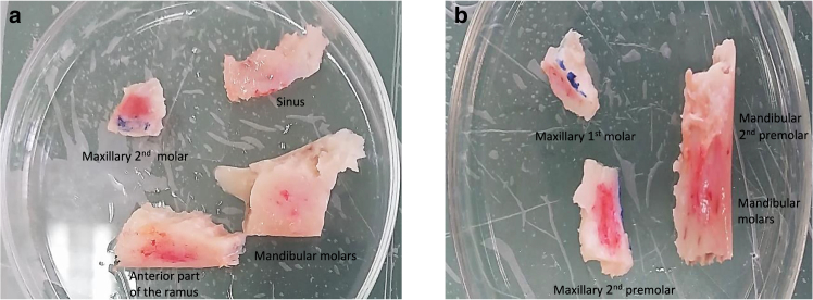 Figure 1
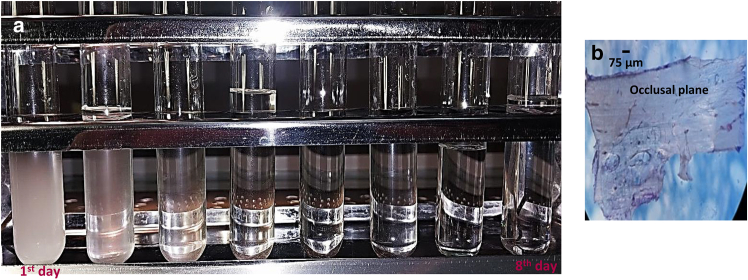 Figure 2
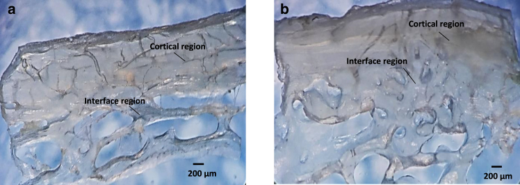 Figure 3
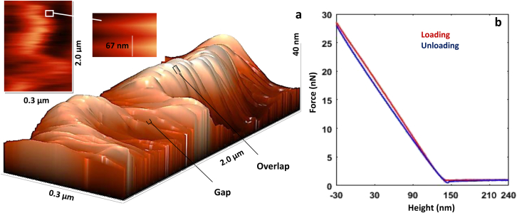 Figure 4
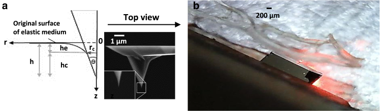 Figure 5
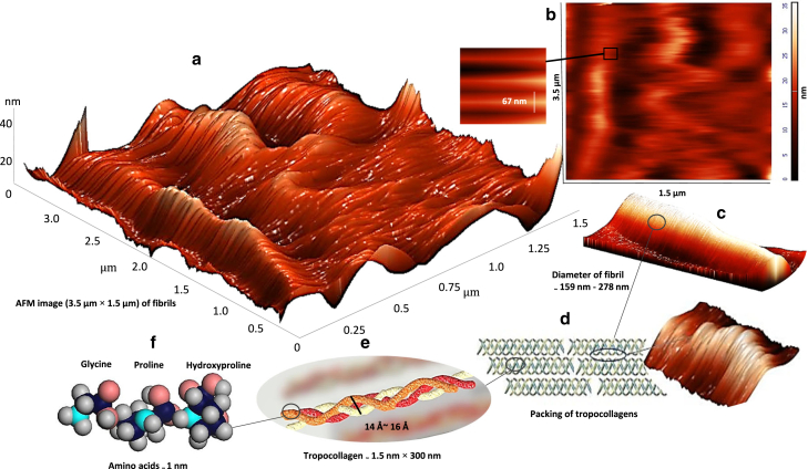 Figure 6
Peer Reviews
No public reviews on file for this paper yet. If you reviewed it on a platform where reviews are public (OpenReview, ICLR, NeurIPS, ICML), you can paste yours below so the community can read it here.
Videos
No videos yet. Explain this paper in a talk, walkthrough, or lecture? Add one.
Taxonomy
TopicsElasticity and Material Modeling · Bone health and osteoporosis research · Tendon Structure and Treatment
Why it matters
In this paper, the mechanical properties of collagen fibrils in the cortical bone and cortical-trabecular bone interface of the human mandible and maxilla have been investigated. So far, the elastic modulus of collagen fibrils has been calculated at macroscopic scale, according to the classical (local) continuum theory, whereas the mechanical properties of the bone collagen fibrils, as a nanostructure, are dependent on the size parameter. In this study, nonlocal elasticity theory as modified size-dependent continuum theory is considered to predict the nanostructure correctly. According to the results, the difference between the nanoscale modulus of collagen fibrils and the elastic modulus at large length scales is significant.
Introduction
Type I collagen, as a structural protein, is the most abundant protein in vertebrates; it is prevalent in organs such as bones, tendon, skin, and the respiratory system (1). Collagen fibrils are responsible for mechanical stability, elasticity, toughness, and strength in tissues. The diameter of a single collagen fibril varies from several tens of nanometers to a micrometer (2). A single collagen fibril is composed of collagen molecules, called tropocollagens. Tropocollagens have a diameter of approximately 1.5 nm and a length of 300 nm; they are staggered in the axial direction of collagen fibrils (3,4,5). Each tropocollagen is made up of a triple helix of left-handed polypeptide chains twisted together into a right-handed triple helix. Each chain contains amino acid residues (6). The general formula for amino acid sequence of collagen is Gly-Pro-X or Gly-X-Hyp, where Pro is a proline and Hyp is a hydroxyproline residue. X can be various other amino acid residues. Glycine is the smallest amino acid without a side chain; it is located in the center of the coiled peptide chain (7,8). Type I collagen fibrils have characteristic periodic patterns of 60–70 nm (D-banding). This periodicity leads to the appearance of the regions called gap (∼0.6 D) and overlap (∼0.4 D) (2,9).
Atomic force microscopy (AFM) nanoindentation has great potential for use in clinical activities; it is recognized as a special technique in the characterization of biological samples at the nanoscale (10). In particular, as a new method, early and accurate diagnosis of cancer and osteoporosis has become possible through nanomechanical characterization of individual collagen fibrils (10,11,12). In addition, the correlation of collagen mutation with some diseases such as osteogenesis, imperfecta, and osteoporosis has been investigated (13,14). Thus, during the last two decades, significant scientific attention has been given to the measurement of the elastic modulus of individual collagen fibrils by applying the AFM nanoindentation technique (10).
At the nanoscale, the dimensions of a system are comparable to the intermolecular and interatomic space of the system. For this reason, the structure cannot be modeled as a continuous medium anymore. Also, at very small scales, the physical properties of the materials change due to the significant effect of intermolecular and interatomic cohesive forces, as well as the discrete nature of the structure. These effects are referred to as the “size” effect (15). Therefore, the mechanical properties of nanostructures such as collagen fibrils are dependent on the size parameter (16). The classical continuum theory has the potential for the analysis of macroscopic structures and is not capable of accounting for the size parameter of nanostructures. Considering the deficiency of classical continuous theories in applying the size effects, using higher-order continuous theories, which can lead to obtaining accurate results by considering size effects, has been highly recommended (17). In this relation, Eringen (18) introduced nonlocal elasticity theory to incorporate nanoscale effects in the classical continuum theory.
Investigating the structure and mechanical behavior of jaw bones is, therefore, important since it leads to the improvement of the function and design of implants. It also minimizes crestal resorption and the possibility of bone-implant junction failure (19). Although the mechanical properties of mineralized fibrils in the jaw bones at the submicron scale have been reported in the literature (20,21), the elastic properties and topographic features of collagen fibrils in the cortical bone and cortical-trabecular bone interface are not available. Also, at the nanoscale, the mechanical properties of collagen fibrils, by employing higher-order continuous theories and indentation parameters, have not been investigated. Furthermore, the difference between the nanoscale properties of collagen fibrils is not known by considering the small-scale effect and elastic properties at the large length scale.
Therefore, in the present paper, attempts have been made, for the first time, to determine the mechanical properties of collagen fibrils in the cortical bone and cortical-trabecular bone interface of the human mandible and maxilla by using the AFM indentation method. The elastic modulus of collagen fibrils is calculated using the Oliver-Pharr method (22). The distribution of stress and strain is determined by considering a semi-infinite elastic solid when its plane surface is deformed under the pressure of a rigid cone (23). By considering the stress and strain tensor components and indentation parameters and by employing the size-dependent nonlocal elasticity theory, the nonlocal parameter is calculated at the nanoscale. Finally, the values of elastic modulus, nonlocal modulus, and the scaling parameter, , in mandibular and maxillary collagen fibrils are compared.
Materials and methods
Specimen preparation
The specimens of mandibular and maxillary bones were obtained from the fresh cadaver of male donors (21 and 37 years old) at autopsy (at Shahrekord Legal Medicine Organization, ethics committee’s approval was obtained, approval no. IR.UIT.REC.1401.001). The times from death to specimen procurement (procurement delay) were 12 and 20 h, respectively. As shown in Fig. 1, bone specimens were harvested from the anterior part of the ramus, from the mandibular second premolars to the distal of the second molars, and the maxillary second premolars to the distal of the second molars regions. Three anatomically characteristic directions, including inferosuperior (Fig. 1, blue), buccolingual (Fig. 1, red), and mesiodistal ones, were marked. Bone specimens were stored in a solution consisting of 95.5% ethanol and 0.9% normal saline in equal proportions (24). Thereafter, bone slices parallel to the occlusal plane were sectioned using an automatic cutting machine on the mandibular and maxillary bone specimens. In all steps, bone slices were kept in a moist state and exposed to fresh deionized water. In general, nine and eight slices were obtained on the mandibular and maxillary bones, respectively. The cutting speed was 10 μm/s. The thickness of each of the slices was 75–200 μm. Also, the dimensions of each of the slices were approximately 3 × 5 mm (width × height). Afterward, in each of the slices, the bone marrow was gently cleaned by a soft brush and water jet.Fig. 1. Different anatomical regions of fresh mandibular and maxillary bone specimens belonging to a 37-year-old-man (a) and a 21-year-old-man (b). Three anatomically characteristic directions, including inferosuperior (blue), buccolingual (red), and mesiodistal ones, are marked.
Collagen fibrils
In the demineralization process, all the slices were soaked in a solution consisting of 22.5% formic acid and sodium citrate solution (100 g/L) in equal proportions. The solution was changed once a day. The decalcification process was completed by verifying the concentration level of free calcium in the decalcifying solution. For this purpose, after each change, 5 mL of decalcifying solution was mixed with 5 mL of 0.5 N sodium hydroxide (NaOH) and 1 mL of 5% ammonium oxalate solution (25,26). As shown in Fig. 2, turbidity in the final mixture indicates the presence of calcium ions. This process was repeated until the final solution became clear. The highest amount of calcium ions was observed on the first day, whereas on the eighth day of decalcification, the mixture was free of calcium ions. The durations of mandibular bone and maxillary bone decalcification were 8–9 and 4–5 days, respectively. After decalcification, to remove traces of acid, the slices were washed five times in a sonic water bath with deionized water. In this step, the temperature of the deionized water did not exceed 30°C. Thereafter, the slices were kept in deionized water at a temperature of 3°C until the AFM nanoindentation test.Fig. 2. Demineralization of mandibular bone slices (a). Bone slice with the thickness of around 75 μm (b). Turbidity in the final mixture indicates the presence of calcium ions. The demineralization process is repeated until the mixture becomes clear. The highest amount of calcium ions is observed on the first day, whereas on the eighth day of decalcification, the mixture is free of calcium ions.
AFM nanoindentation tests
AFM (NT-MDT Company) experiments were carried out. For AFM imaging and the nanoindentation test, an AFM tip (CSG10) with a curvature radius of 6 nm and a spring constant of 0.5 N/m was used (27). Contact mode was then performed in the AFM imaging procedure. The nanoindentation tests were conducted on some random positions selected on different collagen fibrils of the occlusal plane and under the ambient state.
In this study, cantilever deflection signals – force-height curves on the different overlap regions were obtained to measure the elastic modulus. A maximum indentation depth of 30 nm and a velocity of the tip in loading and unloading of 1000 nm/s were considered. 81 AFM nanoindentation tests on collagen fibrils in the cortical bone and cortical-trabecular bone interface were also performed. These regions are shown in the mandibular and maxillary bones in Fig. 3. The substrate material of the AFM test was steel (annealing process). The air at room temperature and humidity was 18°C and 45%, respectively. The maximum duration of indentations in each of the jaw bones and each of the cortical bones and cortical-trabecular bone interfaces was 60 min. A three-dimensional AFM image of the maxillary bone collagen fibril is shown in Fig. 4 A.Fig. 3. Demineralized bone slices in mandible (a) and maxilla (b). The slice thickness is around 200 μm.Fig. 4(a) Three-dimensional (3D) AFM image showing D-periodic banding pattern of the maxillary bone collagen fibril. 2D top-view topography AFM images and their banding patterns (67 nm) and (b) force (nN)- height (nm) curve.
Determination of indentation modulus
According to the Oliver-Pharr method, contact stiffness can be calculated from the unloading part (Fig. 4 B) of the force-indentation curve (22). Then, by applying the indenter force, contact depth, elastic modulus of the indenter, Poisson’s ratio of the sample, and the indenter, the elastic modulus of collagen fibrils is calculated. The Oliver-Pharr analysis is presented in the supporting material.
Nonlocal elasticity theory at small scales
According to this theory, in the domain, the stress at the reference point is a function of the strains at all other points. is the nonlocal modulus, which represents the scale coefficient or nonlocal parameter of the length unit. In fact, this parameter incorporates the effect of the nanoscale on mechanical behavior. and also stand for internal characteristic lengths and external characteristic lengths, respectively. is a constant parameter that is appropriate for each material. In other words, this parameter should be determined for each nanostructure independently (18,28). Collagen fibril is assumed to be a homogenous and isotropic solid. The Poisson’s ratio is also considered to be 0.5 (29,30). The distribution of stress and strain in a semi-infinite elastic medium, when it is deformed by a rigid conical indenter, is determined (23). Fig. 5 A shows the indentation parameters (31). By substituting the contact depth, contact radius, Poisson’s ratio, and elastic modulus of the fibrils, the components of the stress and strain tensors are obtained. Afterward, by applying the nonlocal elasticity theory, the nonlocal parameter of the collagen fibrils is calculated in the elastic medium. Finally, the scaling parameter, , can be obtained at the nanoscale. The components of stress and strain, as well as the nonlocal elasticity theory, are presented in the supporting material.Fig. 5(a) A schematic representation of a conic tip indenting a half-space solid medium (left). The side view of the AFM tip (CSG10) is shown (right). Indentation parameters contain the maximum displacement of the tip , contact depth , elastic displacement of the solid surface at the contact line with the tip , cone angle , and contact radius . (b) The top view of the AFM cantilever during the nanoindentation test on the mandibular collagen fibrils.
Results
The values of elastic modulus (mean ± SD) of the collagen fibrils in mandibular and maxillary bone are shown in Table 1. The value of this parameter varies in cortical bone and the cortical-trabecular bone interface. The elastic moduli measured in the mandibular and maxillary bones were in the range of 2.03 ± 0.14–2.78 ± 0.20 and 3.08 ± 0.04–4.16 ± 0.18 MPa, respectively. According to the results, the elastic modulus of collagen fibrils in the cortical-trabecular bone interface was higher than that of the cortical one. Also, this parameter in collagen fibrils in cortical as well as cortical-trabecular interface regions of the maxillary bone was higher than that of the mandibular bone.Table 1. The elastic modulus values of the collagen fibrils in the mandibular and maxillary boneAge/sexBoneNo. of fibrilsNo. of indentationsNo. of locations per fibrilElastic modulus (MPa) ± SD37-year-old malemandiblecortical bone2105, 52.03 ± 0.14cortical-trabecular bone interface295, 42.15 ± 0.05maxillacortical bone3124, 5, 33.74 ± 0.44cortical-trabecular bone interface3103, 4, 34.16 ± 0.1821-year-old malemandiblecortical bone3124, 4, 42.51 ± 0.36cortical-trabecular bone interface3103, 4, 32.78 ± 0.20maxillacortical bone294, 53.08 ± 0.04cortical-trabecular bone interface294, 53.55 ± 0.03
There was no significant difference in the D-periodic banding pattern between mandibular and maxillary collagen fibrils. The diameter of individual collagen fibrils varied from 159 to 278 nm; additionally, the gap-overlap periodicity was close to 67 nm. Also, it seems that in the upper and lower jaw bones, no association was detected between the diameter of collagen fibrils and their elastic modulus (32). Table 2 presents the values (mean ± SD) of nonlocal parameters, as well as the parameter in the collagen fibrils.Table 2. The values of the nonlocal parameter and scaling parameter, , of collagen fibrils in the mandibular and maxillary boneAge/sexMandibular cortical boneMandibular cortical-trabecular bone interfaceMaxillary cortical boneMaxillary cortical-trabecular bone interface 37-year-old male0.438 ± 0.0170.274 ± 0.0060.430 ± 0.0130.269 ± 0.0060.471 ± 0.0110.294 ± 0.0060.483 ± 0.0110.302 ± 0.00621-year-old male0.459 ± 0.0060.287 ± 0.0030.461 ± 0.0060.288 ± 0.0030.453 ± 0.0040.283 ± 0.0010.464 ± 0.0050.290 ± 0.002
The value of μ in the mandibular and maxillary bones varied from 0.430 ± 0.013 to 0.461 ± 0.006 and 0.453 ± 0.004 to 0.483 ± 0.011 nm, respectively. The highest and lowest values of the nonlocal parameter in collagen fibrils were recorded in the maxillary cortical-trabecular bone interface and mandibular cortical bone, respectively. The difference in the values of the nonlocal parameter in both jaw bones can be attributed to the difference in the values of stiffness and contact depth between the tip and collagen fibrils. The diameter of a tropocollagen is 1.4–1.6 nm (2). The diameter of a tropocollagen is shown in Fig. 6 E. The scaling parameter, , is calculated by assuming that the internal characteristic length is equal to the diameter of a collagen molecule. In the collagen fibrils of maxillary bone, the difference of the constant parameter, , between the cortical bone and the cortical-trabecular bone interface is 3.57%. In the maxillary cortical-trabecular bone interface, the ratio of the elastic modulus to the nonlocal modulus is approximately 8.5.Fig. 6. The structural hierarchy of type I collagen fibrils of mandibular cortical bone. Different stages of fibrillogenesis are illustrated from the primary structure at the nanoscale to fibrils at the submicron scale. (a) Three-dimensional AFM image (3.5 × 1.5 μm) was obtained of collagen fibrils presenting the characteristic banding pattern. (b) Two-dimensional AFM image (right) presenting collagen fibrils and showing the D-periodic banding pattern (67 nm) of the collagen fibril. (c) The range of type I collagen fibril diameters is shown. (d) The collagen fibril is composed of a packing of tropocollagens. (e) A tropocollagen is made up of polypeptide chains. (f) Each chain contains amino acid residues.
Table 3 presents a rigorous statistical analysis, including tests for the significance of differences in the elastic modulus and nonlocal parameters.Table 3. Statistically significant (p < 0.05) results of t-test for differences in elastic modulus and nonlocal parameterAge/sexBonep value of elastic modulusBonep value of nonlocal parameter37-year-old maleMac1 < Mai10.043Mac1 = Mai10.343Mxc1 < Mxi10.034Mxc1 = Mxi10.069Mac1 < Mxc10.001Mac1 < Mxc1<0.001Mai1 < Mxi1< 0.001Mai1 < Mxi1<0.00121-year-old maleMac2 < Mai20.039Mac2 = Mai20.607Mxc2 < Mxi20.048Mxc2 < Mxi20.001Mac2 < Mxc20.012Mac2 = Mxc20.162Mai2 < Mxi20.013Mai2 = Mxi20.288Donor 1 is a 37-year-old male, and donor 2 is a 21-year-old male. For example, Mac1 is the mandibular cortical of a 37-year-old male. Mac, mandibular cortical; Mai, mandibular cortical-trabecular interface; Mxc, maxillary cortical; Mxi, maxillary cortical-trabecular interface.
Discussion
In the present study, the elastic modulus, nonlocal parameter, and constant parameter, , of the collagen fibrils of human mandibular and maxillary bones were obtained. In both the upper and lower jaw bones, at least nine nanoindentation tests were performed on the collagen fibrils in each of the cortical and cortical-trabecular interface regions. More nanoindentation tests were avoided due to the decrease in the humidity of the fibrils, change in the elastic properties, and the reduction of the accuracy of the measurement (33). Force-indentation curves on the overlap regions, on the central line, and in the areas parallel to the long axis of the collagen fibrils were taken. The number of force-displacement curves is equal to the number of nanoindentations. The top view of the AFM cantilever during the nanoindentation test on the mandibular collagen fibrils is presented in Fig. 5 B. The gap region was softer and more deformable than the overlap region. According to a study conducted on bovine Achilles tendon collagen fibrils, the elastic modulus varied in gap and overlap regions (2). In the gap region, due to the increase of the contact area between the collagen fibril and the tip, the stiffness of the fibrils was overestimated (33).
Cortical bone is denser than cancellous bone. The properties of the upper and lower jaws at the submicron scale were examined in one study. Based on the results, the elastic modulus of the cortical bone was reported to be higher than that of the trabecular one. Also, the value of this parameter was higher in the mandibular bone than in the maxillary one (20). Meanwhile, at the nanoscale, in both the upper and lower jaw bones, the elastic modulus of collagen fibrils in the cortical-trabecular interface region was higher than that of the cortical one. The static elastic modulus of mineralized fibrils is directly related to the amount of minerals deposited in the collagen matrix. Specifically, this parameter is increased with raising mineral density (34,35). In addition, the mechanical properties of mineralized tissue depend on the arrangement of the crystals (36).
To preserve tissue and retain the mechanical properties of collagen fibrils, a weak acid was used in the demineralization process. For this reason, acid etching was time consuming (26). Also, the maxillary bone demineralized faster than the mandibular bone. The mineral form of bone is divided into interfibrillar and extrafibrillar parts. The interfibrillar part, which is located in the gap zone between collagen molecules, consists of 25%–30% mineral material. Approximately 70%–75% of the mineral form is etched rapidly, whereas the other remaining mineral is removed at a slow rate (37,38). The characteristic 60–70 nm periodicity of the gap region did not appear on mineralized fibrils, but it was completely revealed with continuous decalcification (29). Therefore, completing the demineralization process of collagen fibrils is strongly recommended because it leads to a more accurate measurement of elastic modulus by applying the AFM nanoindentation technique.
The maximum indentation depth depends on the height of the collagen fibrils and the curvature radius of the tip. In the AFM technique, to avoid the influence of the substrate and underlying and neighboring collagen fibrils on the measurements, the maximum indentation depth is considered small enough. Therefore, the maximum indentation depth should be lower than 5%–15% of the diameter of the fibrils (1,2,33). Considering this range of indentation depth in the force-indentation curve, as well as applying a maximum indentation force lower than 30 nN, the mechanical deformation in collagen fibrils is elastic (2). In the present study, the elastic modulus of collagen fibrils was not changed at loading rates up to 1000 nm/s. In one study, the static nanoindentation measurement was conducted on the collagen fibers of bovine Achilles tendon, showing that the elastic modulus did not change for tip velocities up to 1500 nm/s (6). Employing a relatively fast loading rate and a consistent loading-unloading rate can prevent the potential effects of viscoelasticity on the static nanoindentation measurements (25,32).
Bone specimens have been primarily harvested from older subjects because these specimens are available in donors' cadavers (39). There are limitations to obtaining fresh samples of young donors' cadavers. Also, available maxillary cortical bone is weak and limited in quantity; preparing the specimen and measuring the mechanical properties of this bone are difficult. So far, in a few previous studies, the effect of aging on the elastic properties of collagen fibrils has been reported. The mechanical properties of individual type II collagen fibrils of articular cartilage have been investigated using the nanoindentation test, and variations of the elastic modulus with increasing age and osteoarthritis process have been obtained (40). In the present study, predicting the aging effect on elastic properties could be difficult. According to the results, in the mandibular bone, the elastic modulus of collagen fibrils in the cortical bone and cortical-trabecular bone interface decreased with age. Meanwhile, in maxillary bone collagen fibrils, elastic modulus values increased with age. Therefore, to investigate the aging effect on the mechanical properties of collagen fibrils, future studies with more samples should be planned.
So far, in many studies, the elastic modulus of native collagen fibers has been calculated using the AFM nanoindentation technique at the dehydrated state. In this regard, an elastic modulus of fibrils extracted from sea cucumber of 1–2 GPa was measured (41). In other studies conducted on individual type I collagen fibrils from rat tail tendon, the static elastic moduli were reported to be 3.2 ± 1.1 (1), 5–11.5 (33), and 1–10 (42) GPa. In wild mouse tail tendon fibrils, a value of 7.0 ± 1.5 GPa was recorded (1). Meanwhile, in other studies conducted on bovine Achilles tendon collagen fibrils, 1.9 ± 0.5 (43), 1.2–2.2 (2), 0.5 (44), and 0.9 ± 0.16–1.16 ± 0.27 (45) GPa were reported. Meanwhile, under the hydrated state, the value of the elastic modulus of fibrils prepared from human dentin was in the range of 30–60 MPa (29). This parameter was measured to be 172.5 ± 59 (6) and 5–10 (46) MPa in the individual collagen fibrils from bovine Achilles tendon and porcine sclera, respectively. According to the range of elastic modulus values determined in the literature, the importance of hydration has been highlighted. Collagen fibrils have interstitial water, and drying the fibers increases their stiffness (33). The elastic modulus values in the present study were also in the same order of magnitude as the data obtained under the hydrated state.
In the previous studies, the elastic modulus of collagen fibrils has been calculated at large length scales (1,29). According to the classical (local) continuum theory, in the macroscopic structure, the stress at a point is assumed to be a function of the strain at that point (28). The mechanical properties of the bone collagen fibrils as a nanostructure are dependent on the size parameter (16). In this study, nonlocal elasticity theory as modified size-dependent continuum theory is considered to predict the nano structure correctly. Therefore, at the reference point of the domain, the stress depends not only on the strains at that point but also on the strains at all other points of the domain. In size-dependent nonlocal elasticity theory, a nonlocal modulus indicates small-scale effect intensity (15). The key parameter in this theory at the nanoscale is the constant parameter, . However, there have been few rigorous studies on the estimation of this parameter for different physical problems. The constant parameter, , was proposed by Eringen (47) to be 0.39 in a single-walled carbon nanotube. According to the results, in mandibular and maxillary bone collagen fibrils, there was a significant difference between the elastic modulus at large length scales and the nonlocal modulus at the nanoscale. In the lower jaw, there was no significant difference between the constant parameter, , in the cortical bone and the cortical-trabecular bone interface. Also, there was no significant association between this parameter and the age of subjects.
Conclusion
In this paper, the mechanical properties and topographical features of the collagen fibrils of mandibular and maxillary bones were investigated at the nanoscale. The elastic modulus of wet collagen fibrils in the cortical bone and cortical-trabecular bone interface was calculated using the AFM indentation method, force-indentation data, and the local continuum theory. At the nanoscale, the nonlocal parameter was computed by applying the nonlocal elasticity theory and the components of the stress and strain tensor. No significant difference was observed between the upper and lower jaw bones in the topography of collagen fibrils. Meanwhile, there were differences in the mechanical properties of mandibular and maxillary bones. The elastic modulus of maxillary bone fibrils was higher than that of the mandibular bone. Also, this parameter was found to be more in the cortical-trabecular bone interface than the cortical bone. The range of scaling parameter, , in mandibular and maxillary bones was 0.269–0.302.
Acknowledgments
The authors would like to thank the Legal Medicine center of Chaharmahal and Bakhtiari, Shahrekord, Iran. As well as, Dr. Alireza Tamizifar of the Shahrekord University of Medical Science for providing bone samples.
Author contributions
E.A. conceived and designed the analysis and analysis tool, performed the analysis, wrote the paper, and provided other contributions. A.S. contributed to the design and analysis tool of the work, performed the text mining analysis, and provided supervision, investigation, and final approval of the version to be published. B.E. obtained the samples, performed the text mining analysis, and provided final approval of the version to be published.
Declaration of interests
The authors have declared that no conflict of interest exists.
The reference list from the paper itself. Each links out to its DOI / PubMed record.
- 1Andriotis O.G.Manuyakorn W.Thurner P.J.Nanomechanical assessment of human and murine collagen fibrils via atomic force microscopy cantilever-based nanoindentation J. Mech. Behav. Biomed. Mater.3920149262508199710.1016/j.jmbbm.2014.06.015 · doi ↗ · pubmed ↗
- 2Minary-Jolandan M.Yu M.-F.Nanomechanical heterogeneity in the gap and overlap regions of type I collagen fibrils with implications for bone heterogeneity Biomacromolecules 102009256525701969444810.1021/bm 900519 v · doi ↗ · pubmed ↗
- 3Hay E.D.Cell biology of extracellular matrix 1991 Springer Science & Business Media
- 4Hulmes D.J.Miller A.Woodhead-Galloway J.Analysis of the primary structure of collagen for the origins of molecular packing J. Mol. Biol.791973137148474584310.1016/0022-2836(73)90275-1 · doi ↗ · pubmed ↗
- 5Orgel J.P.Miller A.Wess T.J.The in situ supermolecular structure of type I collagen Structure 92001106110691170917010.1016/s 0969-2126(01)00669-4 · doi ↗ · pubmed ↗
- 6Grant C.A.Brockwell D.J.Thomson N.H.Tuning the elastic modulus of hydrated collagen fibrils Biophys. J.972009298529921994812810.1016/j.bpj.2009.09.010PMC 2784560 · doi ↗ · pubmed ↗
- 7Ottani V.Martini D.Raspanti M.Hierarchical structures in fibrillar collagens Micron 3320025875961247555510.1016/s 0968-4328(02)00033-1 · doi ↗ · pubmed ↗
- 8Szpak P.Fish bone chemistry and ultrastructure: implications for taphonomy and stable isotope analysis J. Archaeol. Sci.38201133583372