Influence of Myosin Regulatory Light Chain and Myosin Light Chain Kinase on the Physiological Function of Inner Ear Hair Cells
Ryohei Oya, Kwang Min Woo, Brian Fabella, R. G. Alonso, Paloma Bravo, A. J. Hudspeth

TL;DR
This study shows that myosin proteins and their regulation affect how inner ear hair cells respond to sound and movement.
Contribution
The study reveals new roles of myosin light chain kinase and myosin regulatory light chain in modulating hair cell physiology.
Findings
NM2 and MLCK inhibitors reduce hair bundle stiffness in bullfrogs.
MLCK inhibition stops spontaneous oscillations and increases channel opening in hair bundles.
MLCK inhibition raises hearing thresholds in mice.
Abstract
In the receptor organs of the inner ear, hair cells detect mechanical stimuli such as sounds and accelerations by deflection of their hair bundles. Myosin regulatory light chain (RLC) and non-muscle myosin II (NM2) are expressed at the apical surfaces of hair cells, and NM2 and the phosphorylation of RLC by myosin light chain kinase (MLCK) have earlier been shown to regulate the shapes of hair cells’ apical surfaces in rodents. The aim of our study was to elucidate the function of myosin molecules on hair cell physiology. We investigated the expression of NM2 and RLC in the bullfrog’s saccule by immunostaining. Using NM2 and MLCK inhibitors, we measured the stiffness, spontaneous oscillation, and resting open probability of frog hair bundles. Six to ten saccules from pleural animals were used in each experiment. In addition, we recorded auditory brainstem responses in ten mice after…
Genes, proteins, chemicals, diseases, species, mutations and cell lines named across the full text — each resolved to its canonical identifier and authoritative record.
Click any figure to enlarge with its caption.
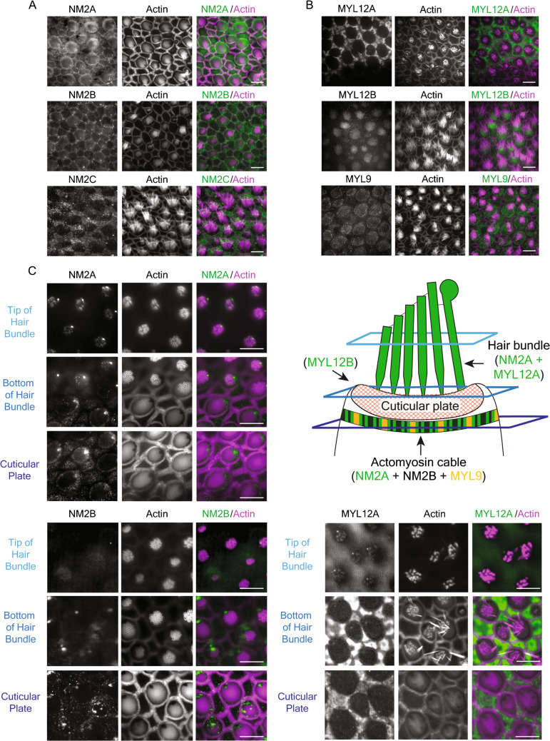 Figure 1
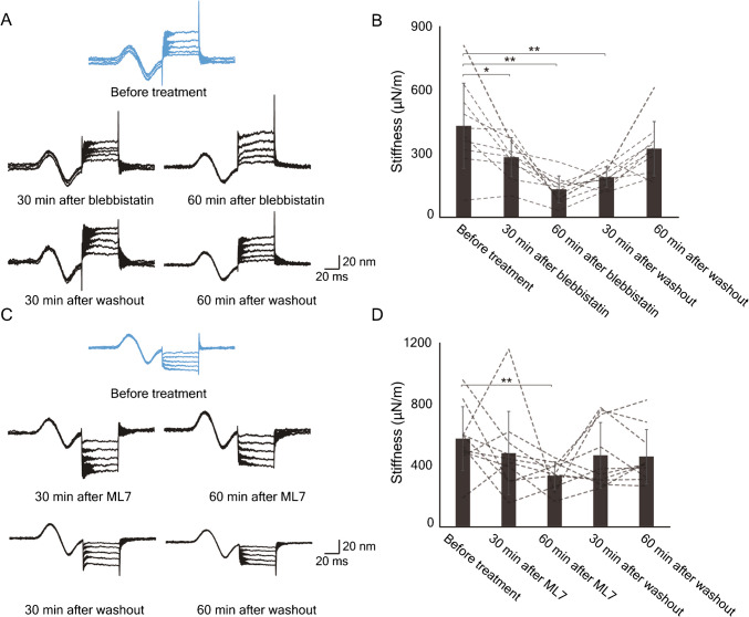 Figure 2
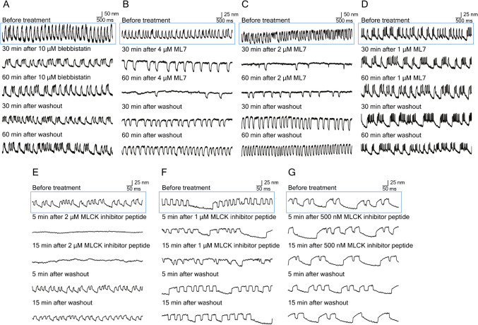 Figure 3
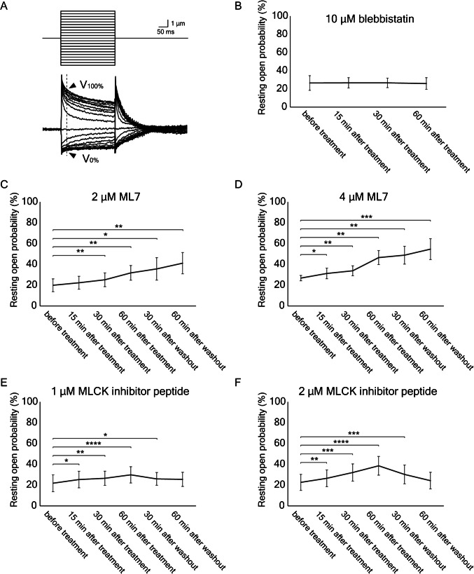 Figure 4
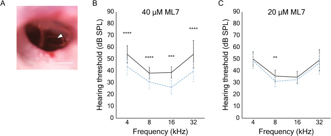 Figure 5
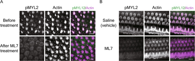 Figure 6
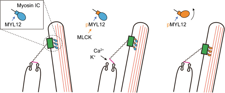 Figure 7
Peer Reviews
No public reviews on file for this paper yet. If you reviewed it on a platform where reviews are public (OpenReview, ICLR, NeurIPS, ICML), you can paste yours below so the community can read it here.
Videos
No videos yet. Explain this paper in a talk, walkthrough, or lecture? Add one.
Taxonomy
TopicsHearing, Cochlea, Tinnitus, Genetics · Acoustic Wave Phenomena Research
Introduction
Sensory hair cells mediate mechanoelectrical transduction in the internal ear and lateral-line system. The site of mechanical sensitivity, which is conserved in the receptor organs of all chordates, is the hair bundle: an erect cluster of actin-filled rods, the stereocilia, protruding from the apical cellular surface. Graded in length across a hair cell’s surface, the stereocilia are conjoined by filamentous tip links that extend from each shorter stereocilium to the longest adjacent one [1]. The lower end of each link is associated with two or more transduction channels that open in response to increased tension in the link and initiate an electrical response in the cell.
Myosin motors play several important roles in the transduction process of a hair cell. For example, a slow adaptation process, which typically exhibits a time constant of 10–30 ms, ensures that the cell’s response does not saturate in response to large, sustained stimuli [1]. The insertional plaque at the upper end of each tip link, also known as the upper tip-link density, includes the proteins harmonin, vezatin, and SANS as well as myosin molecules [2, 3]: myosin 1c (MYO1C) in the frog’s saccule and mammalian vestibular organs [4], and myosin 7a (MYO7A) as well as MYO1C in the mammalian cochlea [5, 6]. In the absence of stimulation, the climbing activity of the myosin molecules maintains in each tip link a tension that biases the channels to a certain resting open probability [6–8]. Moreover, after the bundle has been displaced in the excitatory or inhibitory direction, the myosin molecules at the tip link’s insertion respectively descend or climb to restore an approximately constant tension [9].
Although the elegance, regularity, and complexity of the hair bundle have attracted extensive attention from sensory biophysicists and developmental biologists, the cytoskeletal foundation on which the bundle rests is also of interest. The base of each stereocilium extends an actin rootlet that is anchored in the cuticular plate, a feltwork of actin filaments and spectrin [10]. The cuticular plate is in turn stabilized by various structures. On the apical surface of a cochlear outer hair cell, the cuticular plate is surrounded by a circumferential ring of filamentous actin and non-muscle myosin II (NM2) [11, 12]. The interaction between these proteins mediates contraction of the apical cellular surface [13]. Apical constriction of a similar form occurs in several developmental contexts, for example, in the neural epithelium during closure of the neural tube [13, 14] and in hair-cell morphogenesis [15] such as the developmental alignment of hair bundles [16].
Like other myosin II molecules, NM2 consists of three pairs of proteins: two molecules of a myosin heavy chain, two of an essential light chain (ELC), and two of a regulatory light chain (RLC). The activity of NM2 is regulated through phosphorylation of the RLCs by myosin light chain kinase (MLCK) [12]. When activated by the binding of the Ca^2+^-calmodulin complex, MLCK phosphorylates RLC [17], which in turn causes myosin heads to swing toward actin filaments, increases the duty ratio of myosin-actin interaction, and promotes force generation [18]. Both RLC and MLCK occur in distinct families and in various cell types; for example, the organ of Corti expresses RLCs such as myosin light chain 9 (MYL9) and myosin light chain 12 (MYL12) as well as MYLK1 (also known as smooth-muscle MLCK or smMLCK). The phosphorylation of MYL12 by MYLK1 regulates shape changes in outer hair cells [12]; moreover, deletion of the gene MYLK1 in cochlear hair cells causes hearing loss [19, 20].
Not only NM2 but also non-conventional myosin proteins such as MYO1C and MYO7A can potentially bind RLC [21, 22]. As a result, the phosphorylation of RLC by MLCK might regulate several aspects of cellular shape and hair-bundle motility. In the present study, we therefore investigated the effect of pharmacological inactivation of NM2 and MLCK on physiological parameters of frog hair cells such as stiffness, spontaneous oscillation and resting open probability, as well as the hearing level of mice.
Materials and Methods
Animals
All animal experiments were performed in accordance with institutional guidelines provided by the Institutional Animal Care and Use Committee (IACUC, protocol 22080 USDA, PRV 19085) at The Rockefeller University and ARRIVE guideline. Adult American bullfrogs (Rana catesbeiana) of both sexes were purchased from Frog Pharm (Twin Falls, ID, USA). Female C57BL/6 N mice 7 to 8 weeks of age were obtained from Charles River Laboratories (Wilmington, MA, USA).
Antibodies and Reagents
We used the following antisera and dilutions: anti-MYH9 (ab238131; Abcam, Cambridge, UK; 1:50); anti-MYH10 (ab230823; Abcam; 1:50); anti-MYH14 (20,716–1-AP; Proteintech, Rosemont, IL, USA; 1:50); anti-MYL12A (16,287–1-AP; Proteintech; 1:100); anti-MYL12B (10,324–1-AP; Proteintech; 1:100); anti-MYL9 (ab191393; Abcam; 1:50); anti-phospho-myosin light chain 2 (3671; Cell Signaling Technology, Danvers, MA, USA; 1:50); Alexa Fluor 488-labeled secondary antibody (Thermo Fisher Scientific, Waltham, MA, USA; 1:500). F-actin was labeled with rhodamine-phalloidin (PHDR1; Cytoskeleton, Inc., Denver, CO, USA; 1:167).
We also employed the following reagents: Dako Protein Block Serum-Free (Agilent, Santa Clara, CA, USA); Dako Antibody Diluent (Agilent); VECTASHIELD Antifade Mounting Medium (Vector Laboratories, Newark, CA, USA); blebbistatin (ab120425; Abcam); protease XXIV (Sigma-Aldrich, St. Louis, MO, USA); PeriAcryl90 (GluStitch Inc., Delta, BC, Canada); ML7 (ab120848; Abcam); MLCK inhibitor peptide 18 (19,181; Cayman Chemicals, Ann Arbor, MI, USA).
The concentration of blebbistatin [11], ML7 [12], and MLCK inhibitor peptide 18 [23, 24] was determined according to the previous reports. Blebbistatin and ML7 were stocked in DMSO, and inhibitor peptide was stocked in distilled water. We confirmed that DMSO treatment did not cause any changes in each experiment (data not shown).
Immunofluorescence Labeling
Samples were fixed for 30 min at room temperature with 4% formaldehyde in 0.1 M phosphate-buffered saline solution (PBS). After permeabilization for 30 min at room temperature in 0.2% Triton X- 100 in PBS, they were blocked for 30 min at room temperature with Dako Protein Block Serum-Free. The samples were incubated overnight at 4 ℃ with primary antisera in Dako Antibody Diluent. Sections were incubated for 60 min at room temperature in Alexa Fluor secondary antiserum in blocking solution. VECTASHIELD was used to mount samples. To label F-actin, rhodamine phalloidin was incubated in the blocking solution for 60 min at room temperature. Confocal images were acquired with an IX83 laser scanning microscope (Olympus, Tokyo, Japan) with a UPlanS Apo 60X objective lens (Olympus).
Physiological Recording from Bullfrog Saccules
The frog saccule was dissected as previously described [25]. The dissection was performed in oxygenated artificial perilymph containing 114 mM Na^+^, 2 mM K^+^, 2 mM Ca^2+^, 118 mM Cl^−^, 3 mM D-glucose, and 5 mM HEPES at pH 7.3. The preparation was exposed for 35 min at 26 ℃ to 66 mg/mL protease XXIV in artificial perilymph. After the otolithic membrane had been detached from each specimen by the enzyme treatment, it was removed with fine forceps.
The previously reported protocol to build a two-compartment experimental chamber was optimized for the present study [25, 26]. The saccule was sealed with PeriAcryl 90 cyanoacrylate adhesive over a hole 500–700 µm in diameter in a 10-mm square of aluminum foil with the hair bundles facing upward. Vacuum grease was put on the surface of the lower and upper chambers to prevent the solutions from leaking and mixing with each other. The foil-mounted saccule was then placed atop the lower compartment which was filled with the artificial perilymph. The upper compartment was placed on the foil and filled with artificial endolymph containing 2 mM Na^+^, 118 mM K^+^, 0.25 mM Ca^2+^, 118 mM Cl^−^, 3 mM D-glucose, and 5 mM HEPES at pH 7.3. To adjust the temperature, a temperature controller (TC200; Thorlabs, Newton, NJ, USA) was placed in the upper chamber.
The optical configuration that was previously reported [27] was optimized for the present study. The frog saccule in the two-compartment chamber was placed under an upright microscope (BX51 WI; Olympus, Center Valley, PA, USA) and visualized with a 60X water-immersion objective lens (LUMPlanFL N; Olympus). The microscope was placed on an antivibration table (Micro-g; TMC, Peabody, MA). To record an oscillation and displacement of hair bundles, the specimen was illuminated with a 630 nm light-emitting diode (UHPTSR; Prizmatix, Holon, Israel). The image was projected at a magnification of 1300X onto a dual photodiode, whose calibrated signal represented hair-bundle displacement.
Estimation of Hair-Bundle Stiffness
To measure the stiffness of hair bundles, force was applied by a flexible glass fiber (Figure S1A). After a quartz capillary 1.2 mm in diameter (Q120-60-7.5; Sutter Instruments, Novato, CA, USA) had been tapered with an electrode puller (P2000; Sutter Instrument), its tip was melted and pulled laterally to form a fiber for hair-bundle stimulation. To enhance contrast on the photodiode, the fiber was sputter-coated with gold–palladium (Hummer 6.2; Anatech, Sparks, NV, USA). The stiffness and drag coefficient were estimated by measuring Brownian motion in water with photodiode sampling at 10,000 s^-^^1^ [27, 28]. The stiffness of the fibers (K_f_) was 150-400 μN·m^-^^1^.
A fiber mounted on a piezoelectric actuator (PA 4/12; Piezosystem Jena, Hopedale, MA, USA) was attached to a hair bundle at the kinociliary bulb with a micromanipulator (MP285, Sutter Instrument). The fiber’s displacement (ΔX_f_) was controlled with LabVIEW 2019 software (National Instruments, Austin, TX, USA). The glass probe was displaced in five steps, ranging from 20 to 100 nm, to move the hair bundle in the negative direction such that the transduction channels were largely closed (Figure S1A, left and middle panels). Stimuli 100 ms in duration were presented every 200 ms. The values of ten repetitions at each stimulus magnitude were averaged and recorded. The hair-bundle displacement (ΔX_HB_) was calculated as the average value 30–45 ms after the onset of a stimulus pulse (Figure S1A). Finally, the stiffness of hair bundles of each step (K_HB (single step)) was obtained by the following formula: K_HB (single step) = [(ΔX_f_ − ΔX_HB_)/ΔX_HB_]·K_f_. K_HB (single step)_ for each of the five steps was averaged and used as an estimate of hair-bundle stiffness (K_HB_). To measure the stiffness of hair bundles with broken tip links, we added 2 mM EDTA to endolymph for 30 min, then displaced bundles as mentioned above (Figure S2A,B).
Furthermore, to observe the displacement of the cuticular plate, borosilicate glass microspheres 2 µm in diameter (Duke Scientific, Palo Alto, CA, USA) were placed on the apical cellular surface above the cuticular plate (Figure S3A) and the displacement of the glass beads was measured while the hair bundle was displaced (Figure S3B,C).
The data were obtained from ten hair cells in different animals. The data from each hair bundle were the averages of the ten repetitions of the recording.
Observation of Spontaneous Hair-Bundle Oscillation
Hair-bundle oscillation is readily observable with the optical system described above. To quantify the displacements, 10 s segments of spontaneous hair-bundle oscillation were measured with the photodiode and recorded (LabVIEW 2019). A high-speed camera (Phantom VEO710L; Vision Research, Wayne, NJ, USA) was also used to record spontaneous oscillation. The video data were extracted as consecutive TIF images and the movement of hair bundles was tracked by the TrackMate plugin [29] of Fiji (ImageJ) [30]. Oscillations were recorded before and after treatment with inhibitors and after washout.
Recording of Microphonic Potentials
For the recording of microphonic potentials, which provide a means of assessing the average response of an ensemble of hair cells, a frog’s saccule was placed in a two-compartment experimental chamber [31, 32]. The saccule was sealed with cyanoacrylic adhesive (PeriAcryl 90) across a hole 500–700 µm in diameter through a circular plastic coverslip 10 mm in diameter and 200 µm in thickness (12,547, Thermo Fisher Scientific). The mounted saccule was placed between gaskets of thermoplastic material (Parafilm, Amcor, Zurich, Switzerland) to provide electrical isolation and prevent the mixing of solutions. The coverslip was placed over the lower compartment, which was filled with artificial perilymph containing 99 mM Na^+^, 19 mM K^+^, 2 mM Ca^2+^, 118 mM Cl^−^, 3 mM D-glucose, and 5 mM HEPES at pH 7.3. By partially depolarizing the hair cells, this high-K^+^ perilymph minimized voltage-, time-, and Ca^2+^-sensitive currents and thus simplified the analysis of microphonic responses [31]. The upper compartment was then added to the chamber and filled with artificial endolymph.
To elicit responses from a group of hair cells with similarly oriented hair bundles, we partially removed the otolithic membrane [31]. Displacements produced by a piezoelectric actuator (PA 4/12, Piezosystem Jena) were applied to the remaining otolithic membrane by means of a glass probe produced by melting and roughening the tip of a micropipette. The displacement of the glass probe was controlled by LabVIEW 2019 software, and the maximal amplitude was adjusted to the value that evoked a saturating response. The glass probe was displaced by ten positive and ten negative steps with equal separations in their magnitudes; an unstimulated control was also recorded. The responses were averaged across three repetitions. To estimate the resting open probability, we first determined the maximal positive response, V100%, and the greatest negative response, V0%, that were recorded between 10 and 20 ms after the onset of stimulation (Fig. 4A). Around V100% or V0%, the response was well saturated. Although it cannot be said that all channels were opened or closed at V100% or V0%, we calculated the resting open probability POR as POR = − V0%/(V100% − V0%).
The data were obtained from six different saccules. The data from each represent the average of three repetitions.
Measurement of Auditory Brainstem Responses
To assess the effect of inhibitors on hearing, we performed transtympanic injection of blockers prior to recording of ABRs from mice. Each animal was anesthetized with isoflurane delivered through a face mask at a concentration of 3–5% during induction and of 1–2% to maintain a surgical level of anesthesia. The tympanic membrane was visualized under a microscope and incised with a 30-gauge needle (Fig. 5A). After 5 µL of inhibitor solution or a saline control solution had been gently injected into the middle ear [33], the animal was allowed to awake and kept on a heating pad for 2 hr.
After a mouse had been anesthetized with an intraperitoneal injection of 100 mg/kg ketamine and 10 mg/kg xylazine, the ABR was recorded [34]. Computer-generated stimuli were amplified by a stereo amplifier (SA1, Tucker-Davis Technologies, Alachua, FL, USA) and delivered by a free-field speaker (MF1, Tucker-Davis Technologies). Tone-burst stimuli with rise and fall times of 1 ms and durations of 5 ms were delivered 1000 times apiece at frequencies of 4 kHz, 8 kHz, 16 kHz, and 32 kHz. Subcutaneous electrodes were inserted in three positions: a recording electrode behind the pinna, a reference electrode in the scalp, and a ground electrode in the back near the tail. The electrical signals were amplified 10,000X and bandpass filtered between 300 and 3000 Hz by a preamplifier (P55; Grass Instruments, West Warwick, RI, USA). The averaged responses were reviewed by two authors, R.O. and B.F., and hearing thresholds were determined by their agreement. In total, ten treated ears and ten control ears from ten animals were used for each analysis.
Results
Expression of NM2 and RLC in the Frog’s Saccule
Immunofluorescent labeling of the apical surfaces of the bullfrog’s saccular hair and supporting cells demonstrated NM2A, with MYH9 heavy chains, and NM2B, with MYH10 heavy chains (Fig. 1A). The stereocilia were visualized by rhodamine phalloidin, confirming that the stained cells were hair cells. These proteins occurred in a circumferential ring around the cuticular plate. Although absent from the apical surfaces, NM2C with MYH14 heavy chains occupied the apical cytoplasm of supporting cells. Immunofluorescent labeling of RLCs, such as MYL12A, MYL12B, and MYL9, on the apical surface of the bullfrog’s saccule is shown in Fig. 1B. MYL12A was expressed in the supporting cells and MYL12B was expressed in the hair cell surface. MYL9 occurred at the apical surfaces of hair cells and co-localized with actin (Fig. 1B). To investigate the protein localization in the hair bundles and on the cell surface, we analyzed the multiple layers of the apical portion of hair cells, including the tip and bottom of hair bundles and the cuticular plate (Fig. 1C). The upper right panel of Fig. 1C indicates the level of each layer. NM2A (upper left panel) was expressed in the tip of hair bundles, the kinocilium and apical surface of hair cells, whereas NM2B (lower left panel) was expressed only on the apical surface of hair cells and the kinocilium. The apical surface of a frog saccular hair cell is therefore bounded by an actomyosin cable composed of NM2A and NM2B with MYL9 as the associated RLC (Fig. 1C, upper right panel). Furthermore, MYL12A is expressed in the tip of hair bundles as the associated RLC of NM2A (Fig. 1C, upper right panel and lower right panel).Fig. 1. The expression of NM2 and myosin light chains in frog saccular hair cells. A Immunofluorescence labeling demonstrates NM2A and NM2B in the apical portions of hair cells and NM2C in supporting cells. Here and in panel B and C, actin labeled with phalloidin marks hair bundles, cuticular plates, and cellular perimeters. Scale bars here and in panel A, B, and C, 10 µm. B Immunofluorescence labeling marks MYL12A in supporting cells, MYL12B and MYL9 in the apical cytoplasm of hair cells. MYL9 is co-localized with actin of the actomyosin cable. C Multiple layers of immunofluorescent labeling show the localization of NM2A (upper left panel), NM2B (lower left panel), and MYL12A (lower right panel). The level of each layer is shown in the upper right panel. NM2A is expressed in the tip of hair bundles, the kinocilium and apical surface of hair cells, whereas NM2B is expressed only on the apical surface of hair cells and the kinocilium. MYL12A is expressed in the tip of hair bundles and supporting cells. In a schematic diagram of the apical portion of a frog’s saccular hair cell (upper right panel), NM2A and MYL12A are expressed in the hair bundles and NM2A, NM2B, and MYL9 are expressed in the actomyosin cable
Effect of Inhibiting NM2 and MLCK on Hair-Bundle Stiffness
To examine the physiological function of NM2 and MLCK, we assessed the effect of their inhibitors, blebbistatin and ML7, respectively, on the stiffness of hair bundles of the frog’s saccule (Figure S1A). Based on the results of control experiments, we performed stiffness measurement at room temperature (Figure S1B,C).
We treated the saccule with 10 µM blebbistatin, a specific inhibitor of NM2 isoforms. Within 60 min, the stiffness declined significantly to 30% of its control value (P = 0.0013, N = 10; Fig. 2A, B). The effect was reversible: by 60 min after washout of the compound, the stiffness was not significantly different from the control value (P = 0.13, N = 10). Because blebbistatin significantly reduced the stiffness of hair bundles with disrupted tip links, the effect did not require intact transduction (Figure S2A).Fig. 2. The effects of inhibitors on the stiffness of frog saccular hair bundles. A Records from a hair bundle before, during, and after treatment with 10 µM blebbistatin portray the average responses from ten repetitions of mechanical pulses of five positive amplitudes. Here and in panel C, the sinusoidal curves at the outset of each record represent displacements of the photodiode and serve as calibrations of ± 20 nm. B Averaged over ten repetitions, the data from panel A indicate that blebbistatin significantly and reversibly reduced bundle stiffness. Here and in subsequent figures: *P < 0.05; **P < 0.01. Note that there was no significant difference between the control records and those acquired after 60 min of washout. C Records from a hair bundle before, during, and after treatment with 2 µM ML7 typify the average responses from ten repetitions of mechanical pulses of five negative amplitudes. D Statistical analysis of the data from panel C demonstrates a significant difference between control and treated conditions but no difference 30 min or 60 min after washout
At a concentration of 2 µM, ML7, an inhibitor of MLCK, also significantly affected the stiffness of saccular hair bundles. Treatment for 60 min lowered the stiffness to 58% of its control level (P = 0.0068, N = 10; Fig. 2C, D). The effect was reversible within 30 min after washout (P = 0.40, N = 10). In addition, ML7 reduced the stiffness of hair bundles with disrupted tip links; the effect, however, was not statistically significant (Figure S2B).
We measured the movement of the hair-cell surface during stimulation to confirm that the stability of the cuticular plate was not affected by inhibitors (Figure S3A). Glass beads placed on the cellular apex above the cuticular plate did not show significant displacements associated with stimulation (Figure S3B,C). In conjunction with the foregoing result, the data imply that NM2 and RLC phosphorylation at an actomyosin cable of the hair cell’s apex contribute to the hair bundle’s stiffness.
Effect of Inhibiting NM2 and MLCK on Spontaneous Hair-Bundle Oscillation
When freed of their connections with an otolithic membrane and maintained in a two-compartment experimental environment, the hair bundles of the frog’s saccule exhibit robust, low-frequency oscillations that reflect the active process of mechanoelectrical transduction [35] (Movies S1 and S4). To examine whether the spontaneous oscillation of hair bundles is affected by NM2 and MLCK, we used high-speed video microscopy or photodiode recording to observe spontaneous oscillations during inhibition of the two proteins. Because temperature affects the frequency and magnitude of oscillation, we performed control experiments to establish 20 ℃ as a suitable level for long-term observations (Figure S4A).
As has been observed previously [36], hair cells showed oscillations with a variety of frequencies and waveforms (Fig. 3). Treatment of active hair bundles with 10 µM blebbistatin did not arrest oscillations, an observation that accords with evidence that the slow adaptation process of hair bundles involves MYO1C and MYO7A rather than NM2. The treatment did alter the characteristics of some oscillations; for example, an oscillation that initially occurred at a frequency near 3 Hz became biphasic and declined to a frequency of 1.5 Hz (Fig. 3A).Fig. 3. The effects of inhibitors on spontaneous oscillation by frog saccular hair bundles. A Treatment with 10 µM blebbistatin reversibly reduced the frequency of oscillation. B Treatment with 4 µM ML7 greatly slowed and almost arrested spontaneous oscillation; the effect was partially reversible after washout. C ML7 at a concentration of 2 µM had a similar but weaker effect than the foregoing; washout led to essentially complete recovery. D The effect of 1 µM ML7 was marginal. E Only 5 min of treatment with 2 µM MCK inhibitor peptide 18 completely blocked spontaneous oscillation, which largely recovered upon washout. F At a concentration of 1 µM, peptide 18 elicited a slight slowing of oscillations. G The effect of 0.5 µM peptide 18 was negligible. In all panels, an upward deflection signifies movement in the positive direction, towards the hair bundle’s tall edge. Blue boxes on these figures indicate the control (before treatment)
We also observed the effect of ML7 on spontaneous oscillation. The motion was completely inhibited at a concentration of 2 µM or 4 µM but showed recovery after washout (Fig. 3B, C). At 1 µM, the compound did not significantly affect oscillation (Fig. 3D). High-speed video microscopy confirmed the observations made with the photodiode (Movies S1–S3; Figure S4B). To test an alternative means of inhibition, we also used MLCK inhibitor peptide 18. At a concentration of 1 µM or 2 µM, oscillation was again inhibited and recovered after washout (Fig. 3E, F); an inhibitor concentration of 500 nM had no effect (Fig. 3G). High-speed video imaging confirmed the inhibition of oscillation by 2 µM of the peptide (Movies S4–S6; Figure S4C). Inhibition by peptide 18 was more rapid than that owing to ML7. Both of the MLCK inhibitors froze hair bundles in the positive direction, that is, while deflected toward their tall edges.
Effect of Inhibiting NM2 and MLCK on Transduction Channels’ Open Probability
We next inquired whether inhibiting NM2 or MLCK would affect mechanoelectrical transduction in frog saccular hair cells. To investigate the roles of the proteins in adjusting the resting open probability of mechanoelectrical-transduction channels, we applied inhibitors while recording microphonic responses [31]. These extracellular signals reflect the total current that traverses transduction channels and provide a convenient means of assessing the averaged responses of hundreds of hair cells (Fig. 4A).Fig. 4. The effects of inhibitors on the resting open probability of transduction channels. A In recordings of microphonic responses, a glass probe displaced the otolithic membrane to 21 levels (upper traces), including zero. The maximal response (V100%) and minimal response (V0%) were estimated from averaged electrical records (arrowheads) 10 ms after the onset of stimulation (lower traces). B Treatment with 10 µM blebbistatin had no effect on the resting open probability. C In six experiments, exposure to 2 µM ML7 slightly increased the resting open probability, but the effect was not reversed after washout. D Treatment with 4 µM ML7 in six experiments had a greater irreversible effect. E At a concentration of 1 µM, MLCK inhibitor peptide 18 significantly increased the resting open probability. F In six experiments, 2 µM MLCK inhibitor peptide 18 significantly and reversibly raised the resting open probability. There was no significant difference between the control recording and that of 60 min after washout. Here and in the subsequent figure: ***P < 0.001; ****P < 0.0001
Consistent with the expectation that tip-link tension is set by MYO1C and MYO7A, treatment with the NM2 inhibitor blebbistatin did not significantly affect the resting open probability (Fig. 4B). By contrast, 30 min of treatment with 2 µM of the MLCK inhibitor ML7 raised the resting open probability significantly, from 19.7 ± 6.2 to 25.0 ± 6.6% (P = 0.009, N = 6; Fig. 4C). The effect was not reversed by 60 min of washout of the inhibitor. The resting open probability also showed a significant and lasting elevation after treatment with 4 µM ML7 (Fig. 4D).
Peptide 18 also had a significant effect on the resting open probability of transduction channels. After 60 min of treatment with 1 µM of peptide 18, the probability increased from 21.6 ± 6.8 to 29.7 ± 7.8% (P < 0.0001, N = 6; Fig. 4E). Following 60 min of washout, the open probability of 25.3 ± 6.8% did not differ significantly from the control value (P = 0.086). Furthermore, treatment with 2 µM of peptide 18 evoked a significant increase of the resting open probability and a washout effect as well (Fig. 4F). This and the foregoing experiment indicate that MLCK inhibition causes a small but significant increase in open probability.
Effect of MLCK Inhibition on the Murine Auditory Brainstem Response
To investigate the effect of MLCK inhibition on hearing, we measured the threshold of the auditory brainstem response (ABR) after the transtympanic injection of ML7 in mice (Fig. 5A). In keeping with the volume of the murine middle ear [37], 5 µL of ML7-containing solution was injected into the middle ear on one side and 5 µL of saline solution was injected into the middle ear on the opposite, control side.Fig. 5. The effect of ML7 on hearing thresholds in the mouse. A In an image of the tympanic membrane after transtympanic injection, the white arrow indicates the incision of the tympanic membrane for drug administration. Scale bar, 0.5 mm. B Following the administration of 40 µM ML7 into the middle ear, averaged auditory brainstem responses displayed significantly elevated thresholds at the four frequencies tested. The solid line marks responses from the ear exposed to the inhibitor, whereas the dotted line shows data for the control ear exposed to vehicle. C A lower concentration of ML7, 20 μM, caused a significant threshold elevation only at a stimulus frequency of 8 kHz
Two hours after transtympanic injection at a concentration of 40 µM, ML7 significantly increased the ABR threshold at stimulus frequencies from 4 to 32 kHz (Fig. 5B). A lower concentration of the compound, 20 µM, yielded a significant threshold elevation only at 8 kHz (Fig. 5C).
Effect of Inhibition of MLCK on Phosphorylation of RLC
We performed immunolabeling to ascertain whether the phosphorylation of RLC was significantly affected by MLCK inhibitors. We used the phospho-MYL2 antibody, which can detect the phosphorylation of any type of myosin light chain 2 including MYL12 and MYL9. Using frog saccules from microphonic recordings after 60 min of treatment with 4 µM ML7, we found that immunolabeling of phosphorylated RLC disappeared from hair bundles but not from the apical surfaces of hair cells (Fig. 6A). Immunolabeling of phosphorylated RLC was also performed in the cochleas of mice after ABR recordings. In animals treated with 40 µM ML7, the immunofluorescence signal for phosphorylated RLC was far weaker in hair bundles on the treated than on the untreated side of an animal (Fig. 6B). These control experiments demonstrate that ML7 treatment inhibited the phosphorylation of RLC in both species and support the inference that the observed physiological effects stemmed from reduced RLC activity.Fig. 6. The effect of ML7 on the phosphorylation of RLC in frog and mouse hair bundles. A Immunofluorescence labeling of the phosphorylated regulatory light chain pMYL2 shows a robust signal in frog hair bundles that is abolished by treatment with 4 µM ML7. Scale bar, 10 µm. B In the mouse organ of Corti, immunofluorescence labeling of phosphorylated light chain is abolished after the transtympanic injection 40 µM ML7. In this instance, the control specimen reflects the result after transtympanic injection of the saline vehicle. Scale bar, 10 µm
Discussion
This study of the physiological function of NM2 and MLCK in inner ear hair cells has yielded several results. Frog saccular HCs express NM2 and MYL9 at their apical surfaces and MYL12 in their hair bundles. The bundles’ stiffness declines after treatment with blebbistatin or ML7. Moreover, spontaneous oscillation of the frog saccular HCs is only slightly affected by treatment with blebbistatin but is inhibited by MLCK inhibitors such as ML7 and MLCK inhibitor peptide 18. The resting open probability of frog saccular HCs was increased by treatment with ML7 or peptide 18. Finally, transtympanic injection of ML7 in the mouse middle ear increases the threshold of auditory brainstem responses.
Although bullfrogs have been used extensively in physiological research, they have played less of a role in biochemical investigations. NM2 and RLC have been shown to occur at the apical surfaces of hair cells in rodents [11, 12]. In the current study, we determined that frog saccular hair cells express NM2A/B and MYL9 at their apical surfaces and NM2A and MYL12A in their hair bundles. Our measurements indicate that NM2 plays a role in regulating the stiffness of frog saccular hair bundles. Because we displaced bundles in the negative direction, which reduced the tension in tip links, the results bear primarily on the stiffness of the stereociliary pivots. Our control study with glass beads indicated that the cuticular plate itself does not slip horizontally in response to glass probe stimulation. Although with our current method we could not record vertical displacements such as sinking or rocking of the cuticular plate, the inhibitors certainly affected the stiffness of hair bundles. Our other control studies also showed that inhibitors decreased the hair-bundle stiffness even after tip links had been broken by EDTA treatment (Figure S2A, B). This result confirmed that the inhibition of NM2 and MLCK affects the stiffness of stereociliary pivots, which reflects the anchorage of stereociliary rootlets into the cuticular plate. In conclusion, NM2 and MLCK activity affect the tightness of the insertion of stereociliary rootlets into the cuticular plate or indeed the stiffness of the cuticular plate itself.
The spontaneous oscillation of hair bundles results from the active process of non-mammalian vertebrates [26]. Extensive evidence suggests that oscillation emerges from the activity of myosin motors at the tops of hair bundles [37]. In the present study, we found that spontaneous oscillation is inhibited by the MLCK inhibitors ML7 and peptide 18. Peptide 18 is highly membrane permeable, probably because it has a sequence similarity to the protein transduction domain of the HIV-1 TAT protein [24], and it acts quickly, with its inhibitory effect being reversible. Although the two inhibitors have distinct inhibitory mechanisms [23, 24], their similar and concentration-dependent effects are consistent with MLCK inhibition. In addition, the spontaneous oscillation was not significantly affected by blebbistatin, despite NM2A being expressed in hair bundles. The results suggest that MYL12A serves as a light chain of MYO1C rather than NM2A, perhaps by binding to this myosin’s highly conserved second IQ domain [38].
We observed that the resting open probability of transduction channels does not change with NM2 inhibition but increases when MLCK is inhibited, which suggests that phosphorylation of MYL12A increases the force for myosin motors to descend, resulting in reduced tip-link tension that facilitates channel closure. MYL12 binds to unconventional myosins including MYO15A [39] and MYO7A, a myosin motor of cochlear hair cells [40]. Both of these unconventional myosins possess IQ motifs to which their light chains bind [22]. MYO1C also has IQ motifs that bind calmodulin [41] and could potentially bind light chains such as MYL12. We therefore hypothesize that MLCK phosphorylates MYL12A bound to MYO1C rather than NM2A in the frog’s hair bundles and that inhibition of this phosphorylation reduces the sliding of MYO1C along the actin filaments of stereociliary cores. Consistent with this hypothesis, MLCK inhibitors arrest spontaneously oscillating hair bundles with a deflection in the positive direction. We hypothesize that a complex of MYO1C and MYL1 A is necessary to produce force for myosin motors to descend and reduce tip-link tension during adaptation and that the phosphorylation of MYL12 enhances this force production (Fig. 7). In addition, we also hypothesized that NM2 and MYL9 are not involved in the adaptation process directly. Because our experiment involved the application of inhibitors to the entire saccule, we cannot exclude the possibility that cells other than hair cells were affected by inhibitors and induced changes in stiffness, oscillation, and resting open probability. More detailed molecular studies should clarify the precise sites of action of the inhibitors.Fig. 7. The hypothesized effect of inhibitors on the adaptation motor of hair bundles. The left schematic diagram shows the control configuration of a hair bundle, with the bundle in its resting position and the transduction channels largely closed. In the middle diagram, deflection of the bundle in the positive direction opens transduction channels, which admit K^+^ and Ca^2+^ into the cell. As shown in the right diagram, phosphorylation of the regulatory light chain MYL12 promotes slippage of the myosin motor along stereociliary actin filaments, which presumably slackens tip links and promotes the consequent reclosure of channels
In addition to our ex vivo experiments, we showed that hearing in intact mammals is inhibited by ML7. The experiments demonstrated dose dependency and effects over a broad range of stimulus frequencies. Previous evidence also supports a role of MLCK activity in hearing: mice exhibiting a specific deletion of MLCK in either inner or outer hair cells display increased ABR thresholds [19, 20]. The phosphorylation of MYL12 is therefore important in normal hearing, perhaps by affecting an adaptation motor such as MYO7A. However, there are two limitations in this experiment. First, we do not know the exact concentration of ML7 in the inner ear. ML7 presumably gains access to the cochlea through the round window, but the efficacy of this infiltration is unknown. The inhibitory constant of ML7 for MLCK is 300 nM [42], so concentrations of 20–40 µM might inhibit other kinases as well. The second reservation is that we did not observe a consistent effect of peptide 18 on the ABR. Because the peptide is highly cell-permeable and easy to wash out, it is difficult to maintain an effective concentration during protracted recordings.
Statistics
Statistical analyses were performed using JMP version 17 statistical software (IBM Japan, Tokyo, Japan). All data are expressed as means ± standard deviations. A paired Student’s t-test was performed for the comparison; a value of P < 0.05 was considered to represent a significant difference.
Supplementary Information
Below is the link to the electronic supplementary material.Supplementary movie 1 (MP4 35619 KB)Supplementary movie 2 (MP4 35769 KB)Supplementary movie 3 (MP4 35139 KB)Supplementary movie 4 (MP4 35608 KB)Supplementary movie 5 (MP4 36778 KB)Supplementary movie 6 (MP4 38979 KB)Supplementary source data Figure 2 (XLSX 13 KB)Supplementary source data Figure 4D (XLSX 13 KB)Supplementary source data Figure 5 (XLSX 13 KB)Supplementary source data Figure S1 (XLSX 12 KB)Supplementary source data Figure S2 (XLSX 12 KB)Supplementary data sheet (XLSX 23.2 KB)Supplementary information (DOCX 911 KB)
The reference list from the paper itself. Each links out to its DOI / PubMed record.
- 1Liu Y, Qi J, Chen X et al (2019) Critical role of spectrin in hearing development and deafness. Sci Adv 5:eaav 7803. 10.1126/sciadv.aav 780310.1126/sciadv.aav 7803 PMC 646994231001589 · doi ↗ · pubmed ↗
- 2Azimzadeh JB, Salvi JD (2017) Physiological preparation of hair cells from the sacculus of the American bullfrog (Rana catesbeiana). J Vis Exp. 10.3791/5538010.3791/55380 PMC 540896428362415 · doi ↗ · pubmed ↗