In vitro evaluation of biofilm formation by Streptococcus mutans and Candida albicans in orthodontic aligners
Carolina Veronez Garbúggio SANCHES, Raquel Sano Suga TERADA, Adilson Luiz RAMOS, Janaina de Cássia Orlandi SARDI, Maria Gabriely Malveiro ESTEVES, Maria Cristina Bronharo TOGNIM, Sheila Alexandra Belini NISHIYAMA

TL;DR
This study shows that orthodontic aligners can accumulate harmful biofilms that may lead to tooth decay.
Contribution
The study experimentally demonstrates biofilm formation on orthodontic aligners by S. mutans and C. albicans.
Findings
Monospecies and mixed biofilms of S. mutans and C. albicans formed robustly on Invisalign® aligners.
Biofilm accumulation could increase the risk of carious lesions in users.
Proper cleaning of aligners is essential to prevent microbial buildup.
Abstract
Aligners have been used by the orthodontic community for approximately 20 years, but little research has been carried out on the accumulation of biofilm on the surface of these aligners, as well as their possible impact on the oral ecosystem. Ten hemi-arches of Invisalign® brand orthodontic aligners were used. The hemi-arches were placed inside sterile flasks containing 25 mL of Gibbons and Nygaard broth, with standardized suspensions of the two microorganisms on the 0.5 MacFarland scale and incubated in aerophily (C. albicans) and microaerophily (S. mutans and mixed biofilm) at 37°C for 72h. The biofilm formed was removed by the multiple rinses method to quantify the microorganisms in the biofilms in CFU/mL. A qualitative analysis with scanning electron microscopy was performed to observe the structure of the formed biofilms. It was observed the accumulation of a monospecies biofilm…
Genes, proteins, chemicals, diseases, species, mutations and cell lines named across the full text — each resolved to its canonical identifier and authoritative record.
Click any figure to enlarge with its caption.
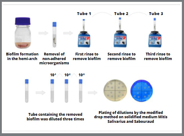 Figure 1
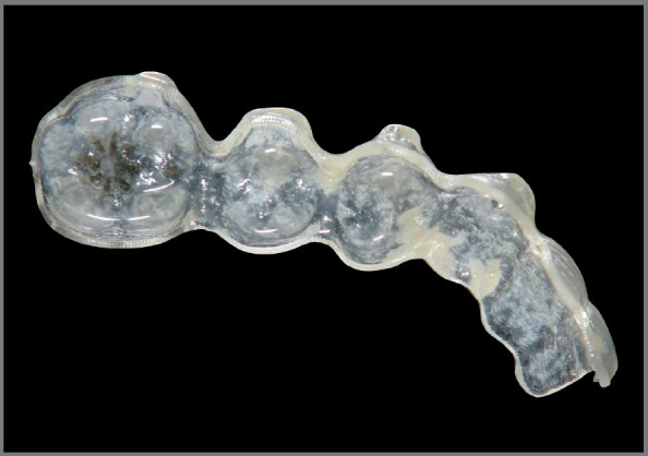 Figure 2
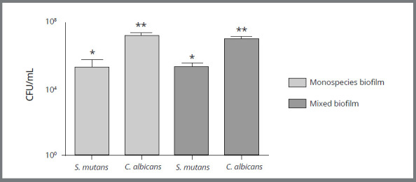 Figure 3
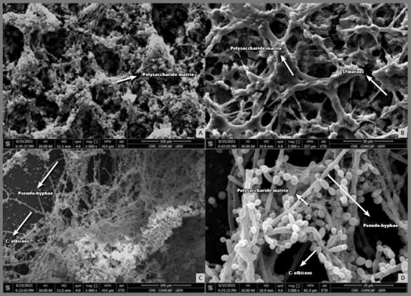 Figure 4
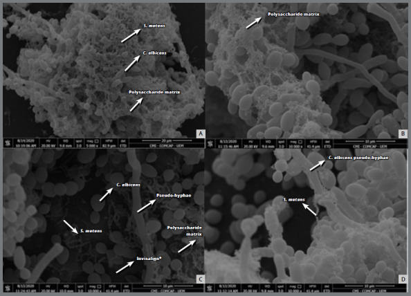 Figure 5
Peer Reviews
No public reviews on file for this paper yet. If you reviewed it on a platform where reviews are public (OpenReview, ICLR, NeurIPS, ICML), you can paste yours below so the community can read it here.
Videos
No videos yet. Explain this paper in a talk, walkthrough, or lecture? Add one.
Taxonomy
TopicsOral microbiology and periodontitis research · Gut microbiota and health · Salivary Gland Disorders and Functions
INTRODUCTION
The number of adult patients seeking orthodontic treatment has increased significantly and, with it, the need to develop more aesthetic orthodontic solutions to meet the growing social and cosmetic demands. Thus, in 1997, the Invisalign^®^ system emerged, which aims to align teeth in a more predictable and comfortable way, using precise “invisible” plastic retainers to achieve tooth movement, thus meeting the aesthetic demands of patients.
Although these aligners have been used by the orthodontic community for almost 20 years, there is still little research conducted on these appliances and how much they can disrupt the oral microbiota and promote biofilm accumulation. It is recognized that the oral cavity is colonized by a very complex microbiota, with several microbial species coexisting in different niches that establish homeostasis with the host.1 However, environmental changes can impact the local microbial composition, resulting in dysbiosis and enabling the development of diseases.2
Many microorganisms present in the oral cavity have the potential to cause diseases in opportune situations. It is known that most oral diseases are associated with microbial biofilms.3 Therefore, maintaining oral health, especially when controlling the most common clinical manifestations associated with biofilms (dental caries, periodontal diseases and oral candidiasis), is essential to understand the formation and composition of dental biofilms in order to find possible strategies that can be adopted for their control.4
The use of removable appliances for relatively short periods can result in bacterial biofilm accumulation on the tooth’s surface and on acrylic bases.5 It has been suggested that removable appliances favor local adherence and colonization of Streptococcus mutans 6 and Candida albicans,7 whose presence has been related to the appearance of caries lesions,6 reddish coloration of the mucosa, discomfort, halitosis and taste alteration.8 Thus, the growth of biofilms on the surface of these appliances can compromise the oral health of patients and compromise the effectiveness of orthodontic treatment.9
In this context, orthodontic aligners are also abiotic surfaces that can facilitate the accumulation of biofilm.10 However, as far as we know, there is still no research aimed at investigating and quantifying the nature of the biofilms adhered to this type of appliance. Considering the long duration of this type of treatment, which can last for up to 42 months,11 there is a concern to satisfy the aesthetic requirements, with the correct alignment of the teeth, but without sacrificing the patient’s health. Thus, the objective of this study was to evaluate the in vitro formation of single and mixed biofilms of Streptococcus mutans and Candida albicans in orthodontic aligners.
MATERIAL AND METHODS
STUDY DESIGN
This is an in vitro study in which triplicate experiments were performed to evaluate the possible formation and quantification of single and mixed biofilms composed of Streptococcus mutans and Candida albicans in orthodontic aligners. A complementary qualitative analysis by Scanning Electron Microscopy (SEM) was performed.
IN VITRO EXPERIMENT FOR BIOFILM QUANTIFICATION
Sample preparation
For the in vitro experiment, 10 hemi-arches of Invisalign^®^ orthodontic aligners were analyzed (Lot 6671479 from the year 2018). The hemi-arches were made using a 7020 double-faced flex diamond disc (KG Sorensen) in a straight piece and Sof-Lex™ Pop-On (3M) abrasive discs on a micromotor. Then, the aligners were sent for ethylene oxide sterilization.
Microorganisms
Streptococcus mutans (ATCC 25175) and Candida albicans (ATCC 10231) strains were used and cultivated in BHI broth at 37°C for 48 hours in microaerophilia for S. mutans and in aerobiosis for C. albicans. After cultivation, an aliquot of each culture was submitted to Gram stain, for the analysis of the morphological and staining characteristics of the microorganisms and pure culture.
Biofilm formation
In a glass jar containing 25 mL of previously sterilized Gibbons and Nygaard broth,12 a hemi-arch of the orthodontic aligner was aseptically introduced for the formation of monospecies and mixed biofilms. Subsequently, standardized inocula of the Streptococcus mutans (which were incubated in microaerophilia) and Candida albicans (which were incubated in aerobiosis) strains were added on a 0.5 McFarland scale (1.5 x 10^8^cells/mL) at 37°C for 72 h. The mixed biofilm was performed under microaerophilia conditions.
Biofilm quantification
After the formation of the monospecies and mixed biofilms, each hemi-arch of the orthodontic aligner was carefully rinsed in a tube containing 10 mL of sterile saline solution (NaCl, 0.9%), to remove the cells that were not adhered.
Then, the biofilm was removed by multiple rinses, where each hemi-arch was inserted into a tube (Tube 1) containing 10 mL of sterile saline solution and vortexed (Scientific Industries Inc., New York, USA) for 60 seconds. The procedure was repeated two more times, with the devices transferred to different tubes (Tubes 2 and 3) in sequence, under the same conditions. After removing the biofilm, each tube was diluted three times (10^-1^, 10^- 2^, 10^-3^) and 20 μL of the obtained dilutions were plated by the modified drop method in triplicate.
For plating and counting the colonies of the monospecies biofilm, Mitis Salivarius Agar (Difco, New Jersey, USA) was used for S. mutans and Sabouraud Agar (Difco, New Jersey, USA), for C. albicans. For the mixed biofilm analysis, 2 μg/mL of amphotericin B were added to Mitis Salivarius Agar and 2 μg/mL of ampicillin, to Sabouraud Agar, to inhibit the growth of C. albicans and S. mutans, respectively. The concentrations of antimicrobials used were based on the European Committee on Antimicrobial Susceptibility Testing (EUCAST - version 10, 2020).
Next, the seeded plates were incubated at 37°C for 48 hours in aerobiosis for C. albicans and in microaerophilia for S. mutans and for the mixed biofilm, for colony count and determination of the CFU/mL of each microorganism (Fig. 1).
Figure 1:Flowchart of the experimental design of the present study.
MORPHOLOGICAL AND STRUCTURAL ANALYSIS OF THE BIOFILM IN SCANNING ELECTRON MICROSCOPE
For the qualitative analysis of the biofilm by SEM, the following were performed: samples with a maximum size of 1 x 1 cm were taken of the aligners, with three samples for the mixed biofilm and three samples of the same size for each microorganism in the monospecies biofilms. For the preparation of such samples, 7020 flexible diamond discs (KG Sorensen) were used in a straight workpiece, and Sof-Lex™ Pop-On (3M) abrasive discs in a micromotor. Once prepared, the aligner samples were sterilized using ethylene oxide before the experiment.
Sample preparation was developed according to Weber et al.13 and Bodelón et al.14, with some modifications. Biofilms were fixed with 1 ml of glutaraldehyde 2.5% in sodium cacodylate buffer 0.2 M (pH 7.2 - 7.4) for 1h at room temperature. The samples were dehydrated with an increasing series of anhydrous alcohol (PA) concentrations: 50%, 70%, 80%, 90%, 95% and 100%, for 15 min in each concentration, and repeated three times in the pure alcohol step, to ensure the complete removal of water.
Subsequently, the samples were transferred to the critical point system (Bal-TEC CPD 030), mounted on aluminum disks (STUB) and glued with double-sided carbon adhesive tape, in order to be coated with a layer of gold (20 nm) using a metallizer (Bal-TEC SCD 050). The analysis was performed using a Quanta 250 scanning electron microscope (Thermo Fisher Scientific, MA, USA), and the images were generated at four levels of magnification: 1,000x, 2,000x, 5,000x and 10,000x.
STATISTICAL ANALYSIS
All data obtained were analyzed by ANOVA test and by Tukey’s post-test. Analyzes were performed in GraphPad Prism 5.0, adopting a confidence level of 95% (p<0.05) for all analyses. The number of microorganisms was compared in the monospecies biofilm and mixed, between species (C. albicans with S. mutans) and intraspecies (C. albicans in the monospecies biofilm with C. albicans in the mixed biofilm and S. mutans in the monospecies biofilm with S. mutans in the mixed biofilm).
RESULTS
BIOFILM QUANTIFICATION
By means of the in vitro experiment using the Invisalign^®^ hemi-arches, it was observed that the microorganisms S. mutans and C. albicans formed a thick layer of monospecies and mixed biofilm (Fig. 2) within 72 h of incubation and the CFU/mL was determined from the average colony count in triplicate. TheS. mutans biofilm presented an average amount of 2.55 x 10^6^CFU/mL in the monospecies biofilm and 2.21 x 10 ^5^CFU/mL when in a mixed biofilm. This was significantly lower ( p<0.05) than C. albicans , which presented an average of 1.62 x 10^7^ CFU/mL in monospecies biofilm and 1.06 x 10 ^7^ CFU/mL when in mixed biofilm. Such a number represents a quantity ten to one hundred times greater than the bacterium ( Fig. 3). The monospecies or mixed biofilm of the isolated microorganisms did not present a statistically significant difference, only when the number of CFU/mL was compared between them.
Figure 2:Mixed biofilm formation of S. mutans and C. albicans in the hemi-arch of the Invisalign^®^ orthodontic aligner.
Figure 3:Quantification of colonies in biofilms formed in hemi-arches of Invisalign^®^ aligner (in vitro study). CFU/mL values of S. mutans and C. albicans organized in monospecies and mixed biofilms. ANOVA test and Tukey post-test. *Different graphic signals represent statistically significant differences (p<0.05).
MORPHOLOGICAL AND STRUCTURAL ANALYSIS OF THE BIOFILM
A qualitative analysis of the biofilm’s morphology for the Invisalign^®^ hemi-arches was performed by SEM. The SEM for the monospecies biofilm of S. mutans showed that this microorganism formed clusters that were incorporated into extracellular polymeric substances (EPS), which were not seen in mixed biofilms, due to the “naked” appearance (without the EPS matrix) of the S. mutans cells (Fig. 4). The photomicrograph of the monospecies biofilm of C. albicans showed that this microorganism formed a much higher number of pseudohyphae when compared to the mixed biofilm. The mixed biofilm revealed a large biomass of microorganisms, in which the presence of the functional matrix formed by extracellular polymers, the adhesion and relationship of microorganisms, and the formation of pseudohyphae in smaller numbers in the yeasts can be observed, when compared to the monospecies biofilm (Fig. 5).
Figure 4:Photomicrographs obtained by scanning electron microscopy of monospecies biofilms formed on the surface of the Invisalign^®^ aligner. A) Overview of the S. mutans biofilm, which has a large amount of polysaccharide matrix (10,000x). B) Close-up view of the S. mutans biofilm, showing the polysaccharide matrix tangle and chains of S. mutans (2,000x). C) C. albicans biofilm overview (1,000x). D) Close-up view of the C. albicans biofilm, showing yeasts, pseudohyphae and polysaccharide matrix (5,000x).
Figure 5:Photomicrographs obtained by scanning electron microscopy of the mixed biofilm formed on the surface of the Invisalign^®^ aligner. A) Overview of the mixed biofilm, which has a large biomass of S. mutans and C. albicans and a polysaccharide matrix (5,000x). B) Polysaccharide matrix (10,000x). C) Relationship of the microorganisms S. mutans and C. albicans with the surface of the Invisalign^®^ aligner (10,000x). D) Close relationship of S. mutans and C. albicans microorganisms (10,000x).
DISCUSSION
In the present study, it was possible to observe the formation of a robust biofilm both in experiments with only one of the microorganisms and in the mixed biofilm of S. mutans and C. albicans in the Invisalign^®^ orthodontic aligners. These results corroborate the findings of Low et al.,10 demonstrating that the surface of the studied orthodontic aligners is susceptible to biofilm formation. We emphasize, however, that the biofilms in this work were developed in vitro with the strains of S. mutans and C. albicans, and the quantification of the attached microorganisms was performed after the formation of the biofilm.
It is recognized that the presence of new abiotic surfaces in the oral cavity, such as clear aligners, facilitates the accumulation of biofilms. The devices can become reservoirs for microorganisms, some with opportunistic characteristics, which represents a greater risk for oral infections and, consequently, to the health of patients.9
Recent molecular studies have modified the conventional view on the pathobiology of dental caries. The predominance of classical cariogenic prokaryotes such as S. mutans has been questioned, suggesting the participation of other microorganisms in the carious process, including eukaryotes such as C. albicans.15 ^,^ 16 Although bacterial biofilms are extensively studied, few studies have addressed fungal-bacterial biofilms and the possible microbial interactions that may determine virulence in mixed biofilms in dental caries. To the best of our knowledge, there are no published studies on biofilm formation of S. mutans and C. albicans in the role of carious lesions in patients using orthodontic aligners.
The participation of S. mutans is recognized in dental caries and is considered the main etiologic agent. Its virulence is also related to the ability to generate an acidogenic niche that goes beyond the salivary buffer capacity, triggering changes in the mineral layer of the tooth’s surface, causing progressive demineralization and initiating the carious process.17 S. mutans is a primary colonizer of dental surfaces, due to its high adhesion capacity associated with the production of a heterogeneous group of proteins, such as glucan-binding proteins (GBPs), that promote its adhesion to tooth surfaces. Furthermore, they produce an extracellular polysaccharide (EPS) matrix, which consists mainly of glucan and fructan from the activity of the exoenzymes glycosyltransferases (Gtfs) and fructosyltransferases (Ftf), respectively, which favor the formation of biofilms.18
The ability to synthesize EPS from S. mutans can be well observed in monospecies biofilm. Cell clusters incorporated into an EPS matrix can be seen in this work in the photomicrographs obtained by SEM. However, in mixed biofilms with C. albicans, it is more common to see “naked” S. mutans cells, that is, without the EPS matrix around them. The rapid consumption of sucrose, observed after 10 h in a biofilm of C. albicans and S. mutans in the study by Sztajer et al.18 may explain the reduction in the synthesis of EPS matrix in the mixed biofilm, due to the substrate depletion and, consequently, inactivity of the Gtf and Ftf enzymes. However, the interactions between these species are more complex. A study carried out by Bachtiar and Bachtiar15 showed that C. albicans contributed to the increase in the expression and concentration of Gtf by S. mutans in mixed biofilms. The Gtf produced by streptococci favors the production of large amounts of EPS in situ using sucrose as a substrate, promoting adhesive interactions and biofilm development between these microorganisms19. There is still a Gtf-mediated cross-feeding mechanism that benefits C. albicans by providing readily metabolizable monosaccharides, promoting fungal growth and acid production. The change in environmental pH, on the other hand, may favor the survival of S. mutans, because it is aciduric.20
It is also suggested that C. albicans develops a symbiotic coexistence with S. mutans in an environment rich in sucrose, increasing the production of lactic acid and reducing the release of mutacins, a protein that interferes with the invasion and proliferation of other bacteria in the biofilm. This could influence the bacterial composition of biofilms formed when C. albicans is present and modulate its pathogenic potential.21
In turn, the cariogenic potential of C. albicans is related to the fact that it is dentinophilic, aciduric and acidogenic, capable of assimilating and fermenting dietary sugars, producing collagenolytic proteases and dissolve hydroxyapatite. A study by Nikawa et al.22 showed that even in lower S. mutans numbers, the yeast was able to dissolve hydroxyapatite at a rate 20 times higher, suggesting a greater cariogenic potential, particularly in dentin and root cementum. Like S. mutans, this yeast has a high ability to adhere to abiotic surfaces, such as denture bases or oral appliances, especially on irregular surfaces with microporosities, cracks and grooves, where it follows the cavity crevices, a property known as thigmotropism.23 Furthermore, the hydrophobicity of its cell surface plays an important role in adhesion to inert surfaces and the formation of robust biofilms.
Biofilm formation and virulence of C. albicans are also related to the transition of the morphotype of this microorganism from yeast to pseudohyphae. Yeast cells predominantly colonize surfaces, while the pseudohyphal form of C. albicans is invasive,19 providing structural integrity to the biofilm, which may hinder the host defense mediated by phagocytic cells and stimulate the production of pro-inflammatory cytokine than the yeast-like form.24 However, in simple biofilm there is a greater number of C. albicans pseudohyphae, as we can see in SEM in this study. According to Jarosz et al.,25 this occurs because during the initial stages of growth, S. mutans is able to secrete a competence-stimulating peptide, a quorum sensing molecule, which inhibits the transition of the pseudohyphal morphotype of C. albicans.
S. mutans is considered an efficient microorganism in adapting to environmental stresses, persisting in the host and competing with other oral microorganisms, particularly when conditions are favorable for the initiation and progression of dental caries, which confers a significant ecological advantage.26 However, we found an amount of C. albicans 100 times greater than that of S. mutans in the mixed biofilm, which was also 10 times greater in the simple biofilm, when compared to the amount of this bacterium in the mixed biofilm. As proposed by Jakubovics,27 the first dental biofilm colonizers can promote the establishment of other species, which become more dominant as the biofilm develops. Brusca et al.28 evaluated the mechanisms of microbial interaction between S. mutans and C. albicans through the growth kinetics of these microorganisms, when cultivated individually or associated for 36 hours. The authors showed that the initial growth rates of both species were positively influenced by their mutual interaction, however, C. albicans prevented bacterial growth at longer incubation times of 36 hours. Thus, during co-colonization, a synergistic beneficial process operates between these microorganisms in short times, followed by competition in longer times, as tested in this study, where S. mutans was finally overcome by C. albicans after cultivation for 72 hours.
When we associate the characteristics of microorganisms to the fact that patients wear aligners for approximately 22 hour a day, that salivary flow becomes limited and that self-cleaning activities of orofacial soft tissues are interrupted, the development of a biofilm under the aligners is of concern.29 This condition can lead to the development of white spots on the enamel and to tooth decay.30 Therefore, with all these findings, we need to consider the possibility of indicating to those patients using Invisalign^®^ and who develop carious lesions, how to perform the correct hygiene during treatment.
CONCLUSION
According to the methodology used and the results obtained in this study, we can conclude that Invisalign^®^ orthodontic aligners are susceptible to the accumulation of S. mutans and C. albicans biofilm. Therefore, it is necessary to consider that patients using Invisalign^®^ can potentially develop carious lesions associated with simple or mixed biofilm formed by these two microbial species. Thus, it is important to guide the patient regarding the correct hygiene of the device during treatment.
The reference list from the paper itself. Each links out to its DOI / PubMed record.
- 1Thomas S Izard J Walsh E Batich K Chongsathidkiet P Clarke G Regulates and maintains human health a primer and perspective for non-microbiologists Cancer Res 2017778178318122829297710.1158/0008-5472.CAN-16-2929 PMC 5392374 · doi ↗ · pubmed ↗
- 2Qi Y Zang S Wei J Yu H Yang Z Wu H High-throughput sequencing provides insights into oral microbiota dysbiosis in association with inflammatory bowel disease Genomics 202111316646763301038810.1016/j.ygeno.2020.09.063 · doi ↗ · pubmed ↗
- 3Sharma S Mohler J Mahajan SD Schwartz SA Bruggemann L Aalinkeel R Microbial biofilm a review on formation, infection, antibiotic resistance, control measures, and innovative treatment Microorganisms 2023116161416143737511610.3390/microorganisms 11061614 PMC 10305407 · doi ↗ · pubmed ↗
- 4Colombo APV Souto RM Silva-Boghossian CM Miranda R Lourenço TGB Microbiology of oral biofilm-dependent diseases have we made significant progress to understand and treat these diseases?Curr Oral Health Rep 201523747
- 5Peixoto IT Enoki C Ito IY Matsumoto MA Nelson-Filho P Evaluation of home disinfection protocols for acrylic baseplates of removable orthodontic appliances A randomized clinical investigation Am J Orthod Dentofacial Orthop 2011140151572172408710.1016/j.ajodo.2009.12.036 · doi ↗ · pubmed ↗
- 6Abeas KA Al-Mahdi Z Merza I Khudir L Mutans streptococci and removable orthodontics Ind J Forens Med Toxicol 2020141587592
- 7Ghazal ARA Idris G Hajeer MY Alawer K Cannon RD Efficacy of removing Candida albicans from orthodontic acrylic bases an in vitro study BMC Oral Health 201919171713104674710.1186/s 12903-019-0765-x PMC 6498590 · doi ↗ · pubmed ↗
- 8Vento-Zahra E De Wever B Decelis S Mallia K Camilleri S Randomized, double-blind, placebo-controlled trial to test the efficacy of nitradine tablets in maxillary removable orthodontic appliance patients Quintessence Int 2011421374321206932 · pubmed ↗