Myasthenia Gravis Presenting With Vocal Cord Paralysis As the Initial Symptom Occurring One Week Following Herpes Zoster Vaccination: A Case Report and Review of the Literature
Shimpei Asada, Hikaru Odera, Koji Morishita, Shusuke Mori

TL;DR
An elderly woman developed myasthenia gravis with vocal cord paralysis shortly after a herpes zoster vaccine, highlighting the rare but possible immune-triggered onset of the disease.
Contribution
This is the first reported case of myasthenia gravis temporally linked to the herpes zoster vaccine, suggesting a potential immune-mediated trigger.
Findings
Vocal cord paralysis was the initial symptom of myasthenia gravis in a patient who recently received the Shingrix® vaccine.
Nine out of 13 reported cases of myasthenia gravis with vocal cord paralysis required airway management due to rapid respiratory compromise.
Despite negative AChR and MuSK antibodies, the diagnosis was confirmed using edrophonium and ice pack tests.
Abstract
Myasthenia gravis (MG) is an autoimmune neuromuscular junction disorder primarily characterized by fluctuating skeletal muscle weakness. While it typically presents with ocular or generalized symptoms, vocal cord paralysis (VCP) as an initial manifestation is exceedingly rare and often leads to diagnostic delays. We report the case of an 86-year-old Japanese woman who developed progressive hoarseness, dysphagia, and respiratory distress approximately one week after receiving the inactivated herpes zoster vaccine (Shingrix®). Initial evaluation revealed unilateral VCP, which progressed to bilateral paralysis, requiring emergency intubation and subsequent tracheostomy. Despite extensive imaging and laboratory investigations, the etiology remained unclear until the patient developed diplopia and ptosis around hospital day 25. Although both anti-acetylcholine receptor (AChR) and…
Genes, proteins, chemicals, diseases, species, mutations and cell lines named across the full text — each resolved to its canonical identifier and authoritative record.
Click any figure to enlarge with its caption.
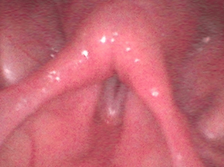 Figure 1
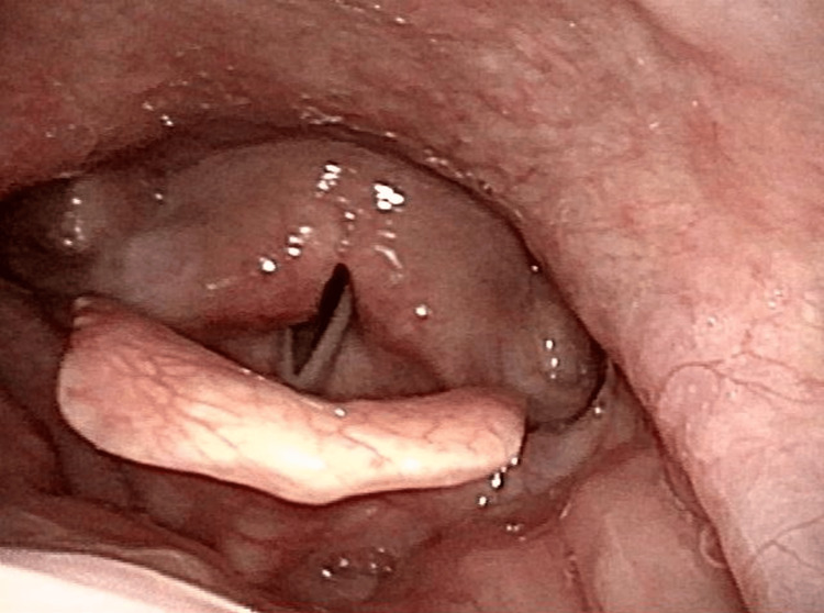 Figure 2
| Year | Author | Sex | Age | Previous MG Diagnosis | Trigger | Diagnostic Tests | Thymoma | Associated Symptoms | Intubation | Treatment | Prognosis |
| 2000 |
Cridge et al [ | M | 88 | None | Not reported | Anti-AChR/MuSK negative | (-) | None | Yes | Not reported | Not improved |
| 2002 |
Teramoto et al. [ | F | 82 | 14 years | Not reported | Anti-AChR/MuSK negative | (-) | None | Yes | Prednisolone, pyridostigmine | Improved |
| 2007 |
Hara et al. [ | M | 56 | None | Not reported | Anti-MuSK positive | (-) | Dysphagia, paralysis, hoarseness | Yes | IVIG, prednisolone, tacrolimus | Not improved |
| 2007 |
Kanemaru et al. [ | M | 76 | None | Not reported | Anti-AChR positive | (-) | None | Yes | Prednisolone, cyclosporine | Not reported |
| 2008 |
Sylva et al. [ | F | 24 | 2 years | Not reported | Anti-MuSK positive | Not reported | Paralysis, stridor | No | Prednisolone, pyridostigmine | Not reported |
| 2010 |
Khan et al. [ | M | 71 | None | Not reported | Anti-AChR positive | (-) | SOB, dysphagia | Yes | Not reported | Improved |
| 2011 |
Kitagawa et al. [ | F | 86 | 5 years | Not reported | Anti-AChR positive | (-) | None | Yes | Methylprednisolone | Deceased |
| 2011 |
Sethi et al. [ | M | 68 | None | Not reported | Anti-AChR/MuSK negative | (-) | None | Yes | Neostigmine | Not reported |
| 2014 |
Sasaki et al. [ | F | 78 | None | Not reported | Anti-AChR positive | (+) | Dysphagia | Yes | Immunosuppressive, thymectomy | Not reported |
| 2020 |
Balabbigari et al. [ | F | 51 | None | Not reported | Anti-AChR/MuSK negative | (-) | Hoarseness | Yes | Prednisone, mycophenolate | Not reported |
| 2020 |
Nelke et al. [ | F | 80 | None | Not reported | Anti-AChR positive | (+) | Dysphagia | Yes | Plasmapheresis, corticosteroids | Deceased |
| 2021 |
Santilli and Stitt [ | F | 45 | None | Not reported | Anti-MuSK positive | (-) | Hoarseness | No | PLEX, rituximab | Not reported |
| 2022 |
Beka et al. [ | F | 58 | None | Upper airway infection | Anti-AChR positive | (+) | None | No | Corticosteroid | Not reported |
| 2023 | Our case | F | 86 | None | Vaccination | Anti-AChR/MuSK negative | (-) | Dysphagia, hoarseness | Yes | Prednisolone, pyridostigmine | Not improved |
Peer Reviews
No public reviews on file for this paper yet. If you reviewed it on a platform where reviews are public (OpenReview, ICLR, NeurIPS, ICML), you can paste yours below so the community can read it here.
Videos
No videos yet. Explain this paper in a talk, walkthrough, or lecture? Add one.
Taxonomy
TopicsMyasthenia Gravis and Thymoma · Peripheral Neuropathies and Disorders · Autoimmune Neurological Disorders and Treatments
Introduction
Myasthenia gravis (MG) is an autoimmune disorder characterized by dysfunction at the neuromuscular junction - the synapse between motor neurons and muscle fibers. The primary pathological mechanism involves autoantibodies targeting acetylcholine receptors (AChR) or muscle-specific kinase (MuSK), impairing signal transmission. Clinically, MG typically presents with fluctuating skeletal muscle weakness, most commonly manifesting as diplopia, ptosis, bulbar weakness, and generalized fatigue. In severe cases, the disease can rapidly progress to respiratory compromise, known as myasthenic crisis [1]. MG diagnosis relies on a combination of serologic, pharmacologic, and electrophysiologic testing. Detection of anti-AChR or anti-MuSK antibodies supports diagnosis in the majority of cases, although a subset remains seronegative. Pharmacological tests, such as the edrophonium (Tensilon) and ice pack tests, offer rapid bedside diagnostic clues, particularly in ocular presentations. Repetitive nerve stimulation (RNS) and single-fiber electromyography (SFEMG) are instrumental for neurophysiological confirmation, especially in seronegative MG.
Vocal cord paralysis (VCP), by contrast, has a broad differential diagnosis. Common etiologies include recurrent laryngeal nerve injury (e.g., post-surgical or neoplastic compression), viral neuropathy, idiopathic causes, and neurodegenerative disorders. In rare instances, MG can present with isolated laryngeal involvement. Laryngeal muscle dysfunction in MG may reflect the selective vulnerability of striated laryngeal muscles innervated by cranial nerves, particularly the recurrent laryngeal branch of the vagus nerve. Such focal involvement underscores the heterogeneous and unpredictable clinical spectrum of MG. Understanding this neuromuscular vulnerability helps contextualize how MG could plausibly present with isolated vocal symptoms prior to the emergence of generalized or ocular features.
Although thymoma, autoimmune comorbidities, and infections are well-recognized triggers of MG, recent studies have also suggested a possible association with vaccination, including reports following administration of human papillomavirus and influenza vaccines [2-4]. VCP as the predominant initial symptom of MG remains exceptionally rare, with only a handful of cases reported in the literature, including instances of bilateral vocal fold involvement necessitating airway management [5]. This clinical pattern underscores the diagnostic challenge and potential severity of laryngeal MG, particularly in seronegative or atypically presenting cases. The present report contributes to this limited body of literature by describing a temporally associated onset of MG following herpes zoster vaccination, warranting further attention to this possible, though rare, association.
Case presentation
The patient was an 86-year-old Japanese woman with no remarkable medical history and previously good functional status without impairment. Fifty-one days prior to admission, she received an inactivated herpes zoster vaccine (Shingrix® intramuscular injection), after which she developed a fever of 38°C the following day. Apart from fever, she exhibited no other accompanying symptoms, and the fever resolved after seven days. However, around the time her temperature normalized, she began to experience episodes of choking, followed by a sense of progressive dysphagia and hoarseness over several days. She consulted an otolaryngologist 20 days post-vaccination, where a laryngeal fiberoptic examination revealed right-sided unilateral VCP. The cause remained unclear, and the decision was made to observe the patient’s condition.
Symptoms continued to progress, and two days before hospital admission, she developed increasing respiratory distress with significant labored breathing, prompting emergency transportation to our facility. Upon arrival, she had a respiratory rate of 30 breaths per minute and SpO₂ of 93%, with marked labored breathing. Physical examination revealed severe hoarseness but no other significant abnormalities. No muscle weakness in the extremities, ptosis, or other neurological deficits were observed, and laboratory tests provided no diagnostic clues. Laryngeal fiberoptic examination showed bilateral vocal cords fixed in a paramedian position (Figure 1).
Laryngeal fiberoptic examination showed bilateral vocal cords fixed in a paramedian position.Laryngoscopic findings on admission showed bilateral vocal cords fixed in the paramedian position. The patient, an 86-year-old woman with no significant medical history, developed fever, dysphagia, and hoarseness following administration of Shingrix®, a recombinant inactivated herpes zoster vaccine. Initial laryngoscopy on day 20 post-vaccination revealed right VCP, which progressed to bilateral involvement and severe respiratory distress by the time of admission.
The patient was diagnosed with respiratory failure due to bilateral VCP, necessitating emergency intubation, after which her respiratory status stabilized.
Head, neck, and chest CT scans, as well as a head MRI, revealed no abnormalities, including the absence of thymoma. Despite inpatient management, the condition did not significantly improve, and tracheostomy was performed seven days later. Repeat fiberoptic examination confirmed persistent bilateral vocal cord fixation in the paramedian position. Her respiratory status remained stable, allowing mechanical ventilation withdrawal by the 10th day of hospitalization, yet the underlying cause remained unknown. The patient did not undergo assessment of Vital Capacity or Negative Inspiratory Force (NIF) because she exhibited no clinical signs of respiratory compromise suggestive of respiratory muscle involvement. On day 31 of hospitalization, the patient reported that she had gradually developed diplopia and ptosis around day 25 of her admission. Retrospective review of her clinical course also suggested fatigability of bulbar symptoms, particularly worsening hoarseness and dysphagia with prolonged vocal effort. Although anti-AChR and anti-MuSK antibodies tested negative, edrophonium (Tensilon) and ice pack tests were positive, leading to the diagnosis of MG; notably, the positive Tensilon test referred to observable clinical improvement, including transient resolution of ptosis and enhanced ocular motility following edrophonium administration. Treatment with pyridostigmine bromide 180 mg/day was initiated, followed by plasma exchange therapy twice weekly for three weeks. Additional immunosuppressive treatment included tacrolimus 2 mg/day and prednisolone 15 mg/day. Two months after initiating therapy, her ptosis improved; however, diplopia and VCP persisted, and she was discharged home with a tracheostomy.
Five months post-discharge, a slight improvement in vocal cord mobility was observed (Figure 2).
Five months post-discharge, laryngeal fiberoptic examination demonstrated a slight improvement in vocal cord mobility.Laryngoscopy five months post-discharge showed slight improvement in bilateral vocal cord mobility. The patient had initially presented with respiratory failure due to bilateral VCP, requiring intubation and tracheostomy. MG was diagnosed based on positive edrophonium and ice pack tests. Despite immunotherapy, VCP persisted at discharge, with gradual improvement noted during follow-up.
However, VCP persisted one year later, making decannulation difficult. Diplopia also persisted, although no further progression of the condition was noted, and she continued to maintain an independent lifestyle.
Discussion
Isolated VCP as the first manifestation of MG is exceedingly rare. In one large series of 1,520 MG patients, only seven cases (∼0.4%) initially presented with dysphonia due to vocal cord involvement [6]. Our review of the literature since 2000 identified 13 additional reported cases of MG with VCP (Table 1), of which 10 had VCP as the initial symptom [5,7-18].
These patients were evenly split by sex and ranged from 45 to 88 years old (median 69.5 years). In 9 of 11 cases, including the present case, bilateral VCP required endotracheal intubation and mechanical ventilation, underscoring the potential for life-threatening respiratory failure.
These cases likely represent a distinct MG subtype -“laryngeal-type” MG-characterized by early bulbar symptoms such as dysphonia, dysphagia, and dysarthria, with minimal or absent ocular involvement. Similar to ocular MG, this subtype shows a lower seropositivity rate for acetylcholine receptor (AChR) antibodies (~45%) compared to generalized MG (~80-90%) [1,19]. Our literature review found a comparable seropositivity rate of 45% among VCP-predominant cases, reinforcing this observation.
Diagnosing laryngeal-onset MG remains challenging due to its atypical presentation, which may mimic primary ENT disorders. Yang X et al. [20] reported that only ~23% of patients were diagnosed with MG at their initial evaluation. Diagnosis is often delayed until classical ocular symptoms, such as ptosis or diplopia, develop. Clinician awareness is essential to facilitate early recognition and prevent respiratory compromise.
Recent studies have improved our understanding of seronegative laryngeal MG. Mullen et al. highlighted key features such as frequent antibody negativity, diagnostic utility of the edrophonium (Tensilon) test, and responsiveness to pyridostigmine [21]. Electrophysiological tests like laryngeal electromyography (EMG) and repetitive nerve stimulation (RNS) were particularly sensitive in these patients. Incorporating these tools is critical in antibody-negative cases.
The Tensilon test has a reported sensitivity of 71-95% and specificity of 97-100% [22,23]. The ice pack test, primarily used for ptosis, has a sensitivity of 80-90% and nearly 100% specificity [23,24]. Though less validated in laryngeal MG, it remains a useful bedside tool when positive.
Electrodiagnostic testing remains a cornerstone of MG diagnosis. RNS detects decremental responses in 75% of generalized MG, while single-fiber EMG offers the highest sensitivity (85-100%) [23,25]. These tests are indispensable when antibody testing is negative and clinical suspicion remains high.
Review of reported cases reveals variable treatments depending on severity and serostatus. Common therapies include pyridostigmine, corticosteroids, IVIG, plasma exchange, and immunosuppressants such as azathioprine, cyclosporine, or tacrolimus [16,18,20]. Acute airway compromise often requires tracheostomy. Prognosis is heterogeneous - some patients recover fully, while others have persistent bulbar dysfunction or require long-term ventilatory support [7,18]. These outcomes underscore the importance of early, individualized treatment.
The present case is notable for symptom onset shortly after varicella-zoster vaccination. Although several case reports suggest temporal associations between vaccination and MG onset [4,26], large-scale reviews have not demonstrated causality. For example, Mailand MT, Frederiksen JL [27] found no consistent evidence linking vaccines to MG or other autoimmune neurologic disorders. Given the widespread use of vaccines, some MG cases will coincide with recent vaccination purely by chance.
Proposed mechanisms for post-vaccination MG include molecular mimicry and immune dysregulation similar to that seen with infections [28]. In our case, the patient developed bulbar weakness immediately after a post-vaccination fever, suggesting a possible immune trigger. However, given limited evidence for a causal link, this association should be interpreted cautiously.
It is also important to acknowledge the limitations of single case reports. While they can highlight rare presentations and hypotheses, they lack generalizability and cannot establish causality. Prospective studies with larger cohorts are essential to determine whether vaccination can meaningfully contribute to MG onset.
This case reinforces the need for early recognition of laryngeal MG, especially when initial symptoms are restricted to the bulbar musculature. A high index of suspicion, timely diagnostic testing, and individualized treatment can prevent airway compromise and improve patient outcomes.
Conclusions
The key lesson from this case is not to alarmingly conclude that vaccines cause MG, but rather to maintain a high index of suspicion for neuro-immunological diseases like MG, in the wake of a recent vaccination. Physicians should be aware that MG can present with atypical features (such as isolated laryngeal symptoms) and, on rare occasions, may coincide with or follow immunizations. This awareness can facilitate timely diagnosis and treatment. In a treatable condition such as MG, early recognition and appropriate therapy, whether the disease is ocular, generalized, or a focal laryngeal subtype, are critical for improving patient outcomes. By acknowledging the potential (albeit rare) links between immune triggers (like infections or vaccines) and MG, clinicians will be better prepared to identify unusual presentations and intervene before the disease progresses to a life-threatening stage.
The reference list from the paper itself. Each links out to its DOI / PubMed record.
- 1Myasthenia gravis: Past, present, and future J Clin Invest Conti-Fine BM Milani M Kaminski HJ 2843285411620061708018810.1172/JCI 29894 PMC 1626141 · doi ↗ · pubmed ↗
- 2Myasthenia gravis following human papillomavirus vaccination: A case report BMC Neurol Chung JY Lee SJ Shin BS Kang HG 2221820183059327010.1186/s 12883-018-1233-y PMC 6309058 · doi ↗ · pubmed ↗
- 3Myasthenia gravis after the third dose of human papillomavirus 9-valent vaccine: A case report Hum Vaccin Immunother Ding Y Fu T Zhou W Zhang X Wang R Liao H 22522521920233764374710.1080/21645515.2023.2252252 PMC 10467512 · doi ↗ · pubmed ↗
- 4Laryngeal myasthenia gravis following influenza vaccination: A case report and literature review Hum Vaccin Immunother Wang F Xiang T He L Wang J 552955311720213455997710.1080/21645515.2021.1977580 PMC 8903928 · doi ↗ · pubmed ↗
- 5Bilateral vocal fold paralysis in myasthenia gravis: A case report and literature review Front Neurol Nelke C Labeit B Meuth SG Warnecke T Dziewas R Ruck T 5810601120203317812410.3389/fneur.2020.581060 PMC 7593483 · doi ↗ · pubmed ↗
- 6Dysphonia as a primary manifestation in myasthenia gravis (MG): a retrospective review of 7 cases among 1520 MG patients J Neurol Sci Liu WB Xia Q Men LN Wu ZK Huang RX 162226020071746633710.1016/j.jns.2007.03.019 · doi ↗ · pubmed ↗
- 7Myasthenic crisis presenting as isolated vocal cord paralysis Am J Emerg Med Cridge PB Allegra J Gerhard H 2322331820001075094310.1016/s 0735-6757(00)90031-7 · doi ↗ · pubmed ↗
- 8Respiratory failure due to vocal cord paresis in myasthenia gravis Respiration Teramoto K Kuwabara M Matsubara Y 2802826920021209777710.1159/000063636 · doi ↗ · pubmed ↗