Cytogenetic Alterations Observed in Exfoliative Cells of the Tongue and Oral Mucosa of SARS-CoV-2-Vaccinated Patients: Report of Two Cases and a Brief Literature Review
Lucas Alves da Mota Santana, Michelly Kierkegaard Campos de Oliveira, Maria Vitória Conceição Carvalho, Pedro Henrique Macedo Moura, Marina dos Santos Barreto, Marcos Antônio Lima dos Santos, Pedro Lima dos Santos, Dalmo Correia, Virgínia Kelma dos Santos Silva

TL;DR
This paper reports cytogenetic changes in oral cells of vaccinated individuals and suggests the need for monitoring such cases after the pandemic.
Contribution
The paper presents two cases of cytogenetic alterations in vaccinated individuals and highlights the need for post-pandemic monitoring.
Findings
Cytogenetic alterations were observed in exfoliative cells of the tongue and oral mucosa of SARS-CoV-2-vaccinated patients.
The presence of ACE2 and TMPRSS2 in oral tissues may make the oral cavity a target for SARS-CoV-2-related lesions.
The paper emphasizes the importance of monitoring vaccinated individuals for potential long-term effects.
Abstract
The wide distribution of angiotensin-converting enzyme 2 (ACE2) and transmembrane protease serine 2 (TMPRSS2) in oral tissues, especially in the salivary glands, which are natural reservoirs of severe acute respiratory syndrome coronavirus 2 (SARS-CoV-2), contributes to the classification of the oral cavity as a potential target for the development of lesions. Despite the effective response produced by next-generation immunizers, the possibility of immune escape by new lineages of SARS-CoV-2 cannot be refuted. Therefore, we describe here the occurrence of cytogenetic alterations in orally exfoliated cells of immunized individuals and, based on the literature review, call attention to the need to monitor these cases in the post-pandemic period.
Genes, proteins, chemicals, diseases, species, mutations and cell lines named across the full text — each resolved to its canonical identifier and authoritative record.
Click any figure to enlarge with its caption.
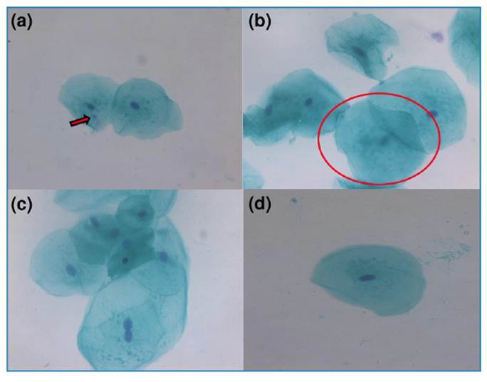 Figure 1
Peer Reviews
No public reviews on file for this paper yet. If you reviewed it on a platform where reviews are public (OpenReview, ICLR, NeurIPS, ICML), you can paste yours below so the community can read it here.
Videos
No videos yet. Explain this paper in a talk, walkthrough, or lecture? Add one.
Taxonomy
TopicsDermatological and COVID-19 studies · Parvovirus B19 Infection Studies · Oral Health Pathology and Treatment
INTRODUCTION
The coronavirus disease (COVID-19) pandemic has been considered the most sanitary crisis in the recent decades, being directly responsible for a large number of hospitalizations and the collapse of health systems throughout the world1. From the first reports of the disease appearing in December 2019 in Wuhan (Hubei province, China) until November 2024, it is estimated that the outbreak caused by severe acute respiratory syndrome coronavirus 2 (SARS-CoV-2), a novel coronavirus type and the main pathogen associated with the emergence of the pandemic, provoked approximately 776.8 million confirmed COVID-19 cases and over 7 million confirmed deaths2. Pathophysiologically, patients positive for SARS-CoV-2 showed a broad spectrum of clinical manifestations, including respiratory disorders, cardiac output, kidney failure, and orocutaneous alterations3.
Although the rapid development of immunizers has made it possible to control the disease and end the health emergency, some variants of the novel coronavirus (especially subvariants of Omicron) are potential targets of interest because of their capacity to overcome vaccine-induced immunity and their wide geographical distribution2 ^-^ 4. Thus, we describe here the occurrence of cytogenetic alterations in the oral cells of vaccinated patients using a micronucleus assay. A review of pertinent literature was conducted using PubMed and SciELO to reinforce the need to monitor these cases during the post-pandemic period.
CASE REPORT
In September 2024, two individuals from the same family tested positive for SARS-CoV-2, which was confirmed using a rapid test for COVID-19 IgG/IgM. The clinical manifestations of the infection were mild, with a prevalence of coryza, coughing, and myalgia during 10 days. Clinical data of the patients are presented in Table 1. The medical history revealed that approximately 3three years ago, the patients were infected with the novel coronavirus for the first time, showing a symptomatology similar to that observed currently. To mitigate the symptoms of the disease, dipyrone (1 g) and multivitamin/mineral supplements (B-complex vitamins, zinc, calcium, vitamin D, and vitamin C) were administered, and a period of 5five days away from work activities was recommended.
TABLE 1:General epidemiological data and serological profiles of SARS-CoV-2 vaccinated patients.CaseSexAgeCOVID symptomsNumber of doses administeredCOVID-19 vaccination schedule*Orocutaneous manifestationsSaturation level (SpO_2_)Past medical historyTreatmentNeutralizing antibody levels (%)# 1M53 yCoryza, cough, and myalgia4ChAdOx1 nCoV-19 (Vaxzevria®, Oxford-AstraZeneca), BNT162b2 mRNA (Pfeizer/ BioNTech)No98%Hypertension, Crohn's disease, and benign prostatic hyperplasiaAnalgesic and Multivitamin/mineral (MVM) supplement89,2# 2F54 yCoryza, cough, myalgia, and ageusia4ChAdOx1 nCoV-19 (Vaxzevria®, Oxford-AstraZeneca), BNT162b2 mRNA (Pfeizer/ BioNTech)No98%Fibromyalgia, rheumatism, and arrhythmiaAnalgesic and Multivitamin/mineral (MVM) supplement78 M: male; F: female; y: years; *****patients received two doses of each immunizing agent.
Subsequently, patients were investigated for the presence of neutralizing antibodies (Nabs). Laboratory results demonstrated a significant rate of circulating antibodies (89,2% and 78%, respectively; Table 1), which partially explained the mild form of the disease. Moreover, smears of exfoliated cells obtained from the buccal mucosa and tongue of affected individuals were collected using a cytobrush to analyze cytogenetic damage and spread on glass slides. In general, the patients in the present case were non-smokers, no exposure to X-rays was monitored in the last months, and no use of mouthwash or topical medication may have modified the cytological profile of the cells. Subsequently, the oral cells obtained were fixed in a 3:1 methanol/acetic acid buffer solution and stained using the Feulgen/Fast Green method. Slide analysis revealed the presence of cytogenetic alterations, such as micronuclei, karyolysis, binucleation, and pyknosis (Figure 1).
FIGURE 1:Metanuclear alterations observed in cytological smears of exfoliative cells of the tongue and oral mucosa of SARS-CoV-2 vaccinated patients. A: Micronucleus (red arrow); B: Karyolysis (red circle); C: Binucleation**; D:** Pyknosis. 400× magnification. Feulguen-Fast-Green stain.
DISCUSSION
Despite the World Health Organization (WHO) declaring the end of the COVID-19 pandemic a global health emergency in May 2023, SARS-CoV-2 remains a potential pathogen of interest to public health because of the increasing number of mutations that favor the emergence of new lineages1 ^,^ 4. Notably, several authors have reported cases of orocutaneous manifestations in vaccinated individuals3. Due to the high expression of angiotensin-converting enzyme 2 (ACE2) and transmembrane protease serine 2 (TMPRSS2) in the oral tissues (e.g., tongue and mucosa), and because salivary glands are considered reservoirs of SARS-CoV-2, the oral cavity is a potential target for the development of lesions5. In addition to changes at the tissue level induced by the virus, such as chronic inflammation and vascular disorders, morphological changes in cellular parameters have also been described6 ^,^ 7. For instance, Marques et al. (2022)7 demonstrated that the epithelial cells collected from the dorsum of the tongue of infected patients showed a decrease in perimeter and a smaller nuclear diameter when compared to the control group.
Another consequence of SARS-CoV-2 infection in oral cells is the possibility of occurrence of mutagenesis and cytotoxicity. In a pioneering study, Pinto et al. (2021)8 observed an increase in the frequency of micronuclei in cells obtained from the buccal mucosa of patients with COVID-19 as well as a significant rate of cell death. Mutagenesis is associated with the occurrence of micronuclei, and their presence is an important biomarker for the assessment of genomic instability, and consequently, carcinogenesis. On the other hand, cytotoxicity represents the risk of cell death, being characterized by the presence of alterations like karyolysis and karyorrhexis8.
In our study, the main cytogenetic alterations observed were micronuclei formation, karyolysis, binucleation, and pyknosis, as reported by Pinto et al. (2021)8. However, the mechanisms employed by SARS-CoV-2 in the occurrence of these cytogenetic disorders remain poorly understood. Nevertheless, Da Silva et al. (2022)9 argue that cell death may occur in response to apoptosis or necrosis through the release of free radicals, mitochondrial dysfunction, and synthesis of inflammatory cytokines, including TNF-α. According to Ren et al. (2021)10, increase of the frequency of micronuclei in cells with high expression of ACE2 is result of the syncytia formation (i.e., cells infected with SARS-CoV-2 fuse with neighboring cells), with subsequent DNA damage accompanied by alteration of intracellular levels of cGAS and γH2Ax. Thus, our preliminary findings call attention to the monitoring of COVID-19 cases in vaccinated patients, who may demonstrate varying degrees of susceptibility.
Regarding vaccine-induced immunity, several hypotheses have been proposed to explain the different grades of immunization agents11 ^,^ 12. According to Abou-Saleh et al. (2022)^11^,neutralizing antibodies against SARS-CoV-2 produced after mRNA vaccine administration (e.g., Pfizer BNT162b2 and Moderna mRNA-1273) declined over time. Moreover, vaccine immunogenicity is reduced in immunocompromised individuals12. Notably, the patients mentioned here had chronic diseases, such as hypertension, Crohn's disease, and rheumatism. Anti-inflammatory drugs used to treat complications can generate negative feedback by suppressing vaccine-induced immune responses. Furthermore, the omicron variant has a unique ability to escape the immune response compared with other SARS-CoV-2 variants4.
In summary, our findings suggest that SARS-CoV-2 infection can induce significant cytogenetic alterations in the oral tissues of vaccinated patients, highlighting the importance of post-pandemic monitoring. Finally, future rigorous epidemiological studies in large populations should investigate the possible mechanisms underlying SARS-CoV-2-induced cytogenetic alterations, and evaluate the effectiveness of preventive strategies.
The reference list from the paper itself. Each links out to its DOI / PubMed record.
- 1Sarker R Roknuzzaman ASM Hossain MJ Bhuiyan MA Islam MR The WHO declares COVID-19 is no longer a public health emergency of international concern: benefits, challenges, and necessary precautions to come back to normal life Int J Surg 202310992851285210.1097/JS 9.000000000000051337222700 PMC 10498846 · doi ↗ · pubmed ↗
- 2World Health Organization (WHO) COVID-19 epidemiological update 24122024 February 10, 2025 Available from: https://www.who.int/publications/m/item/covid-19-epidemiological-update---24-december-2024
- 3Smarz-Widelska I Grywalska E Morawska I Forma A Michalski A Mertowski S Pathophysiology and Clinical Manifestations of COVID-19-Related Acute Kidney Injury-The Current State of Knowledge and Future Perspectives Int J Mol Sci 202122137082708210.3390/ijms 2213708234209289 PMC 8268979 · doi ↗ · pubmed ↗
- 4Wang Y Ma Y Xu Y Liu J Li X Chen Y Resistance of SARS-Co V-2 Omicron variant to convalescent and Corona Vac vaccine plasma Emerg Microbes Infect 202211142442710.1080/22221751.2022.202721935001836 PMC 8803103 · doi ↗ · pubmed ↗
- 5Ma L Liu Q Wang M Li L Hu Z Zhou Y Severe acute respiratory syndrome coronavirus 2 pathology and cell tropism in tongue tissues of COVID-19 autopsies Front Cell Infect Microbiol 2024141394721139472110.3389/fcimb.2024.139472138975331 PMC 11224463 · doi ↗ · pubmed ↗
- 6Santana LADM Vieira WA Gonçalo RIC Lima Dos Santos MA Takeshita WM Miguita L Oral mucosa lesions in confirmed and non-vaccinated cases for COVID-19: A systematic review J Stomatol Oral Maxillofac Surg 20221235 e 241-e 25010.1016/j.jormas.2022.05.00535550190 PMC 9085350 · doi ↗ · pubmed ↗
- 7Marques BBF Guimarães TC Fischer RG Tinoco JMM Pires FR Lima JC Junior Morphological alterations in tongue epithelial cells infected by SARS-Co V-2: A case-control study Oral Dis 202228 Suppl 22417242210.1111/odi.1398834342110 PMC 8447065 · doi ↗ · pubmed ↗
- 8Pinto TG Alpire MES Ribeiro DA Cytogenetic Biomonitoring in Buccal Mucosa Cells of COVID-19 Patients: Preliminary Findings In Vivo 20213563495349910.21873/invivo.1265134697187 PMC 8627778 · doi ↗ · pubmed ↗