Seroepidemiological study of Toxoplasma gondii in equines in Northern Egypt
Ehab Kotb Elmahallawy, Marwa F. Hassan, David Cano-Terriza, Nada Oudah Albalawi, Tomás Fajardo, Asmaa Aboelabbas Gouda, Ayman Atiba, Ahmed Hendawy, Isabelle Villena, Ashraf Mohamed Barakat, Hind Alzaylaee, Sonia Almería, Ignacio García-Bocanegra

TL;DR
This study found a high rate of Toxoplasma gondii infection in equines in Northern Egypt, with donkeys being more affected than horses.
Contribution
The study is the largest serosurvey of T. gondii in equids in Egypt and includes previously unexplored risk factors.
Findings
The overall seroprevalence of T. gondii in equines was 41.11%.
Donkeys had a significantly higher seroprevalence (51.23%) compared to horses (28.03%).
Abstract
Toxoplasmosis, caused by the intracellular protozoan Toxoplasma gondii (T. gondii), continues to be a widespread parasitic zoonotic disease globally. The seroepidemiology of T. gondii infection in Egyptian equids, particularly donkeys, remains insufficiently explored. The present study was designed to assess the seroprevalence of T. gondii in equines from Northern Egypt. A total of 360 serum samples from two equine species (157 horses and 203 donkeys) were obtained during 2023. The Modified Agglutination Test (MAT, cut-off of 1:25) was used to screen for the anti-T. gondii antibodies. The study also analyzed potential risk factors that could contribute to the exposure of the animals to the parasite, including species, breed, sex, age, and the specific location of each animal. The overall seroprevalence of T. gondii among examined equines was 41.11% (95% Confidence Interval [CI]:…
Genes, proteins, chemicals, diseases, species, mutations and cell lines named across the full text — each resolved to its canonical identifier and authoritative record.
Click any figure to enlarge with its caption.
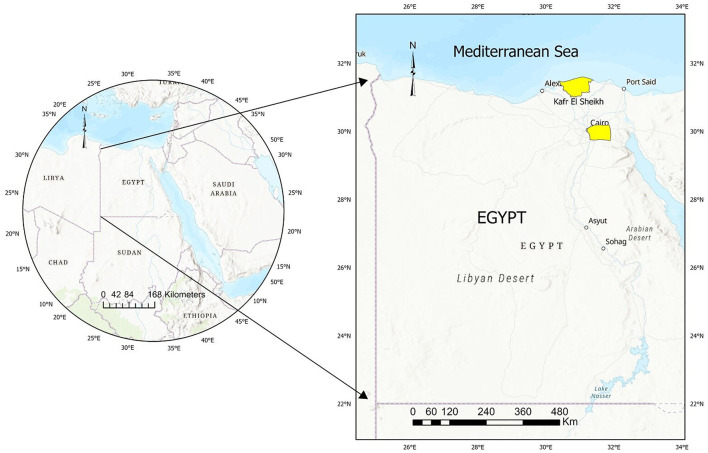 Figure 1
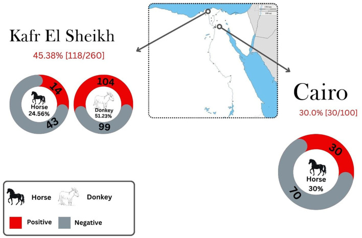 Figure 2
|
|
|
|
|
|
|
|
|---|---|---|---|---|---|---|
| Donkeys | Rural areas | Menoufiya | ELISA | 65.3 (79/121) | NS | ( |
| Individual owners | Menoufiya | LAT, ELISA | 30.2(13/43), 25.6 (11/43) | 1:32; 1:100 | ( | |
| Zoo abattoir | Giza | *MAT (1:25) | a44.5 (89/200), b52.0 (104/200), c36.0 (72/200), d39.0 (78/200) | NS | ( | |
| Individual owners | Giza | LAT, ELISA | 27.6 (16/58), 37.9 (22/58) | 1:32; 1:100 | ( | |
| Individual owners | Cairo | ELISA | 45.0 (45/100) | 1:200 | ( | |
| NS | Dakahlia | LAT, IHA, ELISA | 44.3 (35/79), 67.1 (53/79), 68.4 (54/79) | NS | ( | |
| Individual owners | Matrouh | LAT, ELISA | 22.2 (10/45), 20.0 (9/45) | 1:32; 1:100 | ( | |
| Horses | Farms | NS | **ELISA-LA, ELISA-LAunb, ELISA-Lab, IFAT, MAT (1:25) | a38.1 (160/420), b31.7 (133/420), c51.7 (217/420), 40.5 (170/420), 48.1 (202/420) | NS | ( |
| Individual owners | Cairo | ELISA | 25.0 (25/100) | NS | ( | |
| Main farm | Cairo | LAT, MAT(1:25), ELISA | 52.1 (125/240), 50.8 (122/240), 39.2 (94/240) | NS | ( | |
| NS | Dakahlia | LAT, IHA, ELISA | 50.0 (27/54), 72.2 (39/54), 72.2 (39/54) | NS | ( | |
| Farms | Giza | ELISA | 20.9 (23/110) | NS | ( | |
| Farms | Qalyubia | ELISA | 12.0 (12/100) | NS | ( | |
| Farms | Gharbia | ELISA | 14.0 (14/100) | NS | ( | |
| Farms | Kafr El Sheikh | ELISA | 17.3 (19/110) | NS | ( |
|
|
|
|
|
|
|---|---|---|---|---|
| Species | Donkey | 104/203 | 51.23 | < 0.001 |
| Horse | 44/157 | 28.03 | ||
| Breeds | Pure breed | 37/124 | 29.84 | 0.002 |
| Mixed (Baladi) | 111/236 | 47.03 | ||
| Age | Foal (< 1 year) | 9/23 | 39.13 | 0.005 |
| Young (1–4 years) | 75/144 | 52.08 | ||
| Adult (4–15 years) | 58/170 | 34.12 | ||
| Geriatric (≥15 years) | 6/23 | 26.09 | ||
| Sex | Male | 78/176 | 44.32 | 0.226 |
| Female | 70/184 | 38.04 | ||
| Location | Cairo | 30/100 | 30.00 | 0.008 |
| KFS | 118/260 | 45.38 |
|
|
|
|
|
|
|---|---|---|---|---|
| Species | Donkey | < 0.001 | 2.99 | 2.35–3.81 |
| Horse | a | a | a |
Peer Reviews
No public reviews on file for this paper yet. If you reviewed it on a platform where reviews are public (OpenReview, ICLR, NeurIPS, ICML), you can paste yours below so the community can read it here.
Videos
No videos yet. Explain this paper in a talk, walkthrough, or lecture? Add one.
Taxonomy
TopicsToxoplasma gondii Research Studies · Parasitic Infections and Diagnostics · Rabies epidemiology and control
1 Introduction
Toxoplasma gondii, the causative agent of toxoplasmosis, remains one of the most common intracellular protozoa in the world. Sexual reproduction of these protozoa occurs in felids, the definitive hosts, which release oocysts in their feces. Intermediate hosts, which encompass nearly all warm-blooded species, can also become infected (1, 2). Humans and animals most commonly contract T. gondii through several primary routes: consuming undercooked or raw meat containing tissue cysts of the parasite, ingesting food or water contaminated with sporulated T. gondii oocysts, and via blood transfusions or transplacental transmission involving tachyzoites. In relation to its clinical impact, T. gondii infections are typically sub-clinical in immunocompetent persons; nevertheless, in immunocompromised individuals, this opportunistic protozoon may induce fatal conditions and even death as well as cause abortion, congenital malformations, and stillbirth (1).
Horses and donkeys are essential for agricultural work, transportation, and economic support, especially in rural areas, and they hold cultural and historical significance, contributing to traditional practices and local economies. However, equid production faces major challenges from various pathogens, including parasites. Among others, T. gondii infection in equines occurs primarily through food or water contaminated with sporulated oocysts, nonetheless, tachyzoites may also be transferred from the mare to the fetus through the placenta (3, 4). Despite T. gondii infection in horses being usually subclinical, atypical clinical signs of toxoplasmosis such as ataxia, fever, encephalomyelitis, and retinal degeneration have been reported (5). Abortion, fever, stillbirth, and degeneration in retina were also reported in pregnant mares infected with T. gondii (4, 6). Additionally, some previous works reported the potential association between clinical equine protozoal myeloencephalitis in horses and T. gondii seropositivity (7, 8). In the USA, fatal toxoplasmosis was observed in a horse (9). Collectively, T. gondii infection in equines may have a substantial impact, posing animal health risks and contributing to significant economic and reproductive losses (10). Taking this into account, consumption of equine meat is still prevalent in several countries, particularly those of the European Union (EU) (11). Previous studies have established the epidemiological link between the consumption of horse meat and clinical toxoplasmosis in humans (12). Additionally, the rising popularity of raw donkey milk has raised concerns, suggesting that consuming such milk from seropositive donkeys may increase the risk of human toxoplasmosis (13). In Egypt, horses and donkeys play a crucial role in agriculture, transportation, and economic sustainability, particularly in rural areas. Beyond their practical use, these animals also hold cultural and historical significance, contributing to traditional practices and local economies. Furthermore, raw meat of these animals is used as feed for carnivorous zoo animals, which may be represent a potential source of a source of infection if the meat is contaminated (14). In this context, viable T. gondii was detected in tissues of 25 donkeys slaughtered at the Giza Zoo abattoir (15). Clearly, infection by T. gondii might have an important impact on public health (10).
Monitoring exposure to the parasite is pivotal for implementation of effective control measures. Serological tests are the primary methods for diagnosing T. gondii in farm animals, including equids (14–16). These techniques are helpful tools for conducting screening surveys since they allow us to identify Toxoplasma-positive animals and farms as well as to analyze the associated risk factors linked to parasite exposure (1).
Previous literature showed that numerous serological investigations have been assessing T. gondii exposure in equids worldwide (7, 17). In Egypt, seroprevalence rates of T. gondii in horses and donkeys have been reported to vary widely, ranging from 12.0% to 68.4% (Table 1) (15, 18–25). Most previous studies involved only a limited number of animals and were performed over short periods of time. In addition, existing data on the seroprevalence of T. gondii in equines from Northern Egypt, particularly donkeys, remain insufficient, leaving a gap in comprehensive data. Consequently, the current study aimed to evaluate the seroprevalence of T. gondii and identify potential risk factors associated with exposure to this zoonotic protozoan in equids from Northern Egypt.
2 Materials and methods
2.1 Study area
In 2019, Egypt's equid population was reported to be 958.190, with donkeys making up 90.9% of this number, approximately 871.447 donkeys (26). The present study was conducted in Northern Egypt, primarily focusing on two regions: Cairo and Kafr El Sheikh, which are geographically at 30° 02' 30” N, 031° 14' 07” E and 31° 06' 42” N, 30° 56' 45” E respectively (Figure 1). Both regions experience mild to hot weather throughout most of the year, with average summer temperatures ranging from 31 to 34°C (88.5–92°F) and winter temperatures between 13.5 and 18°C (56.3–65°F). Northern Egypt receives an average annual rainfall of only 100–200 mm (4–8 inches), primarily occurring during the winter months.
Map of Egypt showing the studied area and sampling sites.
2.2 Animals and samples
Between January and December 2023, blood samples were collected from 360 equines, across Northern Egypt (Figure 1). Samples were collected randomly from donkeys and horses owned by small stakeholders relying on them for transportation and agriculture, taking advantage of opportunities presented by local veterinary campaigns. Considering the number of equids in the study area (n > 10.000), an estimated prevalence of 36% (based on an estimated national seroprevalence threshold of 35.79% [Table 1]), an accepted error of 5% and a 95% confidence interval (95%CI) resulted in 354 animals to be sampled. A total of 360 equids, including 157 horses, 203 donkeys, were finally included in the study. A total of 10 ml blood samples were collected from each animal by puncturing the jugular vein. Sera were obtained by centrifugation at 3,000 rpm for 10 min and were kept at −20°C until serological assessment. The information on animals was thoroughly documented, whenever possible. Information collected included species, breed, sex, age, and the specific location of each animal. Age of examined animals were classified into four categories: foal animals (< 1 year), young (1–4 years), adult (4–15 years) and geriatric (>15 years) as outlined in prior studies (27, 28). The ages of the examined equines were based on the animal's dentition (29, 30).
2.3 Serological examination
The presence of T. gondii antibodies in the serum of equids was evaluated by the modified agglutination test (MAT). This method has been previously validated in equine and performed according to the protocols established by Dubey and Desmonts (31). Briefly, the test was utilized to detect T. gondii antibodies, employing whole-killed tachyzoites as antigens which is kindly provided by Laboratory of Parasitology, University of Reims Champagne-Ardenne, France. The protocol (31) incorporated 2-mercaptoethanol directly into the antigen rather than treating the serum. Equine serum samples were first diluted at a 1:20 ratio in phosphate-buffered saline (PBS, pH 7.2), and 0.05 mL of the diluted sample was added to U-bottom microtiter plate wells. Serial two- to fourfold dilutions were then performed using PBS. The antigen stock solution was diluted (1:10) in a freshly prepared alkaline buffer containing 2-mercaptoethanol, which was either made fresh or stored for no longer than 2 weeks. The antigen solution (0.05 mL) was then added to each well, and the plates were sealed with cellophane before being incubated overnight at 37°C in a humidified environment. The results of the assay were assessed using a microtiter plate under appropriate lighting conditions. A diffuse mat across the wellthe well indicated a positive reaction, whereas a compact button at the center signified a negative result. To ensure assay accuracy and reproducibility, each run included validated positive and negative serum controls. A titer of 1:25, was applied as a cut-off for T. gondii seropositivity as previously considered for these animal species (32). Additionally, serum that initially tested positive at ≥1:25 dilution was thereafter retested at 1:25 and 1:50.
2.4 Statistical analysis
To establish the seroprevalence of T. gondii, we computed the percentage of seropositive samples relative to the total number of examined equids, with a 95% confidence interval (95% CI). Relation between explanatory variables (species, breeds, sex, age, and region) and serological results was estimated using a Pearson's chi-square or Fisher exact tests to allocate the relevance of these variables in the risk of exposure of animals to T. gondii in a bivariate analysis. Variables with a p < 0.10 were selected for multivariate analysis, considering collinearity through Cramer's V coefficients. Generalized Estimating Equation (GEE) models were utilized to assess the influence of explanatory variables identified in the bivariate analysis (33), with municipality as a random effect. Statistical significance was set at p < 0.05, and analysis was conducted using SPSS 25.0 software.
3 Results
In this work, anti-T. gondii antibodies were detected in 148 of 360 equines (41.11%; 95% CI: 36.03–46.19). Seropositivity by species was 51.23% (104/203) in donkeys and 28.03% (44/157) in horses (Figure 2, Table 2). Among the positive samples, T. gondii titres in donkeys were 1:25 in 43.3% (45/104) and 1:50 in 56.3% (59/104). In horses, 54.5% (24/44) had titers of 1:25, while 45.4% (20/44) of positive animals showed titers of 1:50. Table 2 presents the distribution of T. gondii seropositivity, as determined by the bivariate analysis and the epidemiological questionnaire results. Two out of five explanatory variables associated (p < 0.10) with T. gondii seropositivity in equines were selected after data exploration and bivariate analyses and included in the multivariable analysis. The final GEE multivariable analysis identified species as risk factorpotentially linked with the exposure to T. gondii (Table 3). In this respect, significantly higher seropositivity was found in donkeys (p < 0.001, OR = 2.99; 95% CI: 2.35–3.81) compared to horses.
Seroprevalence of Toxoplasma gondii in horses and donkeys in Northern Egypt by location.
4 Discussion
Toxoplasma gondii remains a highly prevalent zoonotic pathogen worldwide, affecting a broad spectrum of intermediate hosts, including equines. This far-reaching impact emphasizes the global significance of T. gondii infection (15, 19, 34), as equine toxoplasmosis is increasingly recognized as a major potential source of human infection (34). Unfortunately, there is a paucity of current data on the epidemiology of T. gondii infection in equids in Egypt, especially among donkeys. Most existing research is outdated and based on small sample sizes (15, 18, 19). This present study is considered one of the largest investigations into the seroprevalence of T. gondii in equids in Egypt, with a particular focus on donkeys.
The individual seroprevalence noted in horses in the current study (28.03%) is consistent with a previous study conducted in Cairo which reported a similar seroprevalence rate of 25% (25/100) (23). However, the present results are lower than those reported in various previous investigations in Cairo Governorate which showed seroprevalence values of 48.1% (202/420) (22) and 50.8% (122/240) (24). Another previous investigation reported an even higher seroprevalence rate in this species (72.2%; 39/54) in Dakahlia Governorate, Northern Egypt (15). In contrast, previous investigations reported a lower seroprevalences of 20.9% (23/110), 12.0% (12/100), 14.0% (14/100) and 17.3% (19/110) in horse populations in Giza, Qalyubia, Gharbia, and Kafr El Sheikh Governorates, respectively (25).
In the present study, the individual seroprevalence reported in donkeys was 51.23%, which is within the range previously recorded in this species in Egypt (Table 1). A nearly similar seroprevalence rate against T. gondii was observed in donkeys in Cairo Governorate (45%; 45/100) (21). However, our findings were lower than those reported in Menoufiya Governorate, Northern Egypt, where the seroprevalence was 65.3% (79/121) (18). Another study conducted in Dakahlia Governorate, Northern Egypt reported a higher seroprevalence rate of T. gondii, at 68.4% (54/79), among donkey populations in that area (15). In contrast, the current study found higher seroprevalence rates compared to those reported in Giza, Menoufiya, and Matrouh Governorates, Northern Egypt, using ELISA, where the seroprevalence rates were 37.9% (22/58), 25.6% (11/43), and 20.0% (9/45), respectively (19). The variation in T. gondii seroprevalence rates between the current study and previous research on equines could likely be attributed to several factors, primarily differences in farming management and sanitation practices, the specific serological tests used and the cutoff titers used for interpretation, timing of sampling, sample size, and the density of infected definitive hosts and their interaction with the animals (10, 15, 25, 35, 36). Likewise, climate-related factors such as geographic distribution, population density, and the abundance of cats (more prevalent in rural areas compared to urban governorates) would play a crucial role in the maintenance, spread, survival, and transmission of T. gondii, which contribute to the parasite's dynamics (2, 10, 37).
As shown in the present work, the risk of T. gondii seropositivity was 2.9 times greater in donkeys than in horses, indicating that donkeys are more susceptible to T. gondii exposure. These findings are consistent with previously reported data (38, 39). The variations in T. gondii seroprevalence rates between donkeys and horse may be attributed to differences in feeding, management, and sanitation practices including regular stall cleaning, disinfection, pasture rotation, proper manure disposal, and clean water supply (34). Donkeys in Egypt, often kept in extensive management systems under improper management and sanitation practices, and used more frequently for work, are more exposed to the parasite through ingestion of oocysts contaminating their environment, compared to horses (38). A previous investigation Munhoz et al. (39) concluded that donkeys could maintain detectable T. gondii antibody titers for a longer duration than horses. Additionally, the animal species susceptibility can also influence this difference (38, 40–43).
The current study has several limitations that should be noted. Firstly, the sample size from Cairo Governorate was small, and no donkey samples were collected in that area, which may limit the results' applicability to the overall equine population in this region. Cairo is a densely populated area with relatively few donkeys, which further constrains the extrapolation of our findings. Additionally, the study faced some challenges due to the small number of foals and geriatric equids, resulting in sample sizes variations between age groups. Some epidemiological factors, including breeding systems, sanitation conditions, and the presence of cats and rodents, were not assessed in this study. To gain a more comprehensive understanding, future large-scale serological and molecular studies should integrate these variables.
5 Conclusions
The present study constitutes one of the most extensive serosurveys of T. gondii in equines conducted in Egypt. The high level of exposure detected in these species raises animal and public health concerns. Notably, the study identified higher seroprevalence rates in donkeys compared to horses and represents the largest survey of donkeys ever conducted in Egypt so far. The study also offers new insights into the influence of various risk factors associated with the rate of exposure to the parasite. Given its significance for animal and public health, our seroepidemiological findings provide valuable information for Egyptian authorities to enhance control and prevention strategies for toxoplasmosis in equids. The study also underscores the importance of implementing strict measures to prevent potential transmission of the parasite to humans through the consumption of raw or undercooked meat and milk from the investigated species. Further large-scale serosurveys and molecular studies are warranted to genotype circulating T. gondii strains in equids and clarify their role in transmission to humans and animals in Egypt.
The reference list from the paper itself. Each links out to its DOI / PubMed record.
- 1Dubey JP. Toxoplasmosis of Animals and Humans (3rd ed.). Boca Raton, FL: CRC Press (2022).
- 2Elmahallawy EK Elbarbary NK Cano-Terriza D Fajardo T Albalawi NO Jiménez-Martín D. Toxoplasma gondii in dromedary camels (Camelus dromedarius) in Egypt: a comparative seroepidemiological study in Upper and Lower Egypt. Front Vet Sci. (2025) 11:1508496. 10.3389/fvets.2024.150849639902335 PMC 11788408 · doi ↗ · pubmed ↗
- 3Hill D Dubey J. Toxoplasma gondii: transmission, diagnosis and prevention. Clin Microbiol Infect. (2002) 8:634–40. 10.1046/j.1469-0691.2002.00485.x 12390281 · doi ↗ · pubmed ↗
- 4Tassi P. Toxoplasma gondii infection in horses. A review. Parassitologia. (2007) 49:7–15.18412038 · pubmed ↗
- 5James KE Smith WA Packham AE Conrad PA Pusterla N. Toxoplasma gondii seroprevalence and association with equine protozoal myeloencephalitis: a case–control study of Californian horses Vet J. (2017) 224:38–43. 10.1016/j.tvjl.2017.05.00828697873 · doi ↗ · pubmed ↗
- 6Marques LC Costa AD Lopes CW Moraes FD Moraes JD. Experimental toxoplasmosis in pregnant mares: a study of fetuses and placentas. Braz J Vet Res Anim Sci. (1995) 32:246–50. 10.11606/issn.1678-4456.bjvras.1994.52118 · doi ↗
- 7Dubey JP Murata FHA Cerqueira-Cézar CK Kwok OCH. Toxoplasma gondii infections in horses, donkeys, and other equids: the last decade. Res Vet Sci. (2020) 132:492–99. 10.1016/j.rvsc.2020.07.00532799174 · doi ↗ · pubmed ↗
- 8Schale S Howe D Yeargan M Morrow JK Graves A Johnson AL. Protozoal coinfection in horses with equine protozoal myeloencephalitis in the eastern United States. J Vet Intern Med. (2018) 32:1210–14. 10.1111/jvim.1512729633348 PMC 5980325 · doi ↗ · pubmed ↗