Altered Endothelin Receptor Expression in Idiopathic Male Infertility: A Potential Therapeutic Target
Richard Weiten, Melanie V. Brandenstein, Manuel Huerta, Tim Nestler, Axel Heidenreich, Enno Storz, Jan Herden

TL;DR
This study explores how changes in endothelin receptor expression may contribute to male infertility, suggesting new treatment possibilities.
Contribution
The study identifies altered endothelin receptor expression in idiopathic male infertility and suggests potential therapeutic targets.
Findings
ETAR expression was significantly reduced in infertile men compared to fertile controls.
ETBR was undetectable in all OAT syndrome samples.
Two distinct ETAR expression profiles were observed in the OAT group, indicating phenotypic heterogeneity.
Abstract
Idiopathic male infertility is frequently associated with impaired semen quality, particularly in men diagnosed with oligo-astheno-teratozoospermia (OAT) syndrome. Recent studies suggest a potential role of the endothelin (ET) system, particularly ET receptors A (ETAR) and B (ETBR), in male reproductive physiology. Moreover, antiestrogens such as tamoxifen and clomiphene, which are commonly used empirically in this context, may influence ET signaling pathways. However, the relationship between ET receptor expression and specific subtypes of male infertility remains poorly understood. Semen samples were collected from 56 men: 15 fertile controls with normozoospermia and 41 infertile men with abnormal semen parameters, including 22 with OAT syndrome and 19 with isolated teratozoospermia. Men with identifiable female-factor infertility were excluded. Seminal ETAR and ETBR expression were…
Genes, proteins, chemicals, diseases, species, mutations and cell lines named across the full text — each resolved to its canonical identifier and authoritative record.
Click any figure to enlarge with its caption.
 Figure 1
 Figure 2
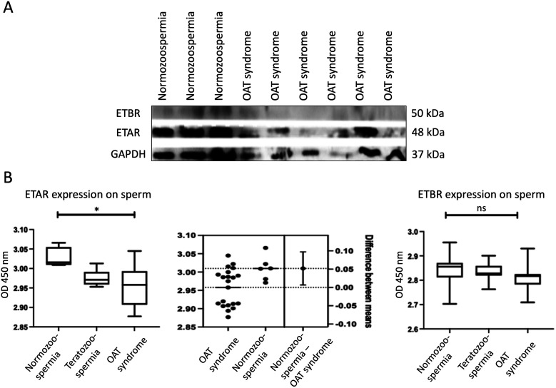 Figure 3
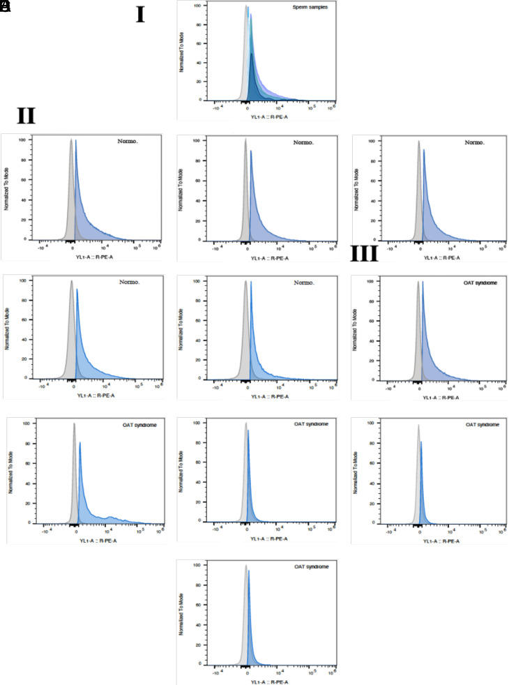 Figure 4
Peer Reviews
No public reviews on file for this paper yet. If you reviewed it on a platform where reviews are public (OpenReview, ICLR, NeurIPS, ICML), you can paste yours below so the community can read it here.
Videos
No videos yet. Explain this paper in a talk, walkthrough, or lecture? Add one.
Taxonomy
TopicsHormonal and reproductive studies · Sperm and Testicular Function · Ovarian function and disorders
Introduction
Infertility is a growing globalhealth concern, affecting a substantial number of couples, with its prevalence increasing steadily over recent decades.1^,^2 Male factors are implicated in approximately 20%-70% of infertility cases, with the global prevalence of male infertility estimated to range between 2.5% and 12%, affecting more than 30 million men worldwide.2 One of the most common diagnostic categories among infertile men is oligo-astheno-teratozoospermia (OAT) syndrome, which is defined by the concurrent presence of oligozoospermia (low sperm concentration), asthenozoospermia (reduced motility), and teratozoospermia (abnormal morphology).1^-^ 3 Although various genetic, endocrine, and environmental factors have been proposed, the underlying etiology of OAT syndrome often remains idiopathic.2^,^3
Empirical medical therapies, particularly antiestrogens such as tamoxifen and clomiphene citrate, are commonly used in the treatment of idiopathic male infertility.4^,^5 These agents exert their effects by antagonizing estrogen receptors in the hypothalamus and pituitary gland, leading to increased secretion of gonadotropin-releasing hormone, which stimulates luteinizing hormone (LH) and follicle-stimulating hormone (FSH) production. This cascade enhances Leydig cell-mediated testosterone synthesis and supports Sertoli cell function, ultimately promoting spermatogenesis.6
Although early studies reported only modest improvements in pregnancy rates with antiestrogen therapy,5^,^7^,^8 more recent meta-analyses, including 11 randomized trials, have demonstrated significant improvements in both sperm parameters and pregnancy rates among men with idiopathic infertility, particularly those with OAT syndrome.4^,^5^,^7 Nevertheless, the clinical utility of antiestrogens remains debated due to heterogeneity in patient selection and variable treatment responses.
Recent research has highlighted the potential role of the endothelin (ET) system in male reproductive physiology. Endothelin-1 (ET-1), a potent vasoconstrictive peptide, has been detected in human seminal plasma, with lower concentrations observed in men with OAT syndrome compared to normozoospermic controls.9^-^ 11 Moreover, antiestrogens such as tamoxifen have been shown to upregulate ET-1 and its receptors in other biological systems, including breast cancer tissue.11 Conversely, the use of ET receptor antagonists (e.g., bosentan) in animal models has been associated with testicular atrophy, reduced sperm production, and impaired fertility, suggesting that intact ET signaling is necessary for normal spermatogenesis.9^-^ 11
These findings suggest a mechanistic link between antiestrogen therapy and ET signaling, with potential implications for male fertility. In particular, ET receptors A (ETAR) and B (ETBR) may serve as downstream effectors or modulators of treatment response. Based on this rationale, it was hypothesized that altered expression of ETAR and ETBR may underlie idiopathic infertility, particularly in men with OAT syndrome, and that characterization of receptor expression patterns could inform treatment stratification. The present study aimed to systematically evaluate the expression of ETAR and ETBR in semen samples from men with idiopathic infertility, including those with OAT syndrome and isolated teratozoospermia, compared to normozoospermic controls.
Material and Methods
Patients’ Samples
Semen samples were collected from 56 men, including 15 fertile controls (normozoospermic) and 41 infertile patients with abnormal semen parameters, comprising 22 with OAT syndrome and 19 with isolated teratozoospermia. Semen analysis and categorization were performed according to the World Health Organization (WHO) laboratory manual for human semen analysis.12^,^13
Inclusion criteria were age ≥18 years and the availability of complete clinical and semen analysis data. Fertile controls were defined as men with documented paternity within the past 2 years and normozoospermic profiles based on WHO reference values. Infertile patients had a history of at least 12 months of unprotected intercourse without conception and met criteria for OAT syndrome or isolated teratozoospermia.
Exclusion criteria included a history of urogenital disorders (e.g., varicocele, orchitis, testicular torsion, malignancy), prior exposure to chemotherapy, radiotherapy, or hormonal therapy, known genetic causes of infertility (e.g., Klinefelter syndrome), systemic illnesses, current infections, and incomplete clinical or laboratory data.
Participants abstained from ejaculation for 2-7 days before sample collection. The study was approved by the Ethics Committee of the Medical Faculty of Medical Faculty of the University of Cologne (Approval No. 22-1178 Date: 18.10.2022) and registered in the German Clinical Trials Registry (DRKS00029986). Written informed consent was obtained from all participants.
Immunofluorescence
Semen samples were centrifuged at 2500 × g for 5 minutes and washed twice with phosphate-buffered saline (PBS; PAN-Biotech GmbH, Germany). A 10 µL aliquot was applied to glass slides (Thermo Fisher Scientific, Germany) and incubated with primary antibodies against ETAR (#sc-33536) and ETBR (#sc-518149; both from Santa Cruz Biotechnology, USA) at 1 : 1000 dilution for 1 hour. Fluorescently labeled secondary antibodies (FITC- or Alexa 594-conjugated anti-mouse IgG; Santa Cruz Biotechnology) were applied for 30-60 minutes. Nuclei were counterstained using DAPI-containing mounting medium (Abcam, UK). Images were captured using an Olympus DP7 fluorescence microscope and analyzed using DISCUS software.
Western Blotting
Sperm lysates (1 × 10^6^ cells) were prepared using RIPA buffer (Biomol, Germany) supplemented with protease inhibitors. Protein concentrations were determined with the Pierce BCA Protein Assay Kit (Thermo Fisher Scientific, USA). Samples were denatured at 95°C in 4 × SDS loading buffer, separated by SDS-PAGE (4% gel), and transferred to 0.45 µm nitrocellulose membranes (Merck, Germany). Membranes were blocked with 5% non-fat milk in TBST and incubated overnight at 4°C with primary antibodies against ETAR, ETBR (both 1 : 500), and GAPDH (1 : 1000; all from Santa Cruz Biotechnology). HRP-conjugated anti-mouse secondary antibodies (Thermo Fisher Scientific, 1 : 5000) were used, and bands were visualized using chemiluminescence (ChemoStar ECL Imager, INTAS, Germany).
Enzyme-Linked Immunosorbent Assay
Enzyme-linked immunosorbent assay was performed as previously described.14 Samples were diluted to 6 × 10^5^ cells per 50 µL. The ETAR and ETBR protein levels were quantified using primary antibodies (Santa Cruz Biotechnology, 1 : 1000) and detected via fluorescent signal using the FLUOstar Omega® microplate reader (BMG Labtech, Germany).
Flow Cytometry
Flow cytometric analysis was conducted using standard immunostaining protocols. Semen aliquots (50 µL) were incubated with anti-human Vim3 (Davids Biotechnologie GmbH, Germany) and Mxi-2 (nanoTools, Germany) primary antibodies (1 : 500). Detection was performed using Alexa 488-conjugated anti-mouse secondary antibodies (Santa Cruz Biotechnology). Data were acquired using the Attune CytPix flow cytometer (Thermo Fisher Scientific, Germany) and analyzed with FlowJo® software (version 10.8, BD Biosciences).
Statistics
Statistical analysis was performed using GraphPad Prism® software (version 9.4.0, GraphPad Software, San Diego, CA, USA). Group comparisons were assessed using the Kruskal–Wallis test. All tests were two-tailed, and a P-value < .05 was considered statistically significant.
Results
Study Cohort
Semen samples from 56 men were included: 15 fertile controls with normozoospermia and 41 infertile patients subdivided into 22 with OAT syndrome and 19 with isolated teratozoospermia. The classification allowed for phenotype-specific analysis of ET receptor expression. Demographic and semen parameter data are summarized in Table 1.
Immunofluorescence Localization of Endothelin Receptors
In normozoospermic controls, immunofluorescence revealed localization of ETAR and ETBR to the sperm head. The ETAR was primarily detected along the plasma membrane adjacent to the post-acrosomal region, whereas ETBR displayed a more diffuse distribution across the post-acrosomal area (Figure 1).
Quantification of Endothelin Receptor Expression
Western blotting and ELISA confirmed reduced ETAR expression in infertile patients compared to controls (P < .05; Kruskal–Wallis test). While ETAR levels varied among OAT patients, 2 distinct expression subgroups emerged: one with ETAR levels similar to controls, and another with markedly reduced expression (Figure 2A-B). The ETBR was undetectable in OAT samples and did not show significant intergroup variation by ELISA. (Figure 2B).
Flow Cytometry Validation
Flow cytometry of samples from 5 controls and 5 OAT patients corroborated prior findings. The ETAR expression was higher in fertile men and showed a bimodal distribution among OAT patients, confirming the presence of 2 phenotypically distinct ETAR expression subgroups within the OAT cohort (Figure 3).
Discussion
This study is the first to systematically investigate the expression patterns of ETAR and ETBR in distinct subgroups of men with idiopathic infertility, specifically those diagnosed with OAT syndrome and isolated teratozoospermia. By stratifying these subgroups, it was aimed to determine whether ET receptor dysregulation is associated with global semen impairment, as seen in OAT, or whether it may also occur in the context of isolated morphological abnormalities.
The results demonstrate that ETAR expression is significantly reduced in both OAT patients and those with isolated teratozoospermia, suggesting that dysregulation of ETAR may occur independently of oligozoospermia or asthenozoospermia. Notably, while patients with isolated teratozoospermia exhibited relatively better sperm morphology than those in the OAT group (1.66% vs. 0.59%, respectively), their ETAR expression remained significantly lower than that of normozoospermic controls. This indicates that impaired morphology alone may be associated with altered ETAR expression and supports the hypothesis that ETAR dysregulation could contribute to infertility even in the absence of more pronounced abnormalities in sperm count or motility.
Previous studies have highlighted the role of ETAR in male reproductive physiology, particularly in regulating sperm motility and the acrosome reaction.9^-^ 11 The data build upon this foundation by revealing inter-individual variability in ETAR expression within the OAT cohort, with 2 distinct ETAR expression patterns. This heterogeneity may have clinical implications, as patients with preserved ETAR expression could potentially benefit more from empiric therapies such as clomiphene citrate or tamoxifen, agents known to influence the ET signaling pathway.4^-^ 7 Thus, ETAR profiling may serve as a basis for individualized therapeutic approaches in male infertility.
In contrast, ETBR expression was consistently undetectable in both OAT and isolated teratozoospermia groups. This observation aligns with preclinical models in which ETBR deficiency is associated with impaired spermatogenesis and compromised testicular function.10 The findings suggest that ETBR may play a limited or absent role in sperm function in the context of idiopathic male infertility, although further investigation is warranted.
A key strength of this study lies in its comprehensive, multimodal methodological approach. By integrating immunofluorescence, ELISA, Western blotting, and flow cytometry, a robust and cross-validated assessment of receptor expression was ensured. Additionally, the phenotypic stratification of infertile patients allowed for a more granular analysis of receptor patterns, uncovering differences that might otherwise be masked in more heterogeneous populations. Compared to previous studies, which often focused on animal models or normozoospermic individuals, the work provides novel insights into receptor expression specifically in human idiopathic infertility subtypes.
Taken together, the findings underscore the potential importance of the ET system, particularly ETAR, in the pathophysiology of idiopathic male infertility. Moreover, the identification of ETAR expression subtypes within the OAT population offers a possible avenue for biomarker-driven stratification and therapeutic targeting. Future studies are warranted to validate these findings and to investigate the mechanistic role of ETAR signaling in male reproductive function.
Limitations
This study has several limitations. First, the sample size for flow cytometry was relatively small (n = 5 per group), which may limit the generalizability and statistical power of subgroup comparisons. Second, although strict inclusion criteria were applied to exclude known confounding factors, unmeasured variables such as environmental exposures, oxidative stress, or epigenetic alterations may have influenced ET receptor expression. Third, the investigation was limited to protein-level analysis; functional assays to assess receptor activity, downstream signaling pathways, or ligand interactions were not performed and would be necessary to establish causality. Fourth, receptor expression was evaluated only in ejaculated spermatozoa. Assessing ETAR and ETBR expression in testicular tissue, Sertoli cells, or seminal plasma could offer a more comprehensive understanding of the ET system’s role in spermatogenesis. Lastly, the cross-sectional design precludes evaluation of longitudinal changes in receptor expression or their potential predictive value in treatment response.
In summary, this study identifies distinct alterations in ET receptor expression in men with idiopathic infertility. Specifically, ETAR expression was reduced in both OAT syndrome and isolated teratozoospermia, suggesting that its dysregulation may contribute to male infertility independently of broader semen quality parameters. The detection of 2 ETAR expression profiles within the OAT subgroup highlights the biological heterogeneity of this condition and supports the potential value of ETAR as a biomarker for individualized treatment planning. The consistent absence of ETBR in infertile men suggests a limited role for this receptor subtype in mature sperm. Future research should include longitudinal studies, functional assays, and larger, well-characterized cohorts to evaluate the prognostic and therapeutic implications of ET receptor expression in male infertility.
The reference list from the paper itself. Each links out to its DOI / PubMed record.
- 1Vander Borght M Wyns C. Fertility and infertility: definition and epidemiology. Clin Biochem. 2018;62:2 10. (doi: 10.1016/j.clinbiochem.2018.03.012) 29555319 · doi ↗ · pubmed ↗
- 2Minhas S Boeri L Capogrosso P European Association of Urology guidelines on male sexual and reproductive health: 2025 update on male infertility. Eur Urol. 2025;87(5):601 616. (doi: 10.1016/j.eururo.2025.02.026) 40118737 · doi ↗ · pubmed ↗
- 3Cavallini G. Male idiopathic oligoasthenoteratozoospermia. Asian J Androl. 2006;8(2):143 157. (doi: 10.1111/j.1745-7262.2006.00123.x) 16491265 · doi ↗ · pubmed ↗
- 4Chua ME Escusa KG Luna S Tapia LC Dofitas B Morales M. Revisiting oestrogen antagonists (clomiphene or tamoxifen) as medical empiric therapy for idiopathic male infertility: a meta-analysis. Andrology. 2013;1(5):749 757. (doi: 10.1111/j.2047-2927.2013.00107.x) 23970453 · doi ↗ · pubmed ↗
- 5Khashaba S Khashaba S Krishan A Efficacy of clomiphene citrate and tamoxifen on pregnancy rates in idiopathic male subfertility: A systematic review and meta-analysis. Asian J Urol. 2025/01/01/ 2025;12(1):15 22. (doi: 10.1016/j.ajur.2024.09.001) 39990067 PMC 11840313 · doi ↗ · pubmed ↗
- 6Patankar SS Kaore SB Sawane MV Mishra NV Deshkar AM. Effect of clomiphene citrate on sperm density in male partners of infertile couples. Indian J Physiol Pharmacol. 2007;51(2):195 198. 18175667 · pubmed ↗
- 7Colpi GM Francavilla S Haidl G European Academy of Andrology guideline Management of oligo-astheno-teratozoospermia. Andrology. 2018;6(4):513 524. (doi: 10.1111/andr.12502) 30134082 · doi ↗ · pubmed ↗
- 8Vandekerckhove P Lilford R Vail A Hughes E. Clomiphene or tamoxifen for idiopathic oligo/asthenospermia. Cochrane Database Syst Rev. 2000;2:Cd 000151. (doi: 10.1002/14651858.Cd 000151) PMC 1086595910796497 · doi ↗ · pubmed ↗