Unmasking vertebral artery stump syndrome in recurrent posterior strokes treated with endovascular therapy
Leonardo Furtado Freitas, Kevin J. Abrams, Márcio Luís Duarte, Guilherme C. Dabus

Abstract
Genes, proteins, chemicals, diseases, species, mutations and cell lines named across the full text — each resolved to its canonical identifier and authoritative record.
Click any figure to enlarge with its caption.
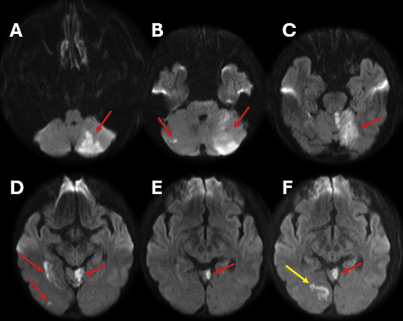 Figure 1
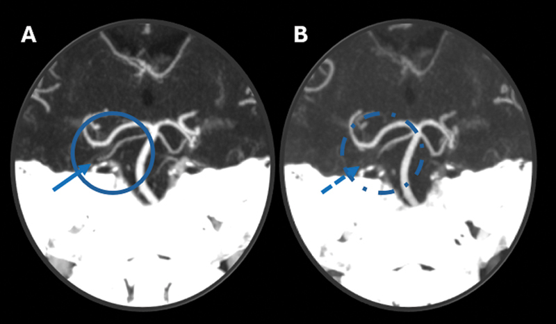 Figure 2
 Figure 3
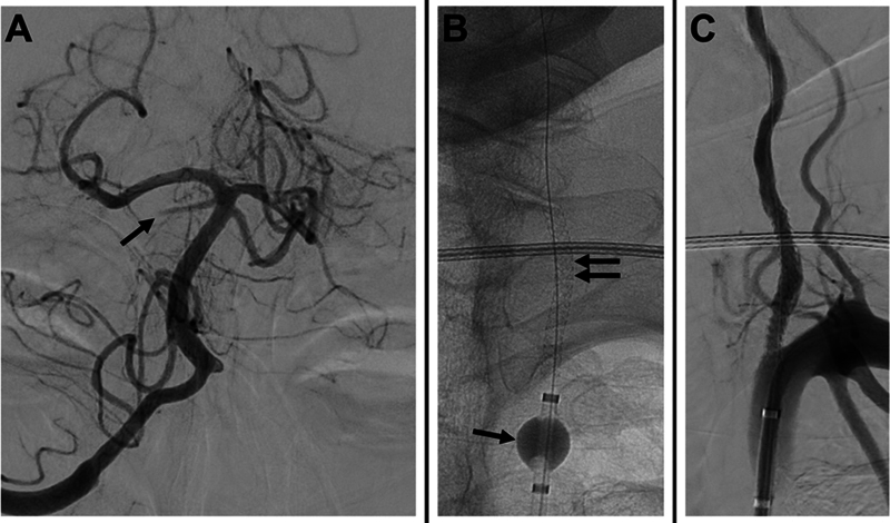 Figure 4
Peer Reviews
No public reviews on file for this paper yet. If you reviewed it on a platform where reviews are public (OpenReview, ICLR, NeurIPS, ICML), you can paste yours below so the community can read it here.
Videos
No videos yet. Explain this paper in a talk, walkthrough, or lecture? Add one.
Taxonomy
TopicsAcute Ischemic Stroke Management · Cerebrovascular and Carotid Artery Diseases · Intracranial Aneurysms: Treatment and Complications
A 64-year-old male patient with hypertension and hyperlipidemia presented with sudden left eye vision loss. Imaging ( Figures 1 2 3 4 ) revealed recurrent posterior circulation infarcts and vessels occlusions. Angiography confirmed vertebral artery stump syndrome (VASS). An endovascular intervention successfully achieved complete recanalization without residual stenosis. The patient was discharged neurologically stable.
Magnetic resonance imaging (MRI) scan of the brain, axial diffusion sequence. ( A–E ) Multiple acute/subacute infarcts in the posterior circulation (red arrows), involving the cerebellum and the right mesial temporo-occipital region. ( F ) Follow-up imaging performed seven days later, showing a new vascular event in the mesial right occipital lobe (yellow arrow).
Computed tomography angiography (CTA) scan of the head, coronal maximum intensity projection (MIP) reconstruction with zoom-in of the vertebrobasilar system. ( A,B ) Images obtained seven days apart , showing an interval occlusion in the mid-to-distal segment of the right superior cerebellar artery (blue circles and arrows).
( A ) Computed tomography angiography (CTA) scan exhibiting normal right vertebral artery (VA). ( B ) Initial intracranial angiography of the right VA reveals normal intracranial flow and retrograde opacification of the intracranial left VA. Note that the right superior cerebellar artery is normal (arrow). ( C,D ) Left subclavian angiography depicts near-occlusion of the left VA origin with occlusion of its proximal V1 segment (arrows). Selective angiography of the left deep cervical artery in the frontal ( E ) and lateral ( F ) projections illustrate collateral supply to the distal V2 and V3 segments of the left VA via muscular anastomotic branches (arrows).
( A ) Repeat cerebral angiography 3 days later revealing a new occlusion of the right superior cerebellar artery (arrow). The decision was then made to attempt recanalization of the left VA. ( B ) A balloon guide catheter was positioned in the proximal left subclavian artery where the balloon was inflated to arrest antegrade flow and prevent distal embolization (single arrow); a balloon-expandable stent was then advanced and successfully deployed (double arrows). ( C ) The balloon of the balloon guide catheter was then deflated, and the final angiography confirms successful recanalization of the left VA.
Vertebral artery stump syndrome is a rare but treatable cause of recurrent posterior strokes, 1 2 3 with key mechanisms including stagnant blood flow leading to thrombus formation, propagation of embolic fragments from the occlusion's distal limit, and emboli introduced via collateral pathways. 4 This case highlights the importance of endovascular treatment in preventing neurological deterioration and improving outcomes.
The reference list from the paper itself. Each links out to its DOI / PubMed record.
- 1Rossi S S Iaccarino G Bonura A Exploring vertebral artery stump syndrome: An overlooked cause of posterior ischemic strokes. A narrative review of current management options J Stroke Cerebrovasc Dis 2024330810781910.1016/j.jstrokecerebrovasdis.2024.107819. Epub 2024 Jun 13. PMID: 3887884538878845 · doi ↗ · pubmed ↗
- 2Ji R Li B Xu Z Retrograde recanalisation for vertebral artery stump syndrome: a case report Stroke Vasc Neurol 202270546246410.1136/svn-2021-001407. Epub 2022 Mar 30. PMID: 35354663; PMCID: PMC 961412535354663 PMC 9614125 · doi ↗ · pubmed ↗
- 3Zhang W Wang S Li CA Case Series and Literature Review of Vertebral Artery Stump Syndrome Front Neurol 20221277084510.3389/fneur.2021.770845. PMID: 35153978; PMCID: PMC 883172635153978 PMC 8831726 · doi ↗ · pubmed ↗
- 4Suzuki M Dembo T Hara W Vertebral artery stump syndrome Intern Med 2018570573373610.2169/internalmedicine.9317-1729151515 PMC 5874350 · doi ↗ · pubmed ↗