Correction: β-catenin-mediated YAP signaling promotes human glioma growth
Yan Wang, Peng Pan, Zhaohao Wang, Yu Zhang, Peng Xie, Decheng Geng, Yang Jiang, Rutong Yu, Xiuping Zhou

Abstract
Genes, proteins, chemicals, diseases, species, mutations and cell lines named across the full text — each resolved to its canonical identifier and authoritative record.
Click any figure to enlarge with its caption.
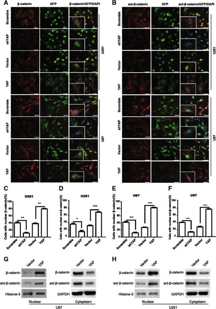 Figure 1
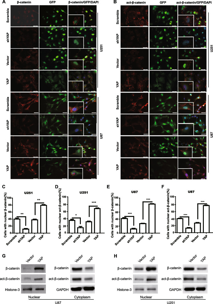 Figure 2
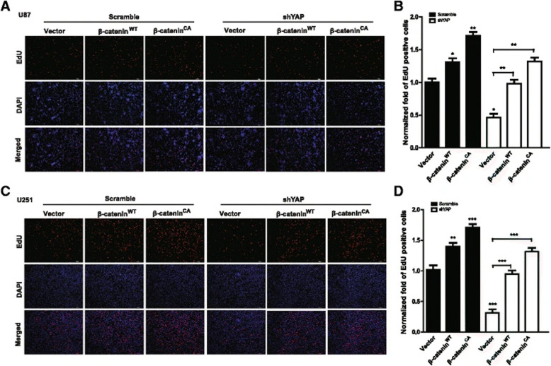 Figure 3
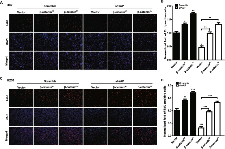 Figure 4
Peer Reviews
No public reviews on file for this paper yet. If you reviewed it on a platform where reviews are public (OpenReview, ICLR, NeurIPS, ICML), you can paste yours below so the community can read it here.
Videos
No videos yet. Explain this paper in a talk, walkthrough, or lecture? Add one.
Taxonomy
TopicsHippo pathway signaling and YAP/TAZ · Cancer-related gene regulation · Kruppel-like factors research
Correction: J Exp Clin Cancer Res 36, 136 (2017)
https://doi.org/10.1186/s13046-017–0606-1
Following the publication of the original article [1], the authors found errors in the figures, specifically, Fig. 4a and Fig. 6c where the IF image of Scramble group in U251 cell and the images of β-catenin(WT) and β-catenin(CA) in shYAP group were incorrect.
Below are the correct figures:
Incorrect Figure 4
Fig. 4YAP modulates the subcellular location of β-catenin. a&b The expression and subcellular location of β-catenin (a) and active-β-catenin (b) were assessed by immunofluorescence in YAP down-regulation or over-expression cells. Scale bar 50 μm. Inset showed the amplified images. c-f Quantification results of the percentage of cells with nuclear β-catenin (c & e) or active-β-catenin (d & f) in U251 and U87 cells. * P < 0.05, ** P < 0.01, *** P < 0.001. g & h Subcellular location of β-catenin or active-β-catenin was detected by using cellular fractionation and immunoblotting. Histone and GAPDH were used as nuclear and cytoplasm loading control respectively
Correct Figure 4
Fig. 4YAP modulates the subcellular location of β-catenin. a&b The expression and subcellular location of β-catenin (a) and active-β-catenin (b) were assessed by immunofluorescence in YAP down-regulation or over-expression cells. Scale bar 50 μm. Inset showed the amplified images. c-f Quantification results of the percentage of cells with nuclear β-catenin (c & e) or active-β-catenin (d & f) in U251 and U87 cells. * P < 0.05, ** P < 0.01, *** P < 0.001. g & h Subcellular location of β-catenin or active-β-catenin was detected by using cellular fractionation and immunoblotting. Histone and GAPDH were used as nuclear and cytoplasm loading control respectively
Incorrect Figure 6 Fig. 6. The effect of YAP down-regulation on glioma cell proliferation was partially mediated by β-catenin. a & c Representative images of EdU assay after over-expression of β-catenin^WT^ and β-catenin^CA^ in U251 (a) and U87 (c) cells with or without YAP down-regulation. The cell proliferation was examined after plating for 48 h. Scale bar, 200 μm. b&d Quantification result of (a & c). * P < 0.05, ** P < 0.01
Correct Figure 6 Fig. 6. The effect of YAP down-regulation on glioma cell proliferation was partially mediated by β-catenin. a & c Representative images of EdU assay after over-expression of β-catenin^WT^ and β-catenin^CA^ in U251 (a) and U87 (c) cells with or without YAP down-regulation. The cell proliferation was examined after plating for 48 h. Scale bar, 200 μm. b&d Quantification result of (a & c). * P < 0.05, ** P < 0.01
The corrections do not compromise the validity of the conclusions and the overall content of the article. The original article [1] has been updated.