Francisella tularensis Subspecies holarctica in Stranded Beluga Whales, Cook Inlet, Alaska, USA
Natalie Rouse, Jeremy Buttler, Kristy Pabilonia, Christina Weller, Laurel Respicio-Kingry, Elizabeth Dietrich, Jeannine Petersen, Ganna Kovalenko, Eric Bortz, Kathy Burek Huntington

TL;DR
Beluga whales in Alaska died from tularemia caused by Francisella tularensis, highlighting concerns for marine mammal health and biosecurity.
Contribution
First report of Francisella tularensis subspecies holarctica in beluga whales using nanopore metagenomics and molecular confirmation.
Findings
Francisella tularensis was detected in stranded beluga whales using nanopore metagenomics.
The subspecies was identified as holarctica through multilocus sequence typing.
The findings suggest implications for marine mammal health and biosecurity in the North Pacific.
Abstract
We report fatal tularemia in stranded beluga whales in Cook Inlet, Alaska, USA. Francisella tularensis was detected by nanopore metagenomics, confirmed by quantitative PCR and immunohistochemistry, and characterized as F. tularensis subspecies holarctica by multilocus sequence typing. Our findings should be considered when assessing biosecurity and marine mammal health in the North Pacific.
Genes, proteins, chemicals, diseases, species, mutations and cell lines named across the full text — each resolved to its canonical identifier and authoritative record.
Click any figure to enlarge with its caption.
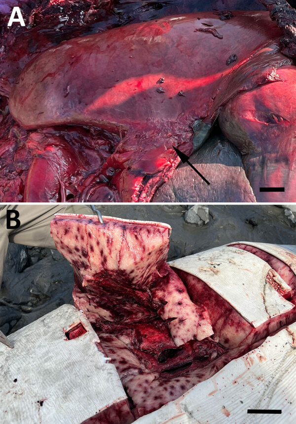 Figure 1
Peer Reviews
No public reviews on file for this paper yet. If you reviewed it on a platform where reviews are public (OpenReview, ICLR, NeurIPS, ICML), you can paste yours below so the community can read it here.
Videos
No videos yet. Explain this paper in a talk, walkthrough, or lecture? Add one.
Taxonomy
TopicsBacillus and Francisella bacterial research · Bacteriophages and microbial interactions · Yersinia bacterium, plague, ectoparasites research
Francisella tularensis is a highly pathogenic gram-negative bacterium that infects a large range of animals and humans, primarily in the Northern Hemisphere, causing the clinical disease tularemia. Human disease manifests with influenza-like symptoms (lymphadenopathy, conjunctivitis, pneumonia, septicemia) and other specific symptoms corresponding to the route of exposure. Two subspecies, F. tularensis subsp. tularensis and holarctica, are known pathogens and can be acquired via multiple routes, including arthropod vector, cutaneous, ingestion, or inhalation (1).
F. tularensis was first documented in Alaska, USA, in 1938 (2) and has been isolated infrequently in ticks, lagomorphs, and rodents. Serologic studies have confirmed exposure in humans, avian species, terrestrial mammals, and polar bears in multiple areas of the state (2). In October 2023, tularemia was diagnosed in a pinniped in Washington, USA, when a biologist was infected during necropsy (3). The same fall, dead stranded beluga whales (Delphinapterus leucas) in Cook Inlet, Alaska, were found to have gross lesions consistent with tularemia. We report the results of an investigation of those deaths.
Necropsies were performed and tissues collected and stored following standard procedures. Samples for histopathology were fixed in 10% neutral buffered formalin (Table). We submitted varied tissues from 2 sufficiently fresh animals (no. 2023279: fetal spleen, mediastinal lymph node, spleen, blowhole swab, heart, liver; and no. 2023288: brain, liver, mammary gland, mediastinal lymph node, spleen) for aerobic culture and testing for known cetacean pathogens, including influenza and Erysipelothrix sp. by PCR, and for harmful algal bloom toxins by ELISA (Table). We analyzed blowhole swab, lung, mediastinal lymph node, and rectal swab samples from animal 2023279 by metagenomic sequencing. In brief, we extracted and amplified total nucleic acids (I.M. Claro et al., unpub. data, https://doi.org/10.12688/wellcomeopenres.17170.2) and sequenced cDNA and metagenomics libraries by SMART9N using an Oxford Nanopore Rapid PCR barcoding and MinION device (https://nanoporetech.com) (Table; Appendix Figure 1) (4). We classified sequence reads by using wf-metagenomics and wf-alignment in epi2melabs v.5.1.3 (Oxford Nanopore), mapped to the F. tularensis genome (GenBank accession no. NC_007880.1) by reference-based assembly using minimap2, and annotated using tbCon and ggplot in RStudio (Posit, https://posit.co) (Table). Subsequently, we tested lung and liver tissue from both animals for F. tularensis by immunohistochemistry and by culture and PCR using Centers for Disease Control and Prevention Laboratory Response Network proprietary protocols (Table). We then typed samples from positive animals by multilocus type sequencing of 6 genes (fabH, tpiA, sdhA, rpoA, groEL, and pgm) (5–7) and sequenced multiplexed amplicon libraries on the MiSeq platform (Illumina, https://www.illumina.com) (Table). We mapped amplicon sequence reads to reference genes from F. tularensis subsp. holarctica live vaccine strain, concatenated, and aligned with corresponding sequences from F. tularensis and other Francisella spp. to construct phylogenetic trees.
Both animals were pregnant adult females with markedly enlarged mediastinal lymph nodes, pleuritis, and pneumonia (Figure, panel A). One animal had severe multifocal random ecchymotic hemorrhage in the blubber (Figure, panel B). Histologic findings included necrosuppurative and histiocytic bronchopneumonia, lymphadenitis, and hepatitis (Appendix Figure 2, panels A–C). Immunohistochemistry demonstrated positive staining in areas of inflammation (Appendix Figure 2, panel D). Domoic acid and saxitoxin were not found, and PCRs and bacterial cultures yielded negative results or mixed organisms believed to be postmortem overgrowth (Appendix Table).
We identified the causative organism by using metagenomics. We mapped sequence reads from animal 2023279 by reference-based assembly and found those reads to be distributed at low read depth (2–21×; 1,181 sequence reads; N50 = 275 nt, quality score = 9) across the 1.89-Mbp F. tularensis genome. We detected F. tularensis DNA in all samples by quantitative PCR with cycle threshold values <25. By multilocus sequence typing, we identified a concatenated sequence of 4,107 bp as F. tularensis subsp. holarctica. Phylogenetic analysis placed this strain in a clade identical to the 2023 pinniped case from Washington, as well as other isolates from the Northern Hemisphere (Appendix Figure 3).
Although Cook Inlet belugas are known to be susceptible to a variety of bacterial pathogens (10), F. tularensis has not been previously detected in this population, or in other cetaceans. The pattern of pathology represents the pulmonary form of tularemia, and the route of exposure was likely inhalation of contaminated water. F. tularensis is primarily a disease associated with freshwater, but the brackish nature of Cook Inlet and nearshore residence of belugas expose them to potentially contaminated freshwater runoff as well as to other reservoirs typically associated with freshwater (e.g., aquatic rodents, mosquito larvae) (1,2). The cause of the infections in a previously unreported host is unknown; however, host factors such as immunosuppression or environmental changes, such as increased runoff, could be considered.
One human case of tularemia was reported in Cook Inlet’s largest adjacent city in 2023 (https://epi.alaska.gov/bulletins/docs/b2024_14.pdf); however, the circumstances of exposure were not reported. The propensity of whales to travel long distances could further disseminate this pathogen, increasing exposure to humans and wildlife. Our findings highlight a new risk to persons working in the marine environment and should be considered when assessing biosecurity and marine mammal health in the North Pacific.
The reference list from the paper itself. Each links out to its DOI / PubMed record.
- 1Hennebique A, Boisset S, Maurin M. Tularemia as a waterborne disease: a review. Emerg Microbes Infect. 2019;8:1027–42. 10.1080/22221751.2019.163873431287787 PMC 6691783 · doi ↗ · pubmed ↗
- 2Smith MM, Van Hemert C, Atwood TC, Sinnett DR, Hupp JW, Meixell BW, et al. A serologic survey of Francisella tularensis exposure in wildlife on the Arctic Coastal Plain of Alaska, USA. J Wildl Dis. 2022;58:746–55. 10.7589/JWD-D-21-0016236302352 · doi ↗ · pubmed ↗
- 3Inouye W, Oltean HN, Mc Millan M, Schnitzler H, Lipton B, Peterson JM, et al. Notes from the field: tularemia associated with harbor seal necropsy—Kitsap County, Washington, October 2023. MMWR Morb Mortal Wkly Rep. 2024;73:731–2. 10.15585/mmwr.mm 73333 a 339173169 PMC 11349381 · doi ↗ · pubmed ↗
- 4Buttler J, Drown DM. Accuracy and completeness of long read metagenomic assemblies. Microorganisms. 2022;11:96. 10.3390/microorganisms 1101009636677391 PMC 9861289 · doi ↗ · pubmed ↗
- 5Ahlinder J, Öhrman C, Svensson K, Lindgren P, Johansson A, Forsman M, et al. Increased knowledge of Francisella genus diversity highlights the benefits of optimised DNA-based assays. BMC Microbiol. 2012;12:220. 10.1186/1471-2180-12-22023009728 PMC 3575276 · doi ↗ · pubmed ↗
- 6Mikalsen J, Olsen AB, Tengs T, Colquhoun DJ. Francisella philomiragia subsp. noatunensis subsp. nov., isolated from farmed Atlantic cod (Gadus morhua L.). Int J Syst Evol Microbiol. 2007;57:1960–5. 10.1099/ijs.0.64765-017766855 · doi ↗ · pubmed ↗
- 7Brett M, Doppalapudi A, Respicio-Kingry LB, Myers D, Husband B, Pollard K, et al. Francisella novicida bacteremia after a near-drowning accident. J Clin Microbiol. 2012;50:2826–9. 10.1128/JCM.00995-1222692740 PMC 3421515 · doi ↗ · pubmed ↗
- 8Pal N, Bender JS, Opriessnig T. Rapid detection and differentiation of Erysipelothrix spp. by a novel multiplex real-time PCR assay. J Appl Microbiol. 2010;108:1083–93. 10.1111/j.1365-2672.2009.04560.x 19840181 · doi ↗ · pubmed ↗