Pharmacokinetics/pharmacodynamics of gamithromycin for treating Pasteurella multocida infection in cattle using a tissue cage model
Qingwen Yang, Xuesong Liu, Yongzhi Lv, Yushen Li, Arifa Mehreen, Arifa Mehreen, Arifa Mehreen, Arifa Mehreen

TL;DR
This study evaluates how well gamithromycin fights Pasteurella multocida in cattle, using a model to measure drug effectiveness and guide dosing.
Contribution
The study identifies optimal PK/PD indices for gamithromycin in treating bovine respiratory infections caused by Pasteurella multocida.
Findings
AUC0-24h/MIC is the best PK/PD index for measuring gamithromycin's antibacterial activity.
Bacteriostatic effects require AUC0-24h/MIC values of 0.27 in serum, 0.17 in transudates, and 0.14 in exudates.
Bacterial eradication in serum requires an AUC0-24h/MIC of 18.46.
Abstract
In this study, gamithromycin, a long-acting azalide antibiotic recently introduced for bovine respiratory disease (BRD) treatment, was evaluated for its effectiveness against Pasteurella multocida using a cattle tissue cage model. Gamithromycin (6 mg/kg) was administered via both intravenous and subcutaneous routes and the gamithromycin contents in sera, transudates, and exudates were measured using HPLC/MS-MS. Non-compartmental methods were utilized for assessing pharmacokinetic parameters and an inhibitory sigmoid Emax model determined associations between the pharmacokinetic/pharmacodynamic (PK/PD) indices and antibacterial activity. The area under the 24-h concentration-time curve/minimum inhibitory concentration (AUC0-24h/MIC) was found to be an optimal measure of antibacterial activity. The AUC0-24h/MIC values over 24 h in sera, transudates, and exudates were 0.27, 0.17, and 0.14,…
Genes, proteins, chemicals, diseases, species, mutations and cell lines named across the full text — each resolved to its canonical identifier and authoritative record.
Click any figure to enlarge with its caption.
 Fig 1
Fig 1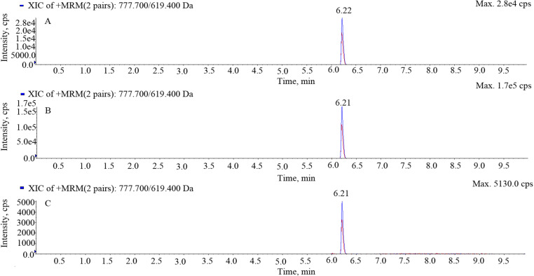 Fig 2
Fig 2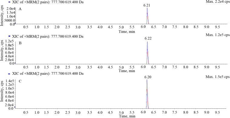 Fig 3
Fig 3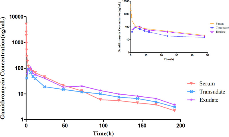 Fig 4
Fig 4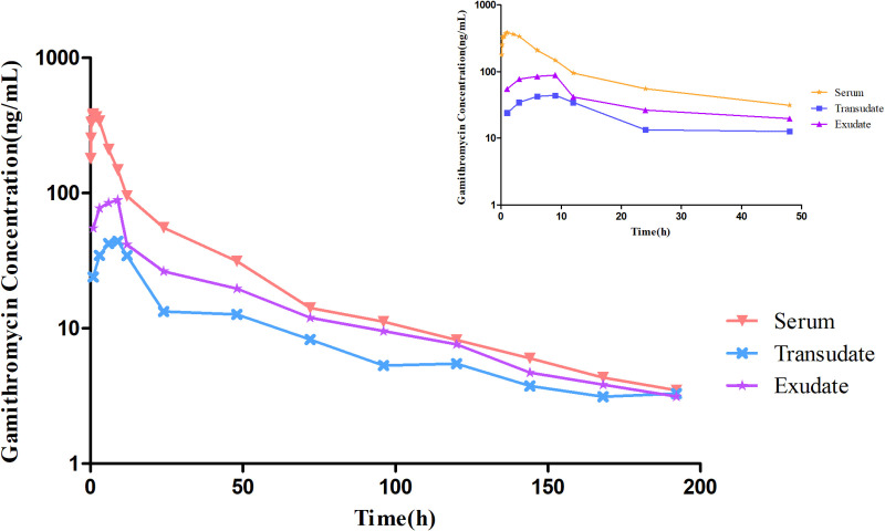 Fig 5
Fig 5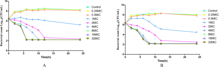 Fig 6
Fig 6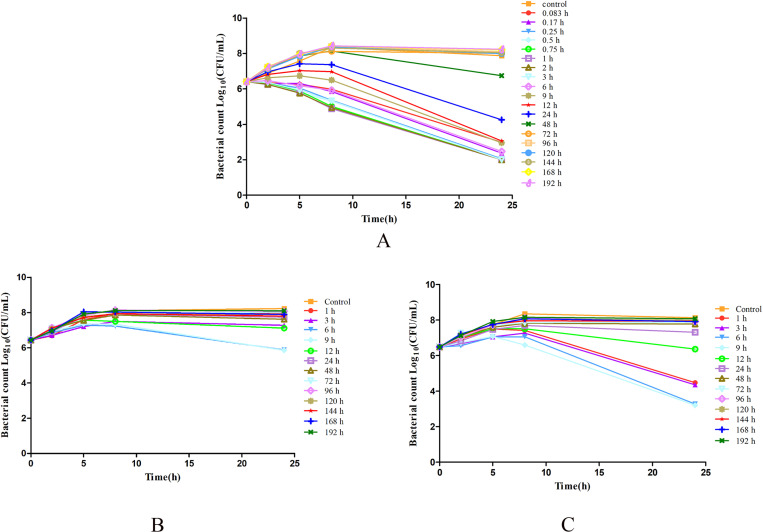 Fig 7
Fig 7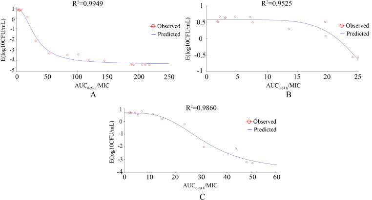 Fig 8
Fig 8- —Science and Technology Research Program of Chongqing Municipal Education Commission
Peer Reviews
No public reviews on file for this paper yet. If you reviewed it on a platform where reviews are public (OpenReview, ICLR, NeurIPS, ICML), you can paste yours below so the community can read it here.
Videos
No videos yet. Explain this paper in a talk, walkthrough, or lecture? Add one.
Taxonomy
TopicsMicrobial infections and disease research · Antibiotics Pharmacokinetics and Efficacy · Antibiotic Resistance in Bacteria
Introduction
Pasteurella multocida is a Gram-negative bacillus of the Pasteurellaceae family and is one of the main pathogens responsible for bovine respiratory disease (BRD) [1,2]. The incidence of BRD has increased with the rapid development of intensive cattle farming, resulting in significant economic losses in the industry[3]. The disease is usually treated with antibiotics. Therefore, it is important to optimize clinical dosages to achieve maximum efficacy and reduce the production of drug-resistant strains.
Gamithromycin was developed for preventing and treating BRD resulting from P. multocida, Mannheimia haemolytica, and Histophilus somni infections [4,5]. This second-generation macrolide is a typical azalide, which contains a 15-member carbon ring with a unique alkylated nitrogen in the 7a position. Like other macrolides, gamithromycin achieves bacteriostatic and bactericidal effects by inhibiting RNA-dependent protein synthesis. Due to its wide-ranging antimicrobial spectrum, strong antibacterial action, and rapid clearance, gamithromycin is widely used for treating and preventing BRD[6,7].
The pharmacokinetics (PK)/pharmacodynamics (PD) model (PK/PD) model integrates PK, describing the ADME characteristics of the drug, with PD, focusing on biological effects and associated mechanisms. The model mathematically characterizes relationships between drug levels and their influence over time[8]. The use of PK/PD models can assist assessments of dosages and regimens for achieving the desired therapeutic effect while minimizing resistance. By simulating various dosing scenarios, researchers can identify the most effective and safe dosing strategies[9]. Specific PK parameters, including the area under the curve (AUC) and the maximum concentration (C_max_), and PD parameters, such as the minimal inhibitory concentration (MIC) and time-kill curves, are commonly integrated into PK-PD models. The indices used are the AUC/MIC ratio, the duration of drug level maintenance above the MIC (%T > MIC), and the C_max_/MIC ratio [10]. When using AUC/MIC value for evaluating breakpoints, a time dimension (in hours) is included, often presenting difficulties for reader comprehension and discussion among scientists. However, division of the AUC/MIC value by the time duration produces more easily understandable values. This approach allows for the assessment of a ratio without dimensions, making the computed numerical value more straightforward and clinically interpretable[11].
Pharmacokinetic studies of gamithromycin in various animal species, such as broiler chickens[12], sheep[13], turkeys[14], and pigs[15], have shown that the drug is rapidly absorbed and widely distributed following subcutaneous (s.c.) administration. However, to the best of our knowledge, there is no information on the combination of in vitro PK data with ex vivo PD values for predicting the various antibacterial activities of gamithromycin in a cattle tissue cage model. Here, the PK/PD values for gamithromycin action on Pasteurella multocida in a tissue cage model in cattle after intravenous (i.v.) or subcutaneous (s.c.) injection were investigated. These data represent a resource for optimizing dose regimens in treating BRD caused by Pasteurella multocida.
Materials and methods
Antibiotics, bacteria and chemicals
Injectable gamithromycin solution (Zactran,^®^ 150 mg/mL) was purchased from Merial (lot number GYR053CB; Toulouse, France). Gamithromycin standard (purity >98%) was from Tiaozhan Biotechnology (Tianjin, China). After isolation from cattle, P. multocida NM-5–7 was identified using a MALDI-TOF/MS system (Axima-Assurance-Shimadzu). Tryptic Soy agar (TSA) and Mueller–Hinton II Broth (MHB) were from Huankai Microbial Technology (Guangdong, China), defibrinated sheep blood was from Ruite Biological Technology (Guangdong, China), and λ-Carrageenan was purchased from Sigma–Aldrich (lot number BCBP8978V; Shanghai, China).
Cattle tissue cage design and surgical implantation
Four tissue cages were implanted as previously described[16]. Briefly, golf practice balls (internal diameter, 42 mm; internal volume, 36.1 mL) and 26 holes (diameter, 5 mm) were used as tissue cages. One cage was implanted in each flank, 8–10 cm from the mid-line, followed by infection protection with 10 000 IU/kg penicillin by intramuscular injection for 3 days, accompanied by simultaneous intramuscular injections of aminopyrine for analgesia. When the wounds had healed and the tissue cages were filled with fluid, 1% sterile carrageenan solution (0.5 mL) was injected into one of the tissue cages. After five weeks, transudates and exudates were harvested from the tissue cages.
Animals and experimental design
Six healthy cattle (mean weight ± SD, 142 ± 18 kg) were randomlyy allocated to two groups (n = 3 per group). The animals received antibiotic-free daily feed, hay, and water ad libitum.
The study design was a two-way crossover, with each animal receiving received gamithromycin (6 mg/kg) both i.v. and s.c. In the first stage of the study, the first group were given 6 mg/kg body weight (bw) gamithromycin i.v. via the left jugular vein, while the second group were given the same gamithromycin dose s.c. into the neck. During the second part of the study, the treatments of the groups were reversed. The interval between the two stages was 28 days. All protocols were approved by the Animal Use and Care Committee of Chongqing Three Gorges Vocational College (Chongqing, China).
Sample collection
The procedures were conducted as described previously[17]. Briefly, blood (10 mL) was sampled from the right jugular vein prior to gamithromycin injection and after 5, 10, 15, 30, and 45 min and 1, 2, 3, 6, 9, 12, 24, 48, 72, 96, 120, 144, 168, and 192 h. Transudate and exudate samples (1.5 mL) were obtained before and at 1, 3, 6, 9, 12, 24, 48, 72, 96, 120, 144, 168, and 192 h after gamithromycin administration. All samples were placed in plastic tubes (without anticoagulant) and were protected from sunlight. The samples were centrifuged (4000 × g, 4°C, 10 min) and retained at -80°C until analyzed.
LC-MS/MS determination of gamithromycin concentrations
The levels of gamithromycin in serum, transudate, and exudate samples were evaluated using analyzed using LC-MS/MS, as described previously[16,18]. MS/MS was conducted in the positive electrospray ionization mode. The reaction transitions of gamithromycin monitored included the protonated molecule to product ion transitions of m/z 777.45 > 619.40 for quantification and m/z 777.45 > 157.70 for identification. The gradient elution was performed with mobile phase A consisting of 1% formic acid in water, and acetonitrile as mobile phase B. The details are provided in Table 1.
Table 1: Mobile phase conditions for HPLC/MS/MS of gamithromycin.
Pharmacokinetic analysis of gamithromycin
The PK parameters used were elimination half-life (t_1/2β_), time of maximum concentration (T_max_), area under concentration-time curve (AUC_0-last_), peak drug concentration (C_max_), mean residence time (MRT_0-last_), the volume of distribution (V_d_), and clearance (CL) of gamithromycin in the sera, transudates, and exudates, determined by non-compartmental analysis (WinNonlin 5.2.1, Pharsight Corporation, USA) and presented as mean±SD.
In vitro susceptibility studies and time-kill curves
The gamithromycin MICs against P. multocida were determined in Mueller Hinton Broth (MHB), sera, exudates, and transudates using the microdilution techniques of the Clinical and Laboratory Standards Institute[19]. The Streptococcus pneumoniae ATCC 49619 served as the MIC quality control strain. The MBC is the lowest concentration that achieves a 99.9% bacterial count reduction. After the determination of the MIC, the in vitro time-kill curves were then evaluated as described[20]. Briefly, a 10 µL aliquot of bacterial suspension containing an estimated 10^8^ CFU/mL was exposed to gamithromycin at 0.25 × , 0.5 × , 1 × , 2 × , 4 × , 8 × , 16 × , or 32 × MIC diluted in MHB and serum (1 mL). The cultures were incubated at 37°C and analyzed at 0, 2, 4, 6, 8, 10, 12 and 24 h post-treatment. One hundred-microliter samples of each culture were serially diluted (10-fold), and 20 μL aliquots were placed on 5% sterile defibrinated sheep blood trypticase soy agar (TSA) for 18–24 h at 37°C, followed by colony counting. The limit of detection (LOD) was 200 CFU/mL.
Ex vivo time-kill curves
The ex vivo time-kill curves were constructed as described [20]. Briefly, 10 µL of bacterial culture in the stationary growth phase was added to 1 mL samples of sera, transudates, and exudates, to about 5 × 10^5^ CFU/mL and cultivated at 37°C with counting at 0, 2, 5, 8, and 24 h post-treatment. Aliquots (100 μL) of the cultures underwent 10-fold serial dilution and 20 μL of each dilution was placed on 5% sheep blood TSA. Colony numbers were determined after incubation for 18–24 h at 37°C. The LOD was 200 CFU/mL.
Integration of PK/PD and modeling
The mean PK parameters were evaluated using WinNonlin software.. The MIC in serum was used for modeling. The PK/PD parameters were determined using the inhibitory effect Emax model as follows:
where E indicates the antibacterial effect, i.e., the changes in log_10_ CFU/mL in the samples (sera, transudates, and exudates) between the initial log_10_ CFU/mL and 24 h post-incubation; Emax denotes the log_10_ CFU/mL change in the control samples; E0 represents the maximum antibacterial activity, i.e., log_10_ CFU/mL changes over a 24-h incubation; EC_50_ indicates the AUC/MIC at 50% of E_max_; C_e_ denotes the PK/PD parameters (*%*T > MIC, C_max_/MIC, AUC/MIC), and N represents the Hill coefficient.
Statistical analyses
Intergroup comparisons were evaluated by t-tests using SPSS software 13.0 and verified by Bonferroni’s correction. *P < *0.05 was considered statistically significant.
Results
Validation of HPLCC analytic methods
The retention time of gamithromycin was approximately 6.21 min with no interfering impurity peaks. The chromatograms of blank serum, transudate, and exudate samples are shown in Fig 1, while those of spiked samples are shown in Fig 2 and those of the separated samples are shown in Fig 3. The LOD and limit of quantification (LOQ) values were 1 and 2 ng/mL, respectively. Recovery of gamithromycin in all samples was over 85%. Variations within and between assays evaluated using the relative standard deviation (%RSD) were all < 10%.
Chromatograms of blank serum, transudate, and exudate samples.A, blank serum; B, blank transudate; C, blank exudate.
Chromatograms of spiked serum, transudate, and exudate samples.A, spiked serum; B spiked transudate; C spiked exudate.
Chromatograms of separated serum, transudate, and exudate samples.A, separated serum; B, separated transudate; C, separated exudate.
Pharmacokinetic analysis
The concentration-time curves for gamithromycin in sera, transudates, and exudates after i.v. and s.c. injection of 6 mg/kg bw are illustrated in Fig 4 and Fig 5. The mean concentration-time profiles were determined using non-compartmental models. The main PK parameters of after i.v. administration of gamithromycin in the different samples are shown in Table 2, while those following s.c injection are shown in Table 3.
Table 2: The pharmacokinetic parameters (mean±SD, n = 6) of gamithromycin in serum, transudate, and exudate after i.v administration of 6 mg/kg.
Table 3: The pharmacokinetic parameters (mean±SD, n = 6) of gamithromycin in serum, transudate, and exudate after s.c administration of 6 mg/kg.
Concentration-time profiles for gamithromycin in sera, transudates, and exudates after i.v. administration of 6 mg/kg body weight.The small graph indicates drug concentrations over 48 h following administration.
Concentration-time profiles for gamithromycin in sera, transudates, and exudates after s.c. administration of 6 mg/kg body weight.The small graph indicates drug concentrations over 48 h following administration.
After a single i.v. injection, gamithromycin demonstrated the following pharmacokinetic parameters. In bovine serum, T_1/2β_ was 35.27 h, AUC_0-last_ was 5.52 μg·h/mL, MRT was 25.86 h, and CL was 1.07 L/h.kg, while in the transudate, the C_max_ was 0.09 μg/mL, T_max_ was 4.50 h, T_1/2β_ was 41.37 h, AUC_0-last_ was 2.65 μg·h/mL, MRT was 55.97 h, and CL was 2.15 L/h.kg, and in the exudate, C_max_ was 0.11 μg/mL, T_max_ was 5.50 h, T_1/2β_ was 38.60 h, AUC_0-last_ was 3.73 μg·h/mL, MRT was 53.30 h, and CL was 1.59 L/h.kg.
The parameters after a single s.c. injection were C_max_ of 0.43 μg/mL, T_max_ of 1.00 h, T_1/2β_ of 48.30 h, AUC_0-last_ of 6.23 μg·h/mL, MRT of 33.87 h, and CL of 0.95 L/h.kg in serum, C_max_ of 0.04 μg/mL, T_max_ of 9.50 h, T_1/2β_ of 63.10 h, AUC_0-last_ of 1.84 μg·h/mL, MRT of 54.87 h, and CL of 2.83 L/h.kg in transudates, and C_max_ of 0.11 μg/mL, T_max_ of 7.00 h, T_1/2β_ of 52.74 h, AUC_0-last_ of 2.99 μg·h/mL, MRT of 49.02 h, and CL of 1.85 L/h.kg in exudates. The absolute bioavailability of gamithromycin after subcutaneous injection was 112.95%.
In vitro susceptibility studies and kill curves
Gamithromycin exhibited antimicrobial activity against P. multocida strain NM-5–7 with varying efficacy across different media. In MHB, the MIC and minimum bactericidal concentration (MBC) were 0.50 and 1.00 μg/mL, respectively. However, the antibiotic demonstrated enhanced potency in physiological fluids. Specifically, in sera, transudates, and exudates, the MIC was consistently lower at 0.03 μg/mL, while the MBC was 0.06 μg/mL. This indicates a notably increased antimicrobial effect of gamithromycin in these biological fluids compared to standard laboratory media.
The in vitro kill curves of gamithromycin against P. multocida NM-5–7 in MHB and serum are presented in Fig 6. The antibacterial trends in broth and serum were similar. The antibacterial activity increased as the drug concentration was raised. At gamithromycin levels below 1 × MIC, there was no inhibitory effect, while at 1 × MIC, there was slight inhibitory activity. Significant bactericidal activity was observed at 2 × MIC, with the rate of killing increasing as the drug concentration rose. At or above 4 × MIC, the bactericidal rate was significantly greater than that at 2 × MIC.
In vitro kill curves of gamithromycin against Pasteurella multocida NM-5-7.A, in MHB; B, in serum.
Ex vivo antibacterial action of gamithromycin
The ex vivo killing curve of gamithromycin against P. multocida NM-5–7 is presented in Fig 7. As illustrated in Fig 7A, serum samples collected during the first 3 hours (0.083, 0.17, 0.25, 0.5, 0.75, 1, 2, and 3 h) after subcutaneous injection exhibited inhibitory activity after 2 h of incubation, while samples obtained at 6 h showed inhibitory activity after 5 h of treatment. After 24 h, serum samples obtained at 24 h displayed inhibitory activity and samples obtained at 0.083, 6, 9, and 12 h showed good bactericidal activity, and those collected at 0.17, 0.25, 0.5, 0.75, 1, 2, and 3 h indicated bacterial clearance. However, samples collected at 48 h or later time points exhibited almost no visible antibacterial activity.
Ex-vivo kill curves of gamithromycin against Pasteurella multocida NM-5-7.A, in serum; B, in transudate; C, in exudate.
As Fig 7A and 7B indicate, the transudate and exudate samples exhibited similar ex-vivo activities against P. multocida NM-5–7. In the transudate samples, the gamithromycin transudate samples obtained at 6 and 9 h showed inhibitory activity after 24 h of treatment, while no bactericidal activity was seen in samples from the other time points. Exudate samples obtained at 1, 3, and 12 h showed slight inhibitory activity after 24 h of incubation, while bactericidal effects could be seen in those collected at 6 and 9 h. The exudate samples collected at other time points showed no visible antibacterial activity.
PK/PD integration and modeling
The inhibitory effect Emax associations between ex-vivo AUC_0-24h_/MIC values for P. multocida NM-5–7 and bacterial counts in the different samples are shown in Fig 8. The PK/PD AUC_0-24h_/MIC values divided by 24 h to evaluate differences in activities are presented in Table 4. In all sample types, the AUC_0–24 h_/MIC was considered the best PK-PD index for assessing antibacterial activity. As a demonstration of bacteriostatic action, the values obtained after dividing the AUC_0-24h_/MIC by 24 h were 0.27, 0.17, 0.14, respectively, in serum, transudate, and exudate samples. To achieve bactericidal action, the AUC/MIC_0-24h_ divided by 24 h values in serum and exudate were 3.76 and 5.31, respectively, while to achieve bacterial eradication action, the value in serum was 18.46.
Table 4: The PK/PD parameter estimates after division of the AUC0-24 h/MIC by 24 h to assess antibacterial effects.
Correlation between AUC0-24 h/MIC for Pasteurella multocida NM-5-7 and bacterial counts after 24 h of treatment.R2 denotes the correlation coefficient. A, in serum; B, in transudate; C, in exudate.
Discussion
Antibiotics are the main methods used for the prevention and treatment of BRD. However, excessive or inappropriate use of antibiotics can result in the development and transmission of drug-resistant strains[21]. The dissemination of drug-resistant strains in animals used for food not only threatens the health of livestock and poultry but also affects human health[22]. Appropriate antibiotic use is essential to reduce the development of drug resistance and maximize the antimicrobial efficacy of the drugs. The combined use of PK and PD for the prediction of the efficacy of antimicrobial agents and to determine effective dosages is useful for optimizing the dosage regimen[8]. Here, the PK/PD of gamithromycin was investigated using an ex vivo tissue cage model. It was found that gamithromycin was rapidly absorbed when delivered at 6 mg/kg s.c., but was eliminated slowly. Furthermore, the AUC/MIC data provided an accurate prediction of gamithromycin efficacy
The recommended dose of gamithromycin is 6 mg/kg. After s.c. administration, the C_max_ value (0.43 μg/mL) found here was similar to the previously reported value in sheep (0.45 μg/mL)[13] and pigs (0.41 μg/mL) [15], but lower than the value found for rabbits (1.64 μg/mL)[5], broiler chickens (0.89 μg/mL)[23], and piglets (0.61 μg/mL)[24]. The t_1*/*2β_ (48.30 h) of cattle serum observed here was consistent with previously reported values (42.5 h) in sheep[13], but higher than those in broiler chickens (11.63 h)[23], rabbits (31.5 h) [5], turkeys (34.9 h) [14], and piglets (29.0h)[24]. These discrepancies may be due to species differences. In this study, after s.c. administration, the AUC_0–last_ was 6.23 μg.h/mL, which was higher the observed values in piglets (5.45 μg.h/mL)[24], pigs (3.48 μg.h/mL)[15], turkeys (5.14 μg.h/mL)[14], and broiler chickens (4.09 μg.h/mL)[23], and lower than the value found in sheep (8.88 μg.h/mL)[13]. Gamithromycin has been reported to have high bioavailability (97.6–112%) after s.c. administration[25]. In this study, the bioavailability following s.c. administration was found to be 112.95%, which was similar to the value found in pigs (117.6%) and higher than those reported in broiler chickens (102.4%) and rabbits (86.7%). These discrepancies may be due to species differences.
Effective clinical outcomes and bacterial eradication using macrolides in both human and veterinary medicine often occur with plasma/serum concentrations significantly below the in vitro MICs. This disparity between in vivo drug levels and MICs has led to frequent questioning of the applicability of traditional PK/PD concepts to macrolides[9]. Pasteurella multocida is an exclusively extracellular pathogen, with the pulmonary epithelial lining fluid (PELF) serving as its primary location. The high levels of the drug observed in the PELF have, however, been considered artificially elevated due to the release of the drug caused by cell lysis during bronchoalveolar lavage used for collecting the PELF[26]. Based on the reasons outlined above, a tissue cage model was utilized here for fitting the PK and PD data.
Earlier research has indicated that MIC values determined in MHB may be artificially elevated relative to those seen in other media and fluids[27]. Therefore, MIC values obtained from biological fluids and matrices are preferred over those from MHB. It was found that the MICs in MHB were 16 times greater than those in sera, transudates, and exudates. It is essential to determine MIC values in the biological matrix to ensure the integrity of the integrated PK-PD model in subsequent steps. Higher MIC values obtained from media can result in relatively lower PK-PD indices. Consequently, lower PD target values may prompt dosage reductions during calculations[28]. Based on the current study, the dose of gamithromycin required to achieve a bactericidal effect via subcutaneous administration is 6.04 mg/kg, which is close to the officially recommended dose. However, this dose calculation is based on the MIC value of a single strain rather than the MIC_90_ value. The MIC_90_ represents the minimum drug concentration required to inhibit the growth of 90% of the tested strains and is generally considered a more reliable and clinically representative pharmacodynamic parameter. Since the susceptibility of different strains to the drug may vary significantly, relying solely on the MIC value of a single strain may lead to limitations in dose recommendations. Therefore, future studies should include more clinical isolates to calculate the MIC_90_ value and integrate population pharmacokinetic models to further optimize dosing regimens and ensure their effectiveness in broader clinical scenarios.
In this study, the Vd value after i.v. injection was 54.33 L/kg while that after s.c. injection was 65.73 L/kg/. These values suggest the likelihood of effective tissue penetration. Examination of the timing and degree of the transfer of gamithromycin into the exudates and transudates showed that this was characterized by a relatively slow T_max_, with 7.00 h observed for the exudate and 9.50 h for the transudate. Similar results have been observed for marbofloxacin in a cattle tissue-cage model[20]. The T_1/2β_ values in the transudates and exudates were higher than those in the sera. The extensive distribution of gamithromycin and its comparable rate and extent of entry into the exudate and transudate highlight its ability to readily penetrate cell membranes, regardless of inflammatory reactions.
The selection of effective PK/PD indices is critical for optimizing dosage regimens[29, 30]. Here, it was found that the PK/PD index AUC/MIC could accurately predict the effectiveness of gamithromycin against P. multocida. However, as the AUC/MIC includes a time dimension (hours), this can present difficulties in comprehension and discussion. To address this, dividing the AUC/MIC by the time period results in a dimensionless ratio, facilitating clearer clinical interpretation of the computed numerical value[11]. In this study, the values obtained by dividing the AUC_0–24 h_/MIC by 24 h were integrated with the decrease in bacterial numbers after 24 h incubation using the inhibitory effect E_max_ model. For serum, these values for achieving bacteriostatic, bactericidal, and eradication were 0.27, 3.76, and 18.46, respectively, while for transudates and exudates, the values were 0.17 and 0.14, respectively, lower than those for sera. The PK/PD indices obtained in this study can then be used for dose calculation.
The model has several limitations. First, only a single strain of P. multocida was used, and further studies should be conducted using more strains. Second, due to differences between ex-vivo and in vivo conditions, the predicted dosages should be evaluated in clinical practice. There are many factors apart from PK, PD, and MICs that contribute to the effectiveness of the treatment regimen, including the breed of cattle, age, physical condition, and environmental conditions, which all play crucial roles in disease outcomes. In addition, there is a suggestion for future development to establish antimicrobial dosage schedules based on population PK data to associate the effectiveness of antimicrobial drugs in disease.
In conclusion, the PK/PD index values determined in this study could assist the development of a dosing regimen aimed at achieving bacteriological cure while mitigating the development of resistance. However, these PK/PD index values should be used cautiously. The involvement of other bacteria in BRD, such as M. haemolytica and H. somni, could affect the efficacy of the dosing regimen. All these factors remain to be evaluated in further studies.
Supporting information
S1 TableIn vitro killing curve in serum.(DOCX)
S2 TableIn vitro killing curve in MHB.(DOCX)
S3 TableThe gamithromycin concentration in serum after intravenous injection.(DOCX)
S4 TableThe gamithromycin concentration in transudate and exudate samples after intravenous injection.(DOCX)
S5 TableThe gamithromycin concentration in serum after subcutaneous injection.(DOCX)
S6 TableThe gamithromycin concentration in transudate and exudate samples after subcutaneous injection.(DOCX)
The reference list from the paper itself. Each links out to its DOI / PubMed record.
- 1Calderón Bernal JM, Fernández A, Arnal JL, Sanz Tejero C, Fernández-Garayzábal JF, Vela AI, et al. Molecular Epidemiology of Pasteurella multocida Associated with Bovine Respiratory Disease Outbreaks. Animals (Basel). 2022;13(1):75. doi: 10.3390/ani 13010075 36611685 PMC 9817976 · doi ↗ · pubmed ↗
- 2Sudaryatma PE, Saito A, Mekata H, Kubo M, Fahkrajang W, Okabayashi T. Bovine Respiratory Syncytial Virus Decreased Pasteurella multocida Adherence by Downregulating the Expression of Intercellular Adhesion Molecule-1 on the Surface of Upper Respiratory Epithelial Cells. Vet Microbiol. 2020;246:108748. doi: 10.1016/j.vetmic.2020.108748 32605748 PMC 7265823 · doi ↗ · pubmed ↗
- 3Blakebrough-Hall C, Mc Meniman JP, González LA. An evaluation of the economic effects of bovine respiratory disease on animal performance, carcass traits, and economic outcomes in feedlot cattle defined using four BRD diagnosis methods. J Anim Sci. 2020;98(2):skaa 005. doi: 10.1093/jas/skaa 005 31930299 PMC 6996507 · doi ↗ · pubmed ↗
- 4Hasoon MF, Jarocki VM, Mohammed MH, Djordjevic SP, Yip HYE, Carr M, et al. Antimicrobial susceptibility and molecular characteristics of Mycoplasma bovis isolated from cases of bovine respiratory disease in Australian feedlot cattle. Vet Microbiol. 2023;283:109779. doi: 10.1016/j.vetmic.2023.109779 37257307 · doi ↗ · pubmed ↗
- 5Wei X-Y, Zhang J, Zhang Y, Fu W-Z, Zhong L-G, Pan Y-D, et al. Pharmacokinetic/pharmacodynamic evaluation of gamithromycin against rabbit pasteurellosis. BMC Vet Res. 2024;20(1):147. doi: 10.1186/s 12917-024-03988-y 38643185 PMC 11031915 · doi ↗ · pubmed ↗
- 6Galecio JS, Escudero E, Corrales JC, García-Romero E, de la Fe C, Hernandis V, et al. Susceptibility of caprine mastitis pathogens to tildipirosin, gamithromycin, oxytetracycline, and danofloxacin: effect of serum on the in vitro potency of current macrolides. World J Microbiol Biotechnol. 2022;38(12):221. doi: 10.1007/s 11274-022-03407-0 36097302 PMC 9468077 · doi ↗ · pubmed ↗
- 7Torres S, Thomson DU, Bello NM, Nosky BJ, Reinhardt CD. Field study of the comparative efficacy of gamithromycin and tulathromycin for the treatment of undifferentiated bovine respiratory disease complex in beef feedlot calves. Am J Vet Res. 2013;74(6):847–53. doi: 10.2460/ajvr.74.6.847 23718652 · doi ↗ · pubmed ↗
- 8Toutain PL, Lees P. Integration and modelling of pharmacokinetic and pharmacodynamic data to optimize dosage regimens in veterinary medicine. J Vet Pharmacol Ther. 2004;27(6):467–77. doi: 10.1111/j.1365-2885.2004.00613.x 15601441 · doi ↗ · pubmed ↗