Optimizing Nutritional Support Through the Retromolar Space
Shikha Yadav, Yugesh Chandra, Navya Gella, Prashanthi Polakonda, Anuvindha JS

TL;DR
This paper discusses how to improve nutrition for patients with maxillofacial injuries by using the retromolar space.
Contribution
The paper introduces the retromolar space as a viable solution for nutritional support during maxillomandibular fixation.
Findings
Maxillofacial trauma affects recovery and requires adequate nutrition.
Nutritional support through the retromolar space is a promising approach.
The method addresses challenges posed by maxillomandibular fixation.
Abstract
Maxillofacial trauma resulting from injuries or surgical procedures significantly impacts patient well-being and recovery outcomes. Adequate nutrition is pivotal in wound healing and overall health during the post-injury phase. This technical report explores the critical relationship between nutrition and maxillofacial trauma management. We discuss the challenges maxillomandibular fixation poses and propose to address nutritional needs effectively through the retromolar space.
Genes, proteins, chemicals, diseases, species, mutations and cell lines named across the full text — each resolved to its canonical identifier and authoritative record.
Click any figure to enlarge with its caption.
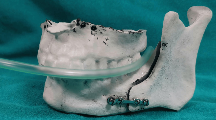 Figure 1
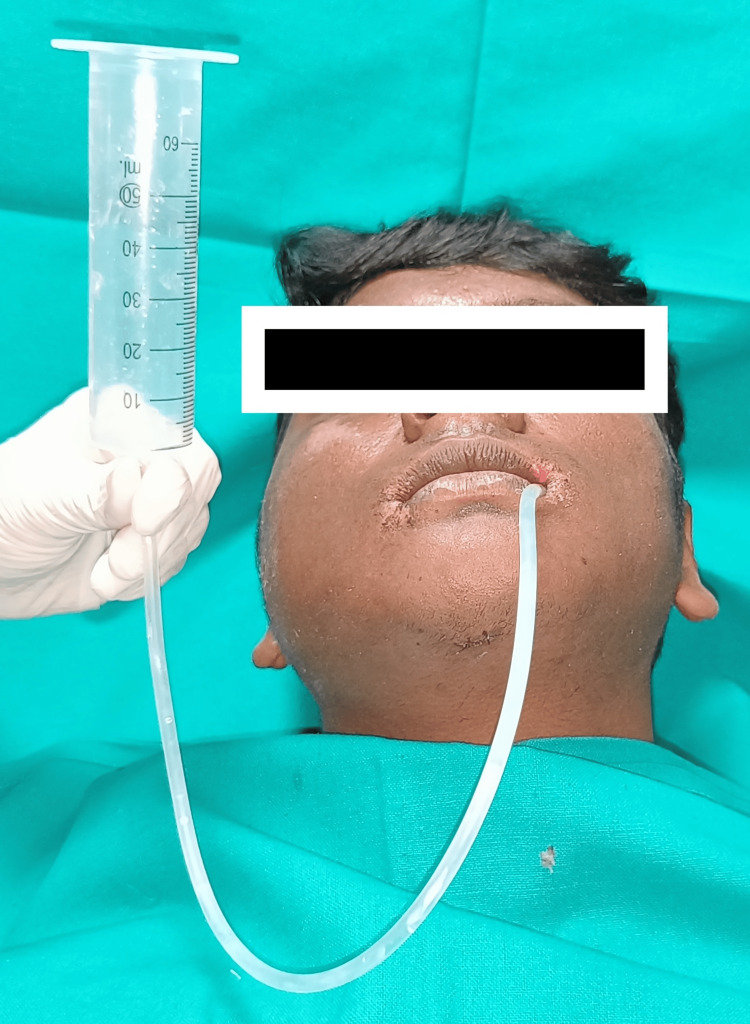 Figure 2
Peer Reviews
No public reviews on file for this paper yet. If you reviewed it on a platform where reviews are public (OpenReview, ICLR, NeurIPS, ICML), you can paste yours below so the community can read it here.
Videos
No videos yet. Explain this paper in a talk, walkthrough, or lecture? Add one.
Taxonomy
TopicsDysphagia Assessment and Management · Nutrition and Health in Aging · Clinical Nutrition and Gastroenterology
Introduction
The relationship between nutrition and wound healing post-injury or surgery has been recognized for centuries, emphasizing its critical role in patient recovery. Malnutrition or inadequate nutrient intake significantly affects outcomes, increasing complication rates and healthcare costs [1]. In the context of maxillofacial trauma management, clinicians face choices between open reduction and internal fixation (ORIF) and conservative methods such as maxillomandibular fixation (MMF). MMF achieved through techniques such as arch bars, Ernst ligatures, or bone-supported devices temporarily immobilizes the maxilla and mandible. However, this immobilization impacts nutritional intake and quality of life (QoL), especially after orthognathic surgery or when managing maxillary mandibular fractures [2]. Addressing prolonged MMF-induced malnutrition is imperative for enhancing patient well-being and optimizing recovery outcomes. Regardless of the choice between MMF with or without ORIF, the functional specificity of the oral cavity may lead to a reduction in physical strength (weight, muscle mass, grip strength, etc.). A team-based approach toward tailored nutrition is essential [3-5].
Technical report
In managing patients with MMF, maintaining oral hygiene and patient comfort can be challenging due to the constraints of a liquid diet. To mitigate these issues, we propose using a suction tube (e.g., F14) inserted through the buccal corridor into the posterior oral cavity via the retromolar space (RMS). This method facilitates the administration of semi-solid/liquid diets (millet/fruit smoothies, lentil/vegetable/chicken soups, milk/water, etc.), ensuring adequate nutritional intake during recovery (Figure 1).
Feeding tube entering via the buccal corridor into the retromolar space.
The proposed technique offers several advantages, including controlled nutrition intake, ease of execution, and maintenance of surgical site hygiene. Compared to direct consumption using spoons/straws, tube feeding provides satiation, ease of administration, and cleanliness without the drawbacks of nasogastric feeding. The rate of administration can be easily controlled by pinching the tube. Post-meal tube cleaning can be easily accomplished by flushing it with water and air drying. The armamentarium, which is economically priced, can be conveniently replaced after a few uses. The learning curve for the patients and caretakers is smooth and short. This cost-effective method has high patient acceptance rates, making it a practical solution for feeding patients with MMF (Figure 2).
Feeding tube with a 50 mL syringe used to feed the patient.
Discussion
The RMS is anatomically bounded superiorly by the maxillary tuberosity and the area posterior to it; inferiorly by the retromolar trigone region; anteriorly by the most posteriorly erupted molar teeth; posteriorly by the anterior border of the ascending ramus of the mandible; medially by the lateral surfaces of the maxillary tuberosity, the last erupted molars, and the adjacent oral cavity; and laterally by the medial surface of the ascending ramus and the buccal vestibule [5]. RMS has been successfully utilized for tracheal tube placement and airway adjunct in adult and pediatric patients undergoing maxillofacial surgeries [6,7]. Despite the potential limitation of a fully erupted maxillary third molar, our experience suggests that the small size of the suction tube negates the need for tooth extraction. In case of paucity of the RMS, the surgeon can take a call concerning third molar tooth extraction.
Significant weight changes during MMF serve as key indicators of patient nutritional status, with studies indicating clinically substantial weight loss [8]. Malnutrition during MMF may necessitate nutritional supplements, underscoring the importance of systematic nutrition support during surgical and conservative treatments [4]. Manifestations of malnutrition include weight loss, neurological changes, skin alterations, and decreased serum proteins and lipids. The resurgence of MMF as a primary treatment during the COVID-19 pandemic, aimed at avoiding aerosol generation, further highlights this need. While nasogastric tube feeding can meet dietary requirements, its prolonged use is discouraged due to associated discomforts such as nausea, vomiting, aspiration pneumonia, ulceration, and faulty tube insertion.
Alternative methods, such as wafer placement between arches, are deemed cumbersome due to the need for fabricating a thicker splint. Moreover, these methods are not required in all cases post-orthognathic surgery and are never needed in maxillofacial trauma/pathology management [9]. A study by Ishikawa et al. demonstrated decreased complications and improved quality of the perioperative period for patients, attributed to the speed of administration and consistency of diet [10]. Consuming food directly using a spoon or straw can accumulate food particles around the teeth, orthodontic appliances, and surgical wounds in areas such as the retromolar and sulcus regions, potentially compromising oral hygiene. Moreover, eating with the jaws wired together can be a challenging experience, adding further difficulties for some patients.
Conclusions
Optimal nutrition is crucial for expedited recovery, emphasizing the importance of meticulous nutritional planning in maxillofacial treatment. Even brief periods of malnutrition can significantly negatively affect wound healing. Hence, dietary deficiencies must be recognized early and repletion initiated as soon as possible. Nutrition significantly impacts feeding duration, comfort, patient satisfaction, and QoL, ultimately influencing wound healing and early discharge.
The reference list from the paper itself. Each links out to its DOI / PubMed record.
- 1Nutritional laboratory markers in malnutrition J Clin Med Keller U 775820193115924810.3390/jcm 8060775 PMC 6616535 · doi ↗ · pubmed ↗
- 2Nutritional intervention during maxillomandibular fixation of jaw fractures prevents weight loss and improves quality of life Br J Oral Maxillofac Surg Popat SP Rattan V Rai S Jolly SS Malhotra S 4784845920213358931110.1016/j.bjoms.2020.10.009 · doi ↗ · pubmed ↗
- 3Change of body composition, physical strength, and nutritional status of patients with mandibular fractures J Craniomaxillofac Surg Hino S Yamada M Iijima Y 2922974920213358933410.1016/j.jcms.2021.01.023 · doi ↗ · pubmed ↗
- 4Evaluation of changes in anthropometric indexes due to intermaxillary fixation following facial fractures J Dent Res Dent Clin Dent Prospects Yazdani J Hajizadeh S Ghavimi MA Pourghasem Gargari B Nourizadeh A Kananizadeh Y 2472501020162809695110.15171/joddd.2016.039PMC 5237672 · doi ↗ · pubmed ↗
- 5Nutritional support team intervention for patients with mandibular fracture treated by intermaxillary fixation J Trauma Treat Horie N Ohmuro M Sato M 562017
- 6An evaluation of the retromolar space for oral tracheal tube placement for maxillofacial surgery in children Anesth Analg Arora S Rattan V Bhardwaj N 1122112510320061705694310.1213/01.ane.0000247852.09732.ec · doi ↗ · pubmed ↗
- 7Does the retromolar area provide adequate space for an oral endotracheal tube without interfering with intermaxillary fixation?J Oral Maxillofac Surg Sittitavornwong S Mostofi P Di Luzio K Kukreja P Deatherage H Kukreja P 245524617920213425602110.1016/j.joms.2021.06.011 · doi ↗ · pubmed ↗
- 8What is the effect of treating mandibular fractures on weight and prealbumin?J Oral Maxillofac Surg Christensen BJ Chapple AG King BJ 1227122677201910.1016/j.joms.2019.01.05930851249 · doi ↗ · pubmed ↗