Endoscopic ultrasonography of a multiseptate gallbladder: differentiation from adenomyomatosis of the gallbladder
Koichi Soga, Fuki Hayakawa, Mayumi Yamaguchi, Takeshi Fujiwara, Ryosaku Shirahashi, Ikuhiro Kobori, Masaya Tamano

Abstract
Genes, proteins, chemicals, diseases, species, mutations and cell lines named across the full text — each resolved to its canonical identifier and authoritative record.
Click any figure to enlarge with its caption.
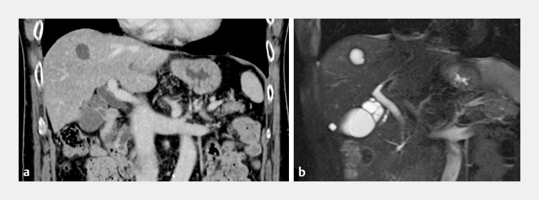 Fig. 1
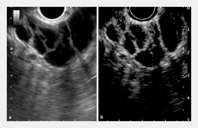 Fig. 2
Peer Reviews
No public reviews on file for this paper yet. If you reviewed it on a platform where reviews are public (OpenReview, ICLR, NeurIPS, ICML), you can paste yours below so the community can read it here.
Videos
No videos yet. Explain this paper in a talk, walkthrough, or lecture? Add one.
Taxonomy
TopicsCholangiocarcinoma and Gallbladder Cancer Studies · Gallbladder and Bile Duct Disorders
A multiseptate gallbladder is a rare gallbladder anomaly, with a recorded occurrence of around 150 documented cases globally 1 . Although multiple theories regarding the pathogenesis of multiseptate gallbladder have been proposed, its rarity precludes definitive management guidelines 2 . It is typically diagnosed using imaging, and ultrasonography is considered the most effective and practical diagnostic modality.
We present the case of a 66-year-old Japanese man who was evaluated at our hospital because of pancreatic enlargement and increased IgG4 levels. The patient had been previously diagnosed with adenomyomatosis of the gallbladder neck and had been undergoing long-term observation ( Fig. 1 ). Endoscopic ultrasonography (EUS) of the pancreas demonstrated diffusely reduced echogenicity in the caudal body, which is consistent with chronic progressive autoimmune pancreatitis. EUS of the gallbladder neck identified multiple septations, with a “honeycomb” pattern of linear internal echoes crossing the gallbladder lumen, which are characteristic of multiseptate gallbladder. The gallbladder had a normal size and thin walls; no evidence of wall thickening, pericholecystic fluid, or cholelithiasis was observed. The biliary tract was not significantly dilated. Enhanced EUS images obtained with perflubutane contrast medium were clearer than standard EUS images ( Fig. 2 ; Video 1 ). Based on the EUS findings, we concluded that the diagnosis was multiseptate gallbladder in the gallbladder neck, rather than adenomyomatosis of the gallbladder.
A multiseptate gallbladder that was found incidentally is seen on: a contrast-enhanced computed tomography (CT), showing increased contrast enhancement in the affected region, and multiple septations, with a “honeycomb” pattern, in the gallbladder neck; b magnetic resonance cholangiopancreatography (MRCP), performed to rule out other abdominal pathologies, showing septations in the gallbladder neck, which are characteristic of a multiseptate gallbladder.
Endoscopic ultrasonography (EUS) images of a multiseptate gallbladder showing: a multiple septations in the gallbladder neck that display the typical “honeycomb” pattern of linear internal echoes crossing the gallbladder lumen; the gallbladder is of normal size and has thin walls, with no evidence of wall thickening, pericholecystic fluid, or cholelithiasis, and no significant dilatation of the biliary tract; b with perflubutane contrast enhancement, a clearer image of the typical “honeycomb” pattern of linear internal echoes crossing the gallbladder lumen, consistent with the diagnosis of multiseptate gallbladder affecting the gallbladder neck.
Endoscopic ultrasonography of a multiseptate gallbladder showing its differentiation from adenomyomatosis of the gallbladder.Video 1
Because of the limited number of cases, no standardized approach for the evaluation or management of multiseptate gallbladder exists. Conservative management is often appropriate; however, surgical intervention may be performed if other biliary pathologies are present 2 .
Multiseptate gallbladder is a rare anatomical anomaly, with minimal clinical significance as it is generally benign. Symptomatic multiseptate gallbladder can be managed conservatively if associated biliary tree abnormalities are not present. In our patient, the multiseptate gallbladder was an incidental finding that did not require surgical intervention. EUS is useful for diagnosing the condition based on its characteristic findings.
Endoscopy_UCTN_Code_TTT_1AS_2AG
The reference list from the paper itself. Each links out to its DOI / PubMed record.
- 1Terkawi RS Qutob D Hendaus MA Understanding multiseptated gallbladder: a systematic analysis with a case report JGH Open 2021598899610.1002/jgh 3.1262134584965 PMC 8454487 · doi ↗ · pubmed ↗
- 2Hsieh YM Hsieh YL Wang NL Multiseptate gallbladder: a case report and literature review Medicine (Baltimore)2021100 e 2799210.1097/MD.0000000000027992 PMC 866382734889244 · doi ↗ · pubmed ↗