Endoscopic ultrasound-guided embolization: Successful management of hemorrhage from gastric artery aneurysm rupture
Beinan Hu, Guilian Cheng, Zhenyun Gong, Duanmin Hu

Abstract
Genes, proteins, chemicals, diseases, species, mutations and cell lines named across the full text — each resolved to its canonical identifier and authoritative record.
Click any figure to enlarge with its caption.
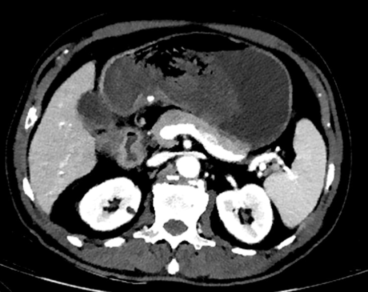 Fig. 1
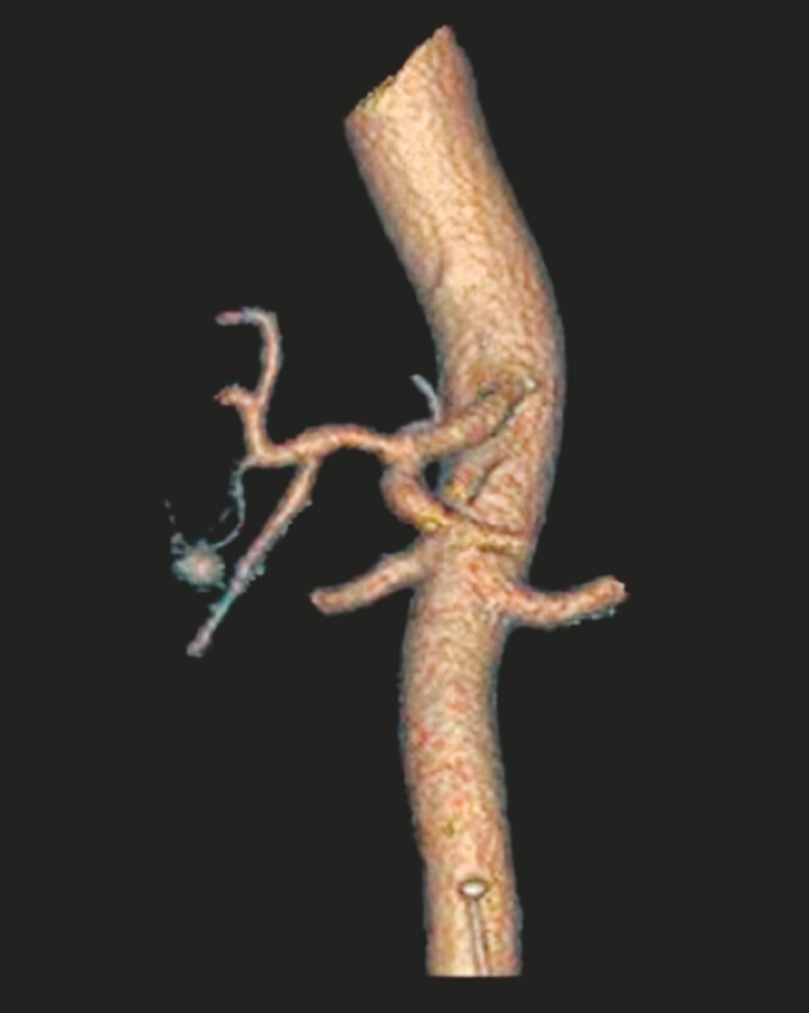 Fig. 2
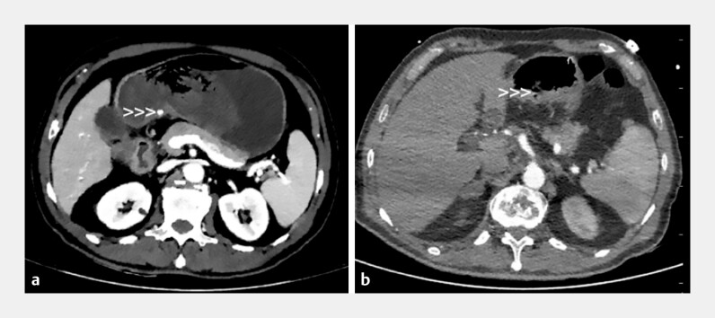 Fig. 3
- —State Key Laboratory of Radiation Medicine and Protection10.13039/501100019643
- —Suzhou Multicenter Clinical Research Project on Major Diseases
Peer Reviews
No public reviews on file for this paper yet. If you reviewed it on a platform where reviews are public (OpenReview, ICLR, NeurIPS, ICML), you can paste yours below so the community can read it here.
Videos
No videos yet. Explain this paper in a talk, walkthrough, or lecture? Add one.
Taxonomy
TopicsAbdominal vascular conditions and treatments · Esophageal and GI Pathology · Gastroesophageal reflux and treatments
Visceral artery aneurysms (VAAs), though uncommon in clinical practice, pose a critical threat due to their life-threatening rupture. We present a case of a ruptured aneurysm arising from a branch of the right gastric artery. Hemorrhage was managed with endoscopic ultrasound (EUS)-guided embolization.
A 69-year-old man was transferred to our department due to recurrent melena for 5 days. The patient had been administered meloxicam for 3 weeks to manage lumbar disc herniation and had not undergone gastroscopy before. Initial contrast-enhanced CT was performed to evaluate the potential for hemorrhage related to gastrointestinal malignancies. A hyperdense spherical lesion was identified on CT ( Fig. 1 ). Subsequent three-dimensional reconstruction demonstrated a saccular structure ( Fig. 2 ), confirming the diagnosis of an aneurysm. Then, the patient received an endoscopy for hemostasis and further examination. Gastroscopy was compromised by active hemorrhage, and EUS and Doppler demonstrated an aneurysm with active bleeding and exhibiting direct continuity with extramural vessels. Spectral Doppler analysis revealed arterial waveforms. Based on the correlative findings from CT and Doppler waveform analysis, considering the aneurysm was arising from a branch of the right gastric artery along the lesser curvature. 1 ml n-Butyl-2-Cyanoacrylate was injected into the aneurysm lumen under EUS guidance, and Doppler confirmed blood flow arrest ( Video 1 ), achieving primary hemostasis. CT performed 3 days after treatment revealed no symptoms of residual perfusion ( Fig. 3 ). No rebleeding episodes or procedure-related complications were observed during the 1-month follow-up.
CT image of the hyperdense spherical lesion located in the lesser curvature.
Three-dimensional reconstruction showed a saccular structure.
CT images before and 3 days after treatment: a CT image before treatment. b CT image 3 days after treatment.
Procedure of the embolization under the guidance of EUS.Video 1
VAAs are rare with incidence rates ranging from 0.01 to 0.2% 1 and 25% of VAAs can present with life-threatening rupture 2 . Current management of ruptured VAAs faces technical limitations in superselective embolization and elevated perioperative risks associated with emergency laparotomy. EUS-guided embolization achieved satisfactory results, circumvented parent vessel occlusion risks and invasive surgery. Our experience suggests EUS-guided embolization may serve as an effective alternative for digestive tract-adjacent aneurysms. Further multicenter studies are warranted to validate its safety profile.
Endoscopy_UCTN_Code_TTT_1AO_2AD
The reference list from the paper itself. Each links out to its DOI / PubMed record.
- 1Al Baghdadi M Yedla P Visceral artery aneurysm: an unusual cause of abdominal pain BMJ Case Rep 202013 e 23801910.1136/bcr-2020-238019 PMC 775462133370948 · doi ↗ · pubmed ↗
- 2Barrionuevo P Malas MB Nejim BA systematic review and meta-analysis of the management of visceral artery aneurysms J Vasc Surg 20207240 S 45S 10.1016/j.jvs.2019.02.02432553135 · doi ↗ · pubmed ↗