Letter to the Editor re: “Systematic review and meta-analysis comparing Manta device and Perclose device for closure of large bore arterial access.” J Vasc Access. 2024 Jan 8:11297298231222314
Angela Di Giorgio, Claudia Carnuccio, Antonio Nesci, Alessia D’Alessandro, Angelo Santoliquido

Abstract
Genes, proteins, chemicals, diseases, species, mutations and cell lines named across the full text — each resolved to its canonical identifier and authoritative record.
Click any figure to enlarge with its caption.
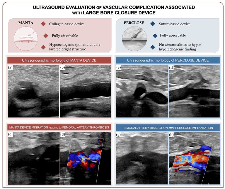 Figure 1
 Figure 2
Peer Reviews
No public reviews on file for this paper yet. If you reviewed it on a platform where reviews are public (OpenReview, ICLR, NeurIPS, ICML), you can paste yours below so the community can read it here.
Videos
No videos yet. Explain this paper in a talk, walkthrough, or lecture? Add one.
Taxonomy
TopicsVascular Procedures and Complications · Central Venous Catheters and Hemodialysis · Ultrasound in Clinical Applications
Dear Editor,
We read with great enthusiasm the article of Tayyab Cheema et al. that compared Manta device and Perclose device for closure of large bore arterial access.^ 1 ^
Vascular closure devices (VCDs) have gained consideration due to the increasing use of large bore arterial accesses and have been shown to be non-inferior to manual compression in achieving successful hemostasis. Actually, there is no consensus on the recommended device of choice^ 2 ^ and, to date, there were no studies comparing their effectiveness, except for the distinguished work of Tayyab Cheema et al. Authors reviewed and performed a meta-analysis focusing on the safety and effectiveness profiles of MANTA and Perclose devices, not revealing significant differences.
Briefly, Perclose ProGlide (Abbott Vascular, Santa Clara, CA) achieves hemostasis with a single 3-0 polypropylene monofilament suture at the vascular access site with a 92% success rate and a 4.4% complication rate.^ 3 ^ MANTA (Teleflex, Wayne, PA) consists of an extravascular hemostatic collagen structure held in place by an endoluminal molded polymer toggle, stainless steel element, and suture that achieves high technical efficacy with a low risk of vascular complications.^ 4 ^
We agree in saying that, bleeding, deployment failure, and device migration are the most common complications associated with VCDs. Color-Doppler ultrasound guides intraoperative VCD deployment and allows early detection of intra- and postoperative adverse events; its use is advocated at each step of the procedure to promptly identify VCD-related complications. In this regard, we need to emphasize that vascular ultrasound is a valuable tool with the advantages of being fast, non-invasive, and readily available, although it requires an experienced operator. In fact, recent meta-analytic data derived from four randomized clinical trials brightly reported that ultrasound guidance for transfemoral access might be an effective and simple choice in order to reduce major vascular complication and major bleeding.^ 5 ^ Vascular ultrasound identifies VCDs immediately after placement and helps in diagnosing complications in the peri-operative follow-up.
Moreover, the normal ultrasonographic appearance of VCDs could resemble the presentation of their complications, hence the need for an evaluation by an experienced operator and our willingness to focus on their uncomplicated appearance through our images (Figure 1). We suggest integrating ECD assessment into procedural planning and performing short-term post-procedural evaluations to promptly identify VCD-related complications. Long-term vascular ultrasound studies are essential to assess the impact of these devices on the arterial wall over time, including potential remodeling and stenosis. It is also critical to evaluate the role of VCDs as potential points of least resistance for future endovascular procedures on the same artery.
The reference list from the paper itself. Each links out to its DOI / PubMed record.
- 1Cheema T Venero C Jr Champaneria S , et al. Systematic review and meta-analysis comparing Manta device and Perclose device for closure of large bore arterial access. J Vasc Access. Epub ahead of print 8 January 2024. DOI: 10.1177/11297298231222314.38189215 · doi ↗ · pubmed ↗
- 2Schulz-Schüpke S Helde S Gewalt S , et al Instrumental Sealing of Arterial Puncture Site- CLOSURE Device vs Manual Compression (ISAR-CLOSURE) Trial Investigators. Comparison of vascular closure devices vs manual compression after femoral artery puncture: the ISAR-CLOSURE randomized clinical trial. JAMA 2014; 312(19): 1981–1987.25399273 10.1001/jama.2014.15305 · doi ↗ · pubmed ↗
- 3Malkawi AH Hinchliffe RJ Holt PJ , et al. Percutaneous access for endovascular aneurysm repair: a systematic review. Eur J Vasc Endovasc Surg 2010; 39(6): 676–682.20185341 10.1016/j.ejvs.2010.02.001 · doi ↗ · pubmed ↗
- 4Van Mieghem NM Latib A van der Heyden J , et al. Percutaneous plug-based arteriotomy closure device for large-bore access: a multicenter prospective study. JACC Cardiovasc Interv 2017; 10(6): 613–619.28335899 10.1016/j.jcin.2016.12.277 · doi ↗ · pubmed ↗
- 5d’Entremont MA Alrashidi S Seto AH , et al. Ultrasound guidance for transfemoral access in coronary procedures: an individual participant-level data meta-analysis from the femoral ultrasound trialist collaboration. Euro Intervention 2024; 20(1): 66–74.37800723 10.4244/EIJ-D-22-00809 PMC 10758987 · doi ↗ · pubmed ↗