The Impact of EPAC2-Associated Junction Plakoglobin on Respiratory Syncytial Virus Infection
Chaitra A. Takle, Eun-Jin Choi, Eun Seok Choi, Devang Deepak, Kashish Khatkar, Jong Min Choi, Ke Zhang, Sung Yun Jung, Tian Wang, Wenzhe Wu, Xiaoyong Bao

TL;DR
This study explores how EPAC2 and junction plakoglobin interact during RSV infection, revealing a potential role in virus replication and immune response.
Contribution
The novel contribution is identifying junction plakoglobin as an EPAC2-interacting protein critical for RSV replication and immune response.
Findings
Junction plakoglobin interacts with EPAC2 and this interaction increases during RSV infection.
Reducing junction plakoglobin decreases RSV particle production by impairing viral budding and gene transcription.
Junction plakoglobin is essential for an effective immune response to RSV.
Abstract
Respiratory syncytial virus (RSV) is a leading cause of lower respiratory tract infections in infants, young children, and immunocompromised individuals. Currently, FDA-approved monoclonal antibody therapies are limited to infants and young children with severe RSV disease. As a result, there is an urgent need for comprehensive studies of RSV pathogenesis to support the development of new therapeutic strategies. Exchange proteins directly activated by cAMP (EPAC) have recently emerged as key regulators in various viral infections. Our previous work identified EPAC isoform 2 (EPAC2) as a critical factor in RSV replication and host innate immune responses. However, the molecular mechanisms underlying EPAC2’s role in RSV infection remain unclear. In this study, we investigated EPAC2-mediated RSV infection by identifying EPAC2-interacting proteins. Proteomics and immunoprecipitation…
Genes, proteins, chemicals, diseases, species, mutations and cell lines named across the full text — each resolved to its canonical identifier and authoritative record.
Click any figure to enlarge with its caption.
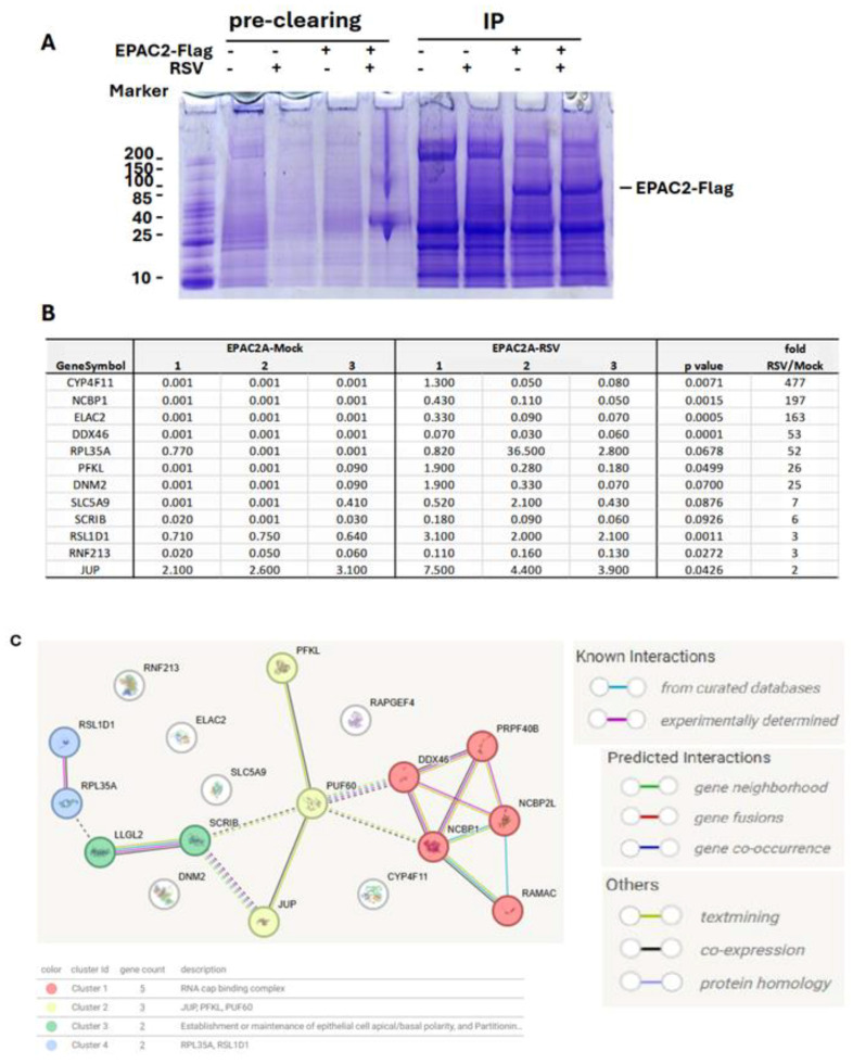 Figure 1
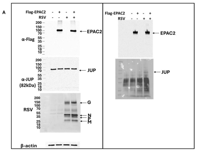 Figure 2
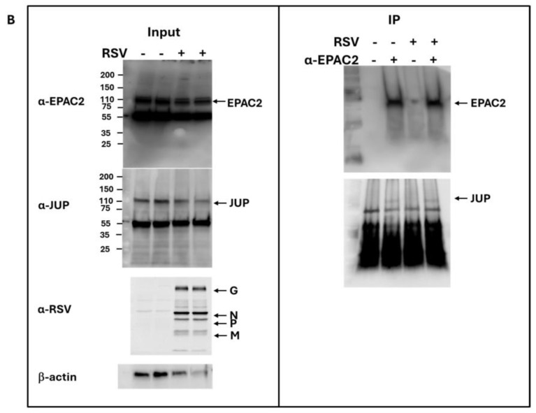 Figure 3
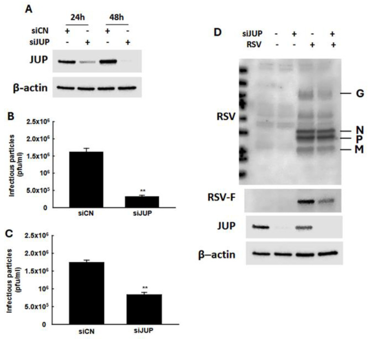 Figure 4
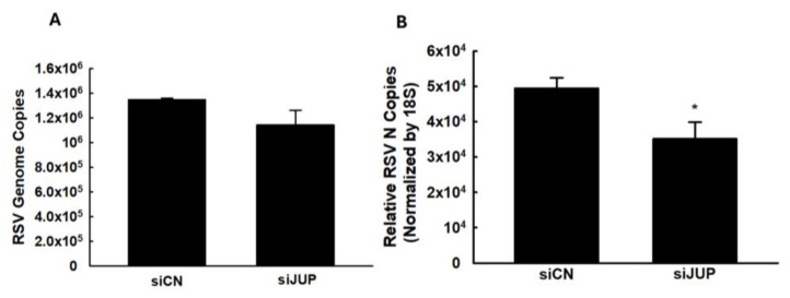 Figure 5
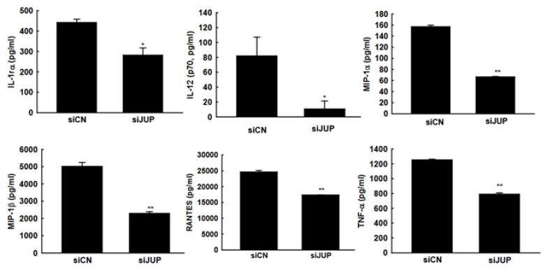 Figure 6
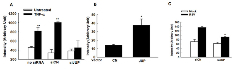 Figure 7
- —National Institutes of Health (NIH)
- —the American Lung Association
Peer Reviews
No public reviews on file for this paper yet. If you reviewed it on a platform where reviews are public (OpenReview, ICLR, NeurIPS, ICML), you can paste yours below so the community can read it here.
Videos
No videos yet. Explain this paper in a talk, walkthrough, or lecture? Add one.
Taxonomy
TopicsRespiratory viral infections research · Neonatal Respiratory Health Research · Hemoglobin structure and function
1. Introduction
Respiratory syncytial virus (RSV) is a negative-strand RNA virus and the most common cause of lower respiratory tract infection (LRTI) in young children and infants [1]. Nearly 90% of infants are infected with RSV within the first two years of life, with the majority of hospitalizations occurring in otherwise healthy infants [1]. While vaccine development has shown promise, current efforts are primarily limited to protecting elderly individuals and pregnant women [2,3]. Nirsevimab remains the only available treatment, and its use is restricted to infants and select young children at increased risk for severe RSV disease [4]. Despite these advances, RSV continues to pose a significant global health challenge due to its considerable economic and clinical burden. This highlights the need for a comprehensive investigation into RSV disease mechanisms to identify therapeutic targets and develop effective strategies to reduce the severity of RSV infection.
Exchange proteins directly activated by cAMP (EPAC) are relatively novel guanine exchange factors (GEFs) that regulate a variety of biological processes [5]. EPAC proteins transduce signals primarily by activating small GTPases, predominantly Rap1 and Rap2, which influence diverse downstream targets [5]. Initially recognized for their roles in cancer, neurological disorders, diabetes, and inflammation [6,7,8,9,10,11,12], EPACs have more recently emerged as key players in viral infections, with essential roles identified in at least seven human viruses [13,14,15,16,17,18]. Two main isoforms of EPAC exist in eukaryotic cells: EPAC1 and EPAC2. EPAC1 has been shown to favor replication of viruses such as Ebola, Middle East respiratory syndrome coronavirus (MERS-CoV), SARS-CoV-2, and influenza [14,15,18]. Our previous studies identified EPAC2 as a promising therapeutic target for RSV infection; knockdown or knockout of EPAC2, as well as treatment with an EPAC2-specific inhibitor, significantly suppressed RSV replication and reduced the expression of pro-inflammatory cytokines and chemokines [13,16]. The critical role of EPAC2 in RSV infection was further validated in mouse models [17]. Beyond RSV, EPAC2 has also been implicated in pulmonary inflammation and tissue remodeling in response to cigarette smoke exposure, highlighting its importance in lung health [19]. However, the downstream targets of EPAC2 in the pulmonary setting remain largely unknown. Identifying these targets could provide valuable insights into RSV pathogenesis and inform the development of novel therapeutic strategies.
Recently, we conducted a proteomics study to identify proteins that interact with EPAC2 and discovered that junction plakoglobin (JUP) was among its associated partners. JUP, also known as γ-catenin, is a homolog to β-catenin and plays a key role in desmosome formation and cell-cell adhesion [20,21]. However, the functions of JUP in viral infection remain largely unexplored. Among the EPAC2-binding proteins identified, JUP exhibited the least enhancement in its interaction with EPAC2 by RSV. We chose to focus on the JUP-EPAC2 interaction based on the rationale that if this modest interaction could be experimentally validated, other EPAC2-associated proteins showing stronger binding might be even more biologically significant. JUP was also of particular interest due to its established functions. Silencing JUP has been shown to disrupt the structure of actin in the cell cytoskeleton^21^, and the cytoskeleton is known to play essential roles in multiple stages of viral infection, including the transport and assembly of viral proteins and particles, viral immune evasion, and cell-to-cell fusion [22,23,24,25]. Although JUP’s role in viral infection has not been defined, its influence on actin dynamics suggests it could impact RSV assembly and budding. Indeed, previous studies have shown that the disruption of actin inhibits RSV release [26], suggesting a potential link between JUP and viral egress. These findings suggest that EPAC2 may regulate RSV assembly and budding, at least in part, through its interaction with JUP.
In this study, we demonstrated that silencing JUP through small-interfering RNA (siRNA) transfection in A549 cells led to a significant decrease in infectious particle production, viral gene transcription, and immune cytokine/chemokine secretion as compared to control samples transfected with non-targeting control siRNA (siCN). In addition, JUP controls replication-independent cellular inflammatory responses. Together, these results indicate that JUP may play a role in EPAC-2-mediated pathways that influence RSV infection.
2. Materials and Methods
2.1. Cell Lines and Virus Preparation
Human alveolar type II-like epithelial A549 cell line and human epithelial type II cell line Hep 2 were purchased from ATCC (Manassas, VA, USA). A549 cells, a common respiratory virus infection model, were cultured and maintained in F12K media with 10% FBS (vol/vol), 100 IU/mL penicillin, and 100 µg/mL streptomycin, as previously described [13,27]. These cell lines were maintained in incubators at 37 degrees Celsius and 5% CO_2_. Viral titer was determined by immunostaining in Hep-2 cells using polyclonal biotin-conjugated goat anti-RSV antibody (7950-0104; Bio-Rad, Hercules, CA, USA) and streptavidin peroxidase polymer (Sigma, St Louis, MO, USA) sequentially, as described [28,29].
2.2. EPAC2-Associated Proteins Preparation
A549 cells in 50% confluence were transfected with Flag-tagged EPAC2 plasmid (a gift from Dr. Susumu Seino, Kobe University Graduate School of Medicine, Japan) using FuGENE 6^®^ Reagent (Promega, Madison, Wi, USA), according to the manufacturer’s protocol. Empty vectors were used as controls. After 30 h post-transfection, the cells were mock-infected or infected with RSV at an MOI of 1 for 15 h. The cells were then lysed by buffer 1 from the Immunoprecipitation Kit (# 11719386001, Roche, Indianapolis, IN, USA), followed by nonspecific cleaning using Protein G-agarose. The proteins in the lysis buffer 1 were then mixed with the monoclonal anti-Flag M2 antibody (F1804, Sigma, Saint Louis, MO, USA) or control and incubated at 4 °C for 4 h on a rocking platform, followed by Protein G resin overnight at 4 °C. The pellet beads by gravity sedimentation were then sequentially washed by Immunoprecipitation Washing buffers 2 and 3, according to the manufacturer’s protocol. An aliquot of the complex was then loaded into an SDS-PAGE gel to validate Flag-tagged EPAC2 overexpression and immunoprecipitation.
The remaining protein complex was analyzed via mass spectrometry to Identify EPAC2-associated proteins. The beads were treated with sample loading buffer and subjected to SDS-PAGE using a 10% Bis-Tris gel. Protein bands were visualized using Coomassie Brilliant Blue staining, and the gel was segmented into four molecular weight fractions. Each gel fragment underwent in-gel digestion with 100 ng of trypsin in 20 µL of 50 mM NH_4_HCO_3_ at 37 °C overnight. Peptides were subsequently extracted with 100% acetonitrile, vacuum-dried, and reconstituted in 10 µL of 5% methanol containing 0.1% formic acid. The samples were then analyzed via nanoHPLC-MS/MS using an EASY-nLC1000 (Thermo Fisher Scientific, Waltham, MA, USA) coupled to an Orbitrap Fusion mass spectrometer (Thermo Scientific) with an electrospray ionization (ESI) source. A custom-built trap column packed with 3 µm Reprosil-Pur Basic C18 beads and a 5 cm × 150 µm capillary column packed with 1.9 µm Reprosil-Pur Basic C18 beads were employed for separation. The instrument, operated under Xcalibur software version 2.2 (Thermo Fisher Scientific), was set to data-dependent acquisition mode. MS/MS spectra were searched against a target-decoy Human RefSeq database (release 2015_06, containing 73,637 entries) using the Proteome Discoverer 1.4 interface (Thermo Fisher) with Mascot algorithm (Mascot 2.4, Matrix Science, Mount Prospect, IL, USA). The precursor ion mass tolerance was set to 20 ppm, with a fragment ion mass tolerance of 0.02 Da, allowing up to two missed cleavages. Peptide identifications were validated using Percolator with a false discovery rate (FDR) threshold of 1% based on q-values. Protein abundance was determined using iBAQ calculations derived from previously published methodologies [30]. Proteins that were not detected and lacked iBAQ values were imputed with half of the minimum detected value to facilitate p-value and fold-change calculations.
2.3. Western Blot
Western blot analysis was performed to validate EPAC-2-associated proteins, identified by the proteomics studies. In brief, Flag-tagged EPAC2 that had undergone immunoprecipitation using an anti-Flag antibody was loaded into an SDS-PAGE gel. The anti-JUP antibody (A303-718A-T, Bethyl, Montgomery, TX, USA) was used to detect JUP in the pull-down complex. The membranes were stripped and reprobed by an anti-Flag antibody to validate the IP. The immunoprecipitation input was also checked by loading the total cell lysates to SDS-PAGE, followed by checking the expression of EPAC2-Flag, JUP, and β-actin.
The endogenous interaction between EPAC2 and JUP was also examined. Briefly, A549 cells were either mock-infected or infected with RSV at a multiplicity of infection (MOI) of 1 for 15 h. Immunoprecipitation was performed as described above, using an anti-EPAC2 antibody (Cat# 19103-1-AP, Proteintech, Rosemont, IL, USA) for pulldown, followed by detection of JUP in the EPAC2 complex using an anti-JUP antibody (Cat# 13-8500, Thermo Fisher Scientific).
2.4. siRNA-Mediated Gene Silencing
A549 cells at 80-90% confluence were transfected with 100 nM siRNA specific for JUP (Table 1, Sigma) using Lipofectamine™ 2000 Transfection Reagent (Thermo Scientific) for 24 h or 48 h. Scrambled siRNAs were used as controls (Cat# SIC001, Sigma). The suppression efficiency of siRNAs was then confirmed by Western blot.
2.5. Cytokine/Chemokine Concentration Measurement
To quantify immune and inflammatory mediators in mock or RSV-infected samples, pellets were removed by centrifugation at 1000 rpm for 5 min. A 50 µL aliquot of the resulting supernatants was analyzed using the Bio-Plex multiplex system (Bio-Plex Pro Human Cytokine 27-plex Assay, Cat #M500KCAF0Y, Bio-Rad) to measure 27 kinds of cytokines and chemokines, including FGF-β, IL-2, IL-10, MIP-1α, Eotaxin, IL-4, IL-12, MIP-1β, G-CSF, IL-5, IL-13, PDGF-bb, GM-CSF, IL-6, IL-15, RANTES, IFN-γ, IL-7, IL-17A, TNF-α, IL-1β, IL-8, IP-10, VEGF, IL-1ra, IL-9, and MCP-1.
2.6. Virus Titration Assay
RSV harvested from A549 cell lysates or culture supernatants was diluted in a 5-fold serial dilution, followed by seeding 150 µL of it to Hep-2 cells, which were grown to confluence in 24-well plates (1.5 × 10^5^ cells/well). Plates were then put in a shaker for 1 h at 37 °C, 5% CO_2,_ and 50 rpm. After 1 h, 1 mL of MEM with 2% serum and 0.75% methylcellulose was added to each well, and the plates were kept in the incubator for 5 days (37 degrees Celsius, 5% CO_2_). After 5 days of incubation, the culturing gel solution was removed from the wells, and 0.5 mL of 10% formaldehyde was added to each well to fix the cells. After 30 min of incubation at room temperature, formaldehyde was removed from the wells, and 1 ml of 1% crystal violet (in EtOH) was added. After 30 min of incubation, the dye was removed, followed by plate washing and plaque counting to calculate the viral titers.
2.7. Quantitative Real-Time PCR (qRT-PCR)
To quantify RSV N gene expression, total cellular RNA was extracted by TRIzol reagents (Thermo Fisher Scientific). CDNA was synthesized with 1 μg of total RNA in a 20-μL reaction mixture using the TaqMan Reverse Transcription Reagents kit from ABI (catalog number N8080234; Applied Biosystems, Foster City, CA, USA). We used RT primer 5′-CTGCGATGAGTGGCAGGCTTTTTTTTTTTTAACTCAAAGCTC-3′. We incorporated a “tag” (underlined letters) as part of the assay due to self-priming exhibited by viral RNA. The tag sequence was derived from the bacterial chloramphenicol resistance (Cm^r^) gene. The sequence with bold letters is complementary to the poly(A) tails of the transcribed RSV N gene. The sequence in italics is N gene-specific. The reaction conditions were as follows: 25 °C for 10 min, 48 °C for 30 min, and 95 °C for 5 min. At a 25 °C annealing temperature, the 8 nucleotides (nt) matching N-specific sequences would not be sufficient for stable efficient priming of cDNA from the antigenome of hMPV. On the other hand, 20 nucleotides matching transcribed N (12 T’s and N gene-specific nucleotides) can attain stable annealing to the transcribed N gene. For Quantitative real-time PCR amplification, we used the RSV tag reverse primer CTGCGATGAGTGGCAGGC and the forward primer ACTACAGTGTATTAGACTTRACAGCAGAAG. The PCR was performed with 1 μL of cDNA in a total volume of 25 μL by using iTaq ^TM^ Universal SYBR Green Supermix (Cat# 1725124, Bio-rad, Hercules, CA, USA). The final concentration of the primers was 300 nM. 18S RNA was used as a housekeeping gene for normalization. PCR assays were run with the ABI Prism 7500 sequence detection system with the following conditions: 50 °C for 2 min, 95 °C for 10 min, and then 95 °C for 15 s and 60 °C for 1 min for 40 cycles. Duplicate cycle threshold (C_T_) values were analyzed in Microsoft Excel by the comparative C_T_ (ΔΔC_T_) method according to the manufacturer’s instructions (Applied Biosystems). The amount of target (2^−ΔΔ^^CT^) was obtained by normalization to the endogenous reference (18S) sample.
To quantify RSV antigenomic copies, synthetic transcripts of the genome were generated from the Topo plasmid containing N-P-M genes, using the T7 MegaScript kit, following the digestion with PmeI. The reaction mixture was then treated with Turbo Dnase and purified using the MegaScript kit. Primers were designed to span the N and P regions of the viral genome and incorporated a Cm^r^ tag. First-strand cDNA was transcribed with a P-specific primer, 5′-CTGCGATGAGTGGCAGGCACTACAGTGTATTAGACTTRACAGCAGAAG-3′. For PCR assays, we used RSV tag primer CTGCGATGAGTGGCAGGC and primer RSV P GCATCTTCTCCATGRAATTCAGG.
2.8. Reporter Gene Assays
To investigate the role of JUP in inflammatory responses, A549 cells in a 24-well plate were transfected in triplicate with 0.05 µg/well luciferase reporter gene plasmids containing multiple copies of NF-κB binding sites (NF-κB-Luc) and 100 nM siRNAs, either scrambled or JUP-specific, using lipofectamine 2000 according to the manufacturer’s protocol. At 24 h post-transfection, the cells were treated with TNF-α at a 20 ng/mL concentration per well. Following 24 h of treatment, the cells were lysed to measure luciferase reporter activity, using a SpectraMax iD3 microplate reader (Molecular Devices, San Jose, CA, USA). Cells without treatment were used as controls.
We also investigated the function of JUP in mediating RSV-induced cellular responses. In brief, A549 cells in 24-well plates were transfected with 0.5 µg/well luciferase reporter plasmids containing multiple copies of IRF-3 binding sites of IFN-β (IRF3-Luc) and 100 nM siRNA, scrambled or JUP-specific, using lipofectamine 2000. A4 24 h post-transfection, the cells were mock-infected or infected with RSV at an MOI of 1 for 15 h, followed by cell lysis and luciferase activity measurement.
2.9. Statistical Analysis
The experimental results were analyzed using GraphPad Prism 5 software. An unpaired two-tailed t-test was employed to compare the difference. A p-value < 0.05 was considered to indicate a statistically significant difference. Single and two asterisks represent p-values of <0.05 and <0.01, respectively. Means ± standard errors (Ses) are shown.
3. Results
3.1. EPAC2-Associated Proteins
The role of EPAC in viral infections is an emerging area of study. In infections caused by MERS-CoV, SARS-CoV-2, or influenza, EPAC1 has been shown to promote viral replication [14,15,18]. Additionally, EPAC1 facilitates Ebola virus uptake into vascular endothelial cells via micropinocytosis. Whether EPAC2 contributes to viral infections remained unclear until our laboratory discovered its significant role in regulating both proinflammatory responses and RSV replication [13]. Most recently, in vivo experiments using RSV-infected mouse models further confirmed the critical involvement of EPAC2 in prompting RSV replication and RSV-induced pulmonary inflammation [17]. We also recently reported a similar role of EPAC2 in hMPV infection [16]. Despite these findings, the molecular mechanisms underlying EPAC2-mediated RSV infection remain largely unknown. To address this, we first employed a proteomics method to identify EPAC2-associated proteins. As shown in Figure 1A, EPAC2-Flag was successfully expressed and enriched using anti-Flag antibodies. Proteomic analysis identified eleven EPAC2-associated proteins; among them, eight exhibited significantly enhanced interaction with EPAC2 following RSV infection, while three showed reduced interaction (Figure 1B). Notably, all listed proteins displayed no detectable binding in cells lacking EPAC2-flag expression. Further STRING network analysis revealed four natural clusters, identified using the MCL clustering method (which detects clusters based on stochastic flow) (Figure 1C).
3.2. JUP-EPAC2 Interaction
As shown in Figure 1B, JUP exhibited the least enhancement in its binding to EPAC2 following RSV infection compared to other EPAC2-associated proteins. Based on this observation, we chose to begin our experimental validation by investigating the JUP-EPAC2 interaction, reasoning that if this modest interaction could be confirmed, the stronger interactions observed for other proteins were likely to be biologically meaningful as well. An additional rationale for selecting JUP as our initial target was its known functional significance. Silencing JUP has been shown to disrupt actin organization within the cytoskeleton [21], and the cytoskeleton plays a critical role in multiple stages of viral infection, including the transport or assembly of viral proteins and particles, immune evasion, and cell-to-cell fusion [22,23,24,25]. As shown in Figure 2A. JUP was detected in the pull-down product using an anti-Flag antibody against Flag-tagged EPAC2, confirming the association of EPAC2 and JUP. This interaction was observed even in the absence of RSV infection (bottom-right panel, second column) and was increased by RSV infection (bottom-right panel, last column).
To further validate this interaction under endogenous conditions, we performed immunoprecipitation using an anti-EPAC2 antibody to pull down native EPAC2 and assess its association with JUP. As shown in Figure 2B, the EPAC2-JUP interaction was confirmed in both mock- and RSV-infected cells.
3.3. The Roles of JUP in RSV Infection
In our previous studies, we demonstrated that EPAC2 facilitates both RSV replication and host inflammatory responses [13,16]. To investigate whether EPAC2-associated JUP contributes to RSV infection, we used JUP-specific siRNA to suppress its expression, followed by an assessment of the impact of JUP silencing on RSV replication. As shown in Figure 3A, treatment with JUP-specific siRNA at a concentration of 100 nM effectively reduced JUP expression, both at 24 h and 48 h post-transfection. We also found that the total number of infectious RSV particles produced by JUP-deficient cells was significantly lower than that of cells treated with scrambled control siRNA (siCN) (Figure 3B). Similarly, the number of infectious particles released into the supernatant was reduced in JUP-siRNA-treated cells compared to siCN-treated cells (Figure 3C). Notably, the reduction in infectious particles released into the supernatant (about 80%) was greater than the reduction observed in the total sample (about 50%), following JUP knockdown. Consistent with these findings, Western blot analysis using anti-RSV antibody confirmed that there was reduced viral protein expression in siJUP-treated cells compared to siCN-treated cells (Figure 3D).
We also investigated whether JUP regulates RSV genome replication and viral gene expression. As shown in Figure 4, silencing JUP did not appear to affect viral genome replication; however, it significantly suppressed the expression of the N gene.
3.4. The Impact of JUP on RSV-Induced Cytokines and Chemokines
In response to RSV infection, infected cells typically activate cellular responses by producing cytokines and chemokines. Our previous studies demonstrated that EPAC2 promotes inflammation. To evaluate whether JUP suppression has a broader impact on RSV-induced secretion of proinflammatory and immunoregulatory molecules, we analyzed the secretion patterns of chemokines and cytokines in A549 cells with or without JUP silencing (Figure 5). Notably, JUP deficiency resulted in significantly lower levels of IL-1rα, IL-12 (p70), MIP-1α, MIP-1β, RANTES, and TNF-α at 15 h post-infection, compared to siCN-treated cells, supporting the importance of JUP in RSV-induced cellular responses. Unlike EPAC2, JUP did not affect the RSV-induced IP-10 and MCP-1 in A549 cells.
3.5. Modulation of Cellular Signaling by JUP
The suppressed cellular responses could be an indirect outcome of the impaired RSV replication by JUP silencing. To investigate whether the JUP knockdown also leads to changes in inflammatory responses independent of viral replication, we used TNF-α to activate NF-κB, a transcription factor that plays an essential role in inducing inflammatory immune mediators in responses to viral infection, and investigated whether silencing JUP impacts NF-κB activation. As shown in Figure 6A, JUP knockdown significantly impaired TNF-α-induced NF-κB activation, in alignment with our previous finding on EPAC2-mediated NF-κB activation and current discovery on EPAC2-JUP interaction.
Many immune mediators also have IRF-3 binding site(s) in addition to the NF-κB binding site. Therefore, we also investigated whether JUP affects cytokine/chemokine expression by regulating IRF-3 activation. As shown in Figure 6B, JUP overexpression led to enhanced IRF-3-mediated luciferase expression. In addition, JUP knockdown significantly suppressed luciferase expression controlled by IFN-β transcription binding sites for NF-κB and IRF-3 (Figure 6C).
4. Discussion
EPAC, a major cellular receptor for cAMP in addition to protein kinase A (PKA), plays major roles in cardiac diseases, cancer, neuronal differentiation, and respiratory inflammation [31,32,33,34]. In non-infectious disease models, EPAC has been reported to promote airway inflammation through the EPAC-activated mitogen-activated protein kinase kinase (MAPKK) pathway [35,36,37,38] or via PLCε [19]. Recently, the significance of EPAC in viral infections was also reported. In MERS-CoV, SARS-CoV-2, or influenza infection, EPAC1 seems to promote viral replication [14,15,18]. Unlike these viruses, our laboratory discovered that it is EPAC2, but not EPAC1, which dominantly affects proinflammatory cellular/pulmonary responses to RSV, human metapneumovirus, and adenovirus [13,17], suggesting that the EPAC isoform evolved in viral infections is pathogen-dependent. However, the molecular mechanisms underlying EPAC-mediated viral infection are not well known, although such information is essential for developing novel therapeutic interventions against viral infections.
By comparing our previous study on EPAC2-mediated and the current study on JUP-mediated cellular responses to RSV infection, we found that EPAC2 deficiency impacts a broader range of cytokines and chemokines than JUP deficiency, supporting the notion that JUP functions as one of the downstream signaling molecules of EPAC2. Unlike EPAC2, JUP did not affect the RSV-induced IP-10 and MCP-1 in A549 cells.
The biological functions of JUP were mostly investigated in the cardiac muscle. In cardiac muscle, JUP is a cytoplasmic component of desmosomes and adherens junctions and is, therefore, essential for its stability [39,40,41]. In cancer, JUP is involved in mediating the cell cytoskeleton actin [20,21]. The role of JUP in viral infection is not well known. We found that JUP deficiency led to a 50% reduction in total infectious particles and an 80% decrease in infectious particles in the supernatant, suggesting that JUP is critical for releasing infectious particles from cells. It has been previously reported that actin controls RSV budding [26]. The importance of actin in mediating virus budding out of cells is also reported for nonlytic rotavirus and SARS-CoV-2 [42,43]. Given the association between JUP and actin, it is not surprising to observe the impact of JUP on viral budding. Combined with our previous finding on viral entry independent of EPAC2 [13], this finding may represent a mechanism by which JUP promotes viral replication via controlling virus budding.
The induction of many cytokines/chemokines is replication-dependent [44,45]. In addition to JUP-enhanced infectious particle production, we also discovered a slight but significant impact of JUP on N gene transcription, possibly leading to increased immune responses to RSV infection. However, we found that JUP could also control cellular responses in a replication-independent manner (Figure 6A, B), similar to our report demonstrating that EPAC2 can control inflammation independent of RSV replication [13] and the report from others showing EPAC2-mediated non-viral respiratory diseases [19].
Based on EAPC2-associated proteins identified by immunoprecipitation/proteomics studies and Cluster analysis by STRING [46], we found that JUP potentially binds to RNA-binding proteins: the nuclear cap-binding protein subunit 1 (NCBP1) and DDX46 (Figure 1C). NCBP1 has been reported to play a crucial role in viral infections by interacting with viral mRNA and influencing antiviral responses [47,48]. DDX46, or DEAD-Box Helicase 46, is a protein that negatively regulates the innate antiviral response to viral infection [49]. Several proteins were not clustered into any groups, including CYP4F11, a cytochrome P450 enzyme. It has been reported previously that the expression of CYP4F11 can be influenced by virus-induced inflammation [50,51]. Some studies have shown that HCV-induced CYP4F12, another important member of P450, is bound to the HCV replication complex to facilitate viral replication [52]. In the future, we will continue to investigate whether these EPAC2-binding proteins are functional in regulating RSV infection.
In summary, this study revealed an important pathway that RSV may use to evade cellular responses, providing a potential therapeutic target for controlling RSV infection.
The reference list from the paper itself. Each links out to its DOI / PubMed record.
- 1Esposito S. Abu Raya B. Baraldi E. Flanagan K. Martinon Torres F. Tsolia M. Zielen S. RSV Prevention in All Infants: Which Is the Most Preferable Strategy?Front. Immunol.20221388036810.3389/fimmu.2022.88036835572550 PMC 9096079 · doi ↗ · pubmed ↗
- 2Walsh E.E. Perez Marc G. Zareba A.M. Falsey A.R. Jiang Q. Patton M. Polack F.P. Llapur C. Doreski P.A. Ilangovan K. Efficacy and Safety of a Bivalent RSV Prefusion F Vaccine in Older Adults N. Engl. J. Med.20233881465147710.1056/NEJ Moa 221383637018468 · doi ↗ · pubmed ↗
- 3Kampmann B. Radley D. Munjal I. Bivalent Prefusion F Vaccine in Pregnancy to Prevent RSV Illness in Infants. Reply N. Engl. J. Med.20233891053105510.1056/NEJ Mc 230772937703563 · doi ↗ · pubmed ↗
- 4Harris E. FDA Approves RSV Monoclonal Antibody for Infants and Young Children JAMA 202333058610.1001/jama.2023.1313737494062 · doi ↗ · pubmed ↗
- 5Sugawara K. Shibasaki T. Takahashi H. Seino S. Structure and functional roles of EPAC 2 (Rapgef 4)Gene 201657557758310.1016/j.gene.2015.09.02926390815 PMC 6636354 · doi ↗ · pubmed ↗
- 6Metrich M. Lucas A. Gastineau M. Samuel J.L. Heymes C. Morel E. Lezoualc’h F. Epac mediates beta-adrenergic receptor-induced cardiomyocyte hypertrophy Circ. Res.200810295996510.1161/CIRCRESAHA.107.16494718323524 · doi ↗ · pubmed ↗
- 7Griggs R.B. Santos D.F. Laird D.E. Doolen S. Donahue R.R. Wessel C.R. Fu W. Sinha G.P. Wang P. Zhou J. Methylglyoxal and a spinal TRPA 1-AC 1-Epac cascade facilitate pain in the db/db mouse model of type 2 diabetes Neurobiol. Dis.2019127768610.1016/j.nbd.2019.02.01930807826 PMC 6776480 · doi ↗ · pubmed ↗
- 8Yang Z. Kirton H. Al-Owais M. Thireau J. Richard S. Steele D. Peers C. EPAC 2-Rap 1 signaling regulates reactive oxygen species production and susceptibility to cardiac arrhythmias Antioxid. Redox. Signal 20162711713210.1089/ars.2015.648527649969 PMC 5510674 · doi ↗ · pubmed ↗