Molecular Prevalence and Phylogenetic Analysis of Anaplasma spp. in Goats from Adana, Türkiye
Tülin Güven Gökmen, Armağan Erdem Ütük, Esra Aslan Tokgöz, Nur Sima Uprak, Afra Sena Tekin, Ufuk Erol, Pınar Ayvazoğlu Demir, Osman Sezer, Elçin Günaydın

TL;DR
This study found a 15.1% prevalence of Anaplasma ovis and a 3% prevalence of A. phagocytophilum in goats in Adana, Türkiye, and identified a new variant of A. phagocytophilum for the first time in the region.
Contribution
The first detection of A. phagocytophilum-like 2 in goats in Türkiye and the identification of new phylogenetic variants.
Findings
Anaplasma ovis was detected in 15.1% of goat samples.
A. phagocytophilum-like 2 was detected for the first time in goats in Türkiye.
Two distinct groups of A. ovis isolates were identified with a single nucleotide difference.
Abstract
Anaplasmosis is a very important disease due to the economic losses it causes in animals and the potential for transmission to humans via ticks. Adana province is a province with a suitable climate and geography for the spread of anaplasmosis. In relation to intensive goat farming, this study aimed to investigate Anaplasma agents and related strains in goats. Blood samples from a total of 364 goats were collected, and the presence of 55 Anaplasma spp. was assessed by PCR. In this study, A. ovis was detected in 55 samples (15.1%) and A. phagocytophilum in 11 samples (3%). Nine A. phagocytophilum isolates were detected as A. phagocytophilum-like 1 and one isolate as A. phagocytophilum-like 2 by DNA sequencing. Anaplasma ovis isolates were categorized into two groups, with a single nucleotide difference between them. Here, the isolate defined as A. phagocytophilum-like 2 was detected in…
Genes, proteins, chemicals, diseases, species, mutations and cell lines named across the full text — each resolved to its canonical identifier and authoritative record.
Click any figure to enlarge with its caption.
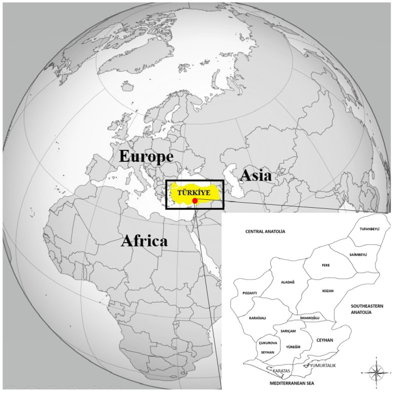 Figure 1
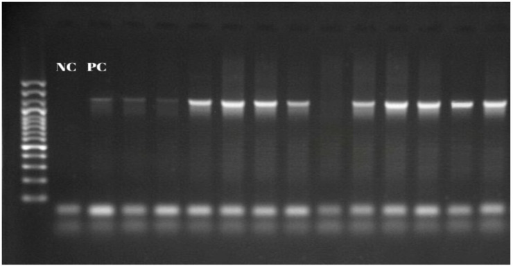 Figure 2
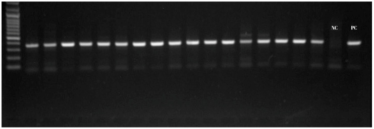 Figure 3
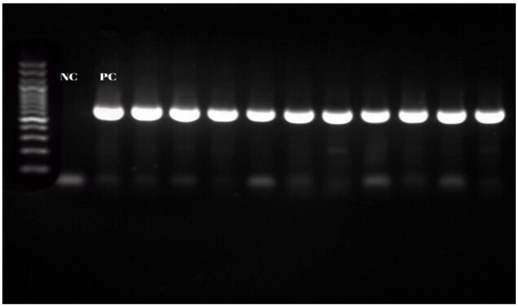 Figure 4
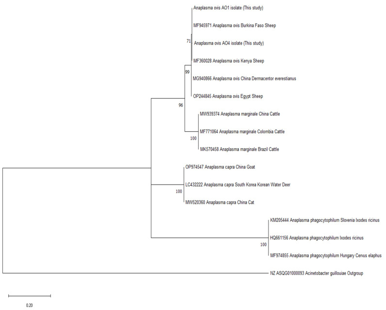 Figure 5
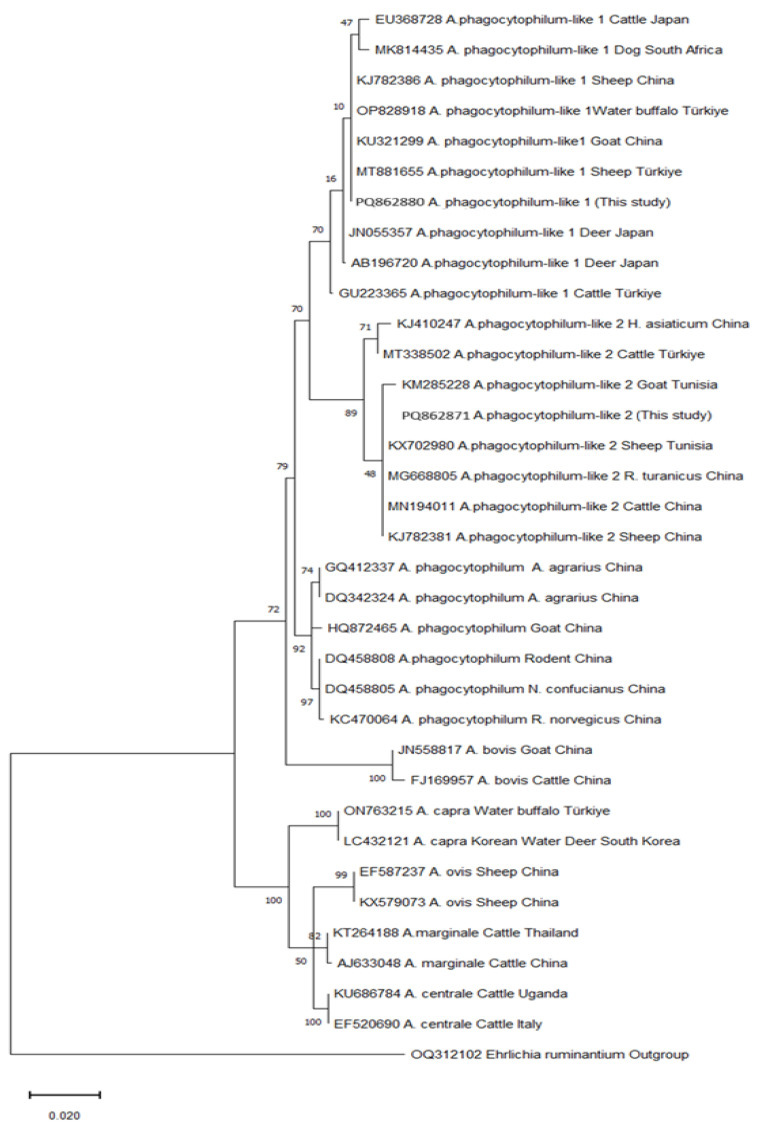 Figure 6
- —Scientific Research Projects Unit of Adana Çukurova University
Peer Reviews
No public reviews on file for this paper yet. If you reviewed it on a platform where reviews are public (OpenReview, ICLR, NeurIPS, ICML), you can paste yours below so the community can read it here.
Videos
No videos yet. Explain this paper in a talk, walkthrough, or lecture? Add one.
Taxonomy
TopicsVector-borne infectious diseases · Viral Infections and Vectors · Vector-Borne Animal Diseases
1. Introduction
Anaplasmosis is an infectious disease caused by Anaplasma species that affects both domestic and wild animals in temperate, tropical, and subtropical regions, and it holds significant economic relevance [1]. Anaplasma species can infect various cell types and are classified as intracellular pathogens. Specifically, Anaplasma phagocytophilum (A. phagocytophilum) infects granulocytes in humans and domestic animals, while Anaplasma marginale (A. marginale), Anaplasma centrale (A. centrale), and Anaplasma ovis (A. ovis) target the erythrocytes of ruminants, and Anaplasma platys (A. platys) infects canine platelets [2]. The transmission of Anaplasma species occurs primarily through ticks from the genera Ixodes, Dermacentor, Rhipicephalus, Haemaphysalis, Hyalomma, Ornithodoros, and Amblyomma [3].
As with other domestic animals, goats are exposed to species in the Anaplasma genus. A. phagocytophilum and A. ovis are the most frequently identified pathogens in goats [4]. In addition to these species, Anaplasma bovis (A. bovis), Anaplasma platys (A. platys), and Anaplasma capra (A. capra) have also been detected in goat herds from various parts of the world [5,6,7,8]. While anaplasmosis generally manifests as a mild clinical condition in goats, it can develop into a more severe form due to environmental factors, the presence of additional infectious agents, or a compromised immune system. Infected goats may present with clinical symptoms such as high fever, anemia, leukopenia, neutropenia, thrombocytopenia, anorexia, decreased milk production, abortion, infertility, depression, weakness, and weight loss [1,4].
Anaplasma phagocytophilum has a broad host spectrum, is usually transmitted by Ixodes (with some exceptions), and poses a threat to public health as a zoonotic pathogen [5]. Recent years have seen the identification of various variants, such as A. phagocytophilum-like 1 and A. phagocytophilum-like 2. These variants mainly infect sheep, cattle, and goats and are spread by ticks of various genera [6].
Some species in the Anaplasma genus are known to cause severe clinical symptoms in animals and humans [1,9]. Therefore, the effective management of Anaplasmosis is of great importance for both veterinary and public health reasons due to the potential economic-related losses in livestock and the zoonotic nature of some Anaplasma species. The effective management of the disease also requires developing treatment and control strategies. Early diagnosis, the determination of endemic areas, and phylogenetic analysis are among the basic components of Anaplasmosis control strategies [10].
Understanding these phylogenetic relationships is important for mapping geographical and ecological distributions and host–species relationships. Adana province is located in a transitional area between two regions of Türkiye with quite different climates, which has a diversity of animals and vectors that can host new variants of many agents [11]. However, data on the population structure and phylogenetic relationships of Anaplasma species isolated from goats in this region are currently insufficient. The aim of this study was to investigate the presence of Anaplasma species in goats obtained from different parts of Adana using Nested-PCR and to determine the phylogenetic relationships of the variants detected through sequencing analyses.
2. Materials and Methods
2.1. Collection of Blood Samples from Goats and DNA Extraction
Blood samples were collected from goat herds in 15 districts of Adana between June and September 2017. A total of 364 blood samples were taken into collection tubes with EDTA. The animals were clinically healthy and at least six months old, and the samples were stored at −20 °C until DNA isolation. Genomic DNA was extracted from the blood samples of the goats using QIAamp DNA Blood Kits (Qiagen, Hilden, Germany). Genomic DNA was stored at −20 °C until use.
2.2. Detection of Anaplasma spp. by 16S rRNA PCR
The 16S ribosomal RNA (16S rRNA) gene region of Anaplasma/Ehrlichia species was amplified by EC9 and EC12A “catch-all” primers (Table 1). In a 25 µL final volume, 45 mM Tris–HCl, pH 8.8; 11 mM (NH_4_)2_SO_4; 2.5 mM MgCl_2_; 0.8 mM each of dATP, dCTP, dGTP, and dTTP; 1 U Taq DNA polymerase (Solis biodyne, Tartu, Estonia); 10 µM EC9 and EC12A; and 2 µL genomic DNA sample were used. The PCR assays were performed as described previously [12]. The thermal cycling protocol used was as follows: an initial denaturation phase of 5 min at 94 °C, followed by 35 cycles of 94 °C for 1 min, 58 °C for 1 min, 72 °C for 1 min, and, finally, an additional extension phase at 72 °C for 7 min. Amplicons were subjected to 1.5% agar gel electrophoresis with ethidium bromide at 100 V for 60 min, and, after electrophoresis, they were visualized with a gel imaging system. A 50–2000 bp marker was used to determine the band size.
2.3. Investigation of A. phagocytophilum by the Nested-PCR Method
The SSAP2F and SSAP2R primers specific to the 16S rRNA region were used for the detection of A. phagocytophilum (Table 1). In the PCR, 45 mM Tris–HCl, pH 8.8; 11 mM (NH_4_)2_SO_4; 2.5 mM MgCl_2_; 0.8 mM each of dATP, dCTP, dGTP, and dTTP; 1 U Taq DNA polymerase (Solis biodyne, Tartu, Estonia); 10 µM primers; and 1 µL EC9/EC12A Nested-PCR amplicon were employed in a final volume of 25 µL. The PCR assays were performed as described previously [13]. The thermal cycling protocol used was as follows: an initial denaturation phase of 5 min at 94 °C, followed by 35 cycles of 94 °C for 35 s, 55 °C for 40 s, 72 °C for 40 s, and, finally, an additional extension phase at 72 °C for 10 min. Amplicons were run on a 1.5% agarose gel at 100 V for 1 h and visualized with a UV transilluminator.
2.4. Investigation of A. ovis by the msp4 Nested-PCR Method
msp4F and msp4R primers were used to detect the msp4 gene region (Table 1). At a final volume of 25 µL, 45 mM Tris–HCl, pH 8.8; 11 mM (NH_4_)2_SO_4; 2.5 mM MgCl_2_; 0.2 mM each of dATP, dCTP, dGTP, and dTTP; 1.25 U Taq DNA polymerase (Solis biodyne, Tartu, Estonia); 0.4 µM primers; and 1 µL 16S rRNA Nested-PCR amplicon were used. The PCR assays were performed as described previously [14]. The thermal cycling protocol used was as follows: an initial denaturation phase of 10 s at 94 °C, followed by 30 cycles of 94 °C for 30 s, 62 °C for 15 s, 72 °C for 30 s, and, finally, an additional extension phase at 72 °C for 5 min. Amplicons were run on a 1.5% agarose gel at a 100 V current for 1 h and visualized with a UV transilluminator.
2.5. Investigation of A. capra by the gltA Nested-PCR Method
The Nested-PCR assays were performed as described before [12]. The gltA gene region with OuterF/R and InnerF/R primers is shown in Table 1. In the first step, PCR reactions were performed with a total volume of 25 μL containing 45 mM Tris–HCl, pH 8.8; 11 mM (NH_4_)2_SO_4; 2.5 mM MgCl_2_; 0.2 mM each of dATP, dCTP, dGTP, and dTTP; 1.25 U Taq DNA polymerase (Solis biodyne, Tartu, Estonia); 0.4 µM OuterF/R primers; and 1 µL DNA. The PCR conditions comprised initial denaturation for 5 min at 94 °C followed by 30 cycles of denaturation for 45 s at 94 °C, annealing for 45 s at 60 °C, and elongation for 45 s at 72 °C. In the second step of the Nested-PCR, 1 µL OuterF/R amplicon and 0.4 µM InnerF/R primers were added to the master mix. The only difference in amplification conditions was the annealing temperature of 55 °C. Amplicons were run on a 1.5% agarose gel at a 100 V current for 1 h and visualized with a UV transilluminator.
2.6. Sequencing and Phylogenetic Analysis
All PCR-positive samples were sequenced. The DNA sequence analyses were performed by a commercial company (Macrogen, Amsterdam, The Netherlands). To perform sequence analysis, the amplicons were purified by use of a clean-up kit (Zymo Research, Irvine, CA, USA). SSAP2F/R and msp4F/R primers were used for the bidirectional sequence analysis. Sequencing was performed using an ABI 3730XL analyzer (Applied Biosystems, Foster City, CA, USA) and a BigDye Terminator v3.1 Cycle sequencing kit (Applied Biosystems, Foster City, CA, USA).
The obtained chromatograms were opened using Geneious Prime 2021.0.3 software (https://www.geneious.com) accessed on 20 September 2024, and their quality scores were examined. A consensus sequence was created by combining the 5′-3′ and 3′-5′ sequences of each isolate using the same program. A local database was created by downloading sequences from the NCBI database (https://www.ncbi.nlm.nih.gov) accessed on 25 September 2024 in order to compare the partial sequences of the msp4 (A. ovis) and 16s rRNA (A. phagocytophilum) genes of Anaplasma agents. Phylogenetic trees were created using the reference sequences in the local database with the help of the Mega-11 program [15]. The consensus sequences generated within the scope of this study using the MUSCLE algorithm of the Mega-11 program were aligned and recorded with the nucleotide sequences downloaded from GenBank.
The best models for use in tree creation were determined using the “Find Best Substitution Models” feature of the Mega-11 program. These models were determined as “Kimura-2 + Gamma Distributed with Invariant Sites (G + I)” for A. phagocytophilum and “Kimura-2 + Gamma distribution (+G)” for A. ovis. Phylogenetic trees were created using the maximum likelihood statistical method in the Mega-11 program. Bootstrap analysis (1000 replications) was performed to determine the reliability of the tree topology.
2.7. Statistical Analysis
The data were analyzed using SPSS Statistics 26.0 (IBM Corp., Armonk, NY, USA). Pearson’s Chi-square test was employed to determine the relationships between categorical variables. Prior to analysis, the assumption that at least 80% of the expected frequencies were greater than 5 was checked in order to ensure the validity of the test. A significance level of p < 0.05 was adopted for all comparisons.
3. Results
In this study, blood samples of 364 goats were collected from 15 districts of Adana province, showing various geographical and climatic differences (Figure 1).
The presence of Anaplasma spp. was investigated by the PCR method with EC9 and EC12A primers and was detected in 55 (15.1%) blood samples. When the distribution of isolates according to districts was examined, the highest prevalence was determined in Tufanbeyli district, at 3.84% (14/364) (Figure 2, Table 2).
The distribution of Anaplasma spp. in the goats was also examined according to demographic information, and statistical analysis was performed using the Chi-square test. Of the 55 goats in which Anaplasma spp. was detected, 51 (15.5%) were female and 4 (11.1%) were male. According to the Chi-square analysis, the association between gender and the Anaplasma spp. rate was not statistically significant (p > 0.05). The goats were divided into four age groups (0–1/2–3/4–5/5<). According to the Chi-square analysis, the association between age and Anaplasma spp. positivity was not statistically significant (p > 0.05) (Table 2).
The goats were examined at the breed level, and the presence of Anaplasma spp. was determined at rates of 17.8% in Turkish Hair goats, 17.2% in Aleppo goats, and 3% in other breeds. The associations were found to be statistically significant (p < 0.05). In addition, the relationship between the distribution of Anaplasma spp. in the goats and the altitude of the districts was investigated. The districts were divided into three groups according to their altitudes (0–50 m, 50–500 m, >500 m). Statistical analysis was performed using the Chi-square test, and the association between altitude and Anaplasma prevalence was not found to be statistically significant (p > 0.05) (Table 2).
The presence of A. ovis was determined by the msp4 Nested-PCR technique. A. ovis was present in all 55 blood samples that had previously tested positive for Anaplasma spp. (Figure 3). When the distribution by districts was examined, the highest A. ovis prevalence was seen in Tufanbeyli, at 3.84% (Table 2). Anaplasma capra was not found in this study.
In the Nested-PCR performed with SSAP2f and SSAP2r primers targeting the 16S rRNA gene region in the samples positive for A. phagocytophilum, 11 (20%) isolates were detected (Figure 4). When the distribution of isolates by districts was examined, the highest prevalence was found in Tufanbeyli, where nine (2.47%—9/364) isolates were detected (Table 2).
In this study, sequence analysis was performed on the msp4 and 16S rRNA gene regions for A. ovis and A. phagocytophilum isolates, respectively, and phylogenetic trees were constructed based on similarities.
In the msp4 gene sequence analysis, 48 of the 55 isolates were identified as A. ovis, and two variants with single nucleotide differences of 46 and 2 members were detected. In the phylogenetic tree comparing the isolates in this study with the isolates in the genebank, it was determined that the 48 A. ovis isolates were in the same clade as sheep isolates from Burkina Faso and Kenya (Figure 5, Table 3).
The 16S rRNA gene region sequence analysis was performed on 10 A. phagocytophilum isolates. Nine isolates were identified as A. phagocytophilum-like 1, and one was identified as A. phagocytophilum-like 2. In the phylogenetic tree, it was determined that nine A. phagocytophilum-like 1 isolates were in the same clade as a buffalo isolate from Türkiye and sheep and goat isolates from China (Table 3). Furthermore, the A. phagocytophilum-like 2 isolate was in the same clade as goat and sheep isolates from Tunisia and sheep and cattle isolates from China (Figure 6).
4. Discussion
Türkiye, which ranks first among European countries and 23rd in the world in terms of its goat population, is a country where goat breeding is intensively carried out in its Mediterranean, Aegean, and Southeastern Anatolia regions due to the economic value of goat meat and milk [16]. Adana province, where this study was conducted, is among the provinces in Türkiye where the highest numbers of goats are raised [17]. This province borders the Antalya and Mersin provinces, wherein we find 46% of the Anaplasma in goats in Türkiye [18].
Goats may encounter more plants and microhabitats compared to sheep and other small ruminants due to their roaming and climbing behaviors. Ticks may cling to goats more easily as their fur is shorter, and the risk of tick infestation may be higher because goats feed on the upper leaves of plants. For this reason, goats are useful for the analysis of Anaplasmosis—a tick-borne disease—in our province. In addition, Adana, which is at the easternmost border of the Mediterranean basin, where the Mediterranean climate transitions into the continental climate, has an altitude of 0–1474 m and is an important location both ecologically and epidemiologically, is a region likely to host new and different variants of Anaplasma species in goats.
Anaplasma ovis and A. phagocytophilum are particularly common in goats and are often unnoticed due to subclinical carriage. However, Anaplasma spp. can cause coinfections with hemoparasites, such as Erlichia, Babesia, and Theileria, in goats. These coinfections can increase the severity of the disease and make diagnosis and treatment more difficult. Mixed infections or coinfections put more pressure on the immune system and can ultimately cause a loss of productivity and increased mortality rates [19]. In anaplasmosis, the vector is as important as the presence of Anaplasma spp., as it is a tick-borne disease. It is usually carried by ixodidae ticks. In Türkiye, A. ovis is mostly carried by Boophilus, Dermacentor, Rhipicephalus, Hyalomma, Ixodes, and Ornithodorus species, while A. phagocytophilum has been detected in Ixodes. Adana province has a subtropical climate, forests, and pastures. For this reason, it is a suitable location representing the habitat and seasonal activities of ticks [20]. Anaplasma phagocytophilum causes a disease in humans commonly known as human granulocytic anaplasmosis (HGA), which is particularly notable in immunocompromised individuals. HGA can range from an asymptomatic or self-limited infection to a life-threatening infection with multiorgan involvement. Common symptoms include fever, gastrointestinal complaints (most commonly diarrhea), headaches, myalgia, and arthralgia. However, it can also present more seriously with pneumonia, sepsis, myocarditis, peripheral neuropathy, splenic rupture, trigeminal neuralgia, orchitis, cerebral infarction, dysarthria, and hemophagocytic lymphohistiocytosis (HLH) [21]. Following assessments of the agent, vector, and host characteristics, determining the transmission dynamics of the agent, as well as the geographical distribution, prevalence, and phylogenetic relationships of the vector and the agent, is of critical importance for the implementation of effective control strategies, and this requires regular epidemiological studies [22]. Studies on Anaplasma species in goats from this region are limited. Therefore, in order to provide epidemiological data from our region and contribute to control strategies, blood samples were collected from 364 goats from 15 districts of Adana province, and the presence of A. ovis, A. phagocytophilum, and A. capra species was investigated.
Among all blood samples, 55 (15.1%) Anaplasma spp. isolates were detected by PCR with primers specific to the Anaplasma/Ehrlichia spp. 16S rRNA gene region. The distribution of Anaplasma spp. in goats was examined according to breed, age, sex, and the altitude of the districts, and statistical analysis was performed with a Chi-square test. According to the Chi-square analysis, the differences between the rates of Anaplasma spp. in goats according to sex and age were not statistically significant (p > 0.05). However, when goats were examined at the breed level, it was determined statistically that Anaplasma spp. was more prevalent in Turkish Hair goats (p < 0.05). In addition, the relationship between the distribution of Anaplasma spp. in goats and the altitude of the districts was investigated, but it was not found to be statistically significant (p > 0.05). Interestingly, the highest number of goats from which Anaplasma was isolated was seen in Tufanbeyli district, and A. phagocytophilum-like 2 isolates were also detected there. Additionally, 9 (81.8%) of the 11 A. phagocytophilum isolates were detected in Tufanbeyli. Although no relationship was found in terms of altitude, it was estimated that the reason for this higher rate in Tufanbeyli could be related to animal management systems, climate, and the presence and distribution of tick species. The fact that Tufanbeyli district is located on the border of the Central Anatolia Region, where the tick population and diversity are higher, and that its climate is continental, unlike in other Adana districts with a Mediterranean climate, represents important differences (Figure 1). In addition, when investigating satellite maps, it was thought that northern Adana could represent an ecological area where tick populations are dense, as it is a district located on the forest–steppe border.
When Anaplasma isolates were investigated at the species level, the presence of A. ovis (100%) was determined in all 55 Anaplasma spp. isolates with a primer specific to the msp4 gene region. The prevalence of A. ovis was 15.1% (55/364). In countries bordering the Mediterranean, the prevalence rates in goats were 33% in Egypt, 65–70% in Tunisia, and 54% in Algeria [23,24,25,26]. In the neighboring countries of Iran and Iraq, the prevalence in goats was 54% and 66.6%, respectively [27,28]. In Türkiye, A. ovis prevalence was determined as 15–50% in various studies [18,29,30,31,32]. In a study conducted in Adana in 2018, A. ovis was found in 7 (18.9%) of 37 goat blood samples, which is consistent with this study’s results. However, the most comprehensive sampling in Adana was performed in this study [30].
Of the 55 isolates identified as A. ovis, 48 were identified by msp4 gene sequence analysis, and two variants of A. ovis, one containing 46 isolates and the other containing 2 isolates, were identified. Only a single nucleotide was detected to be different between them. This mutation was a silent mutation as it did not cause any change in the amino acid sequence of major surface protein 4 (Serine: TCT→TCC) (Table 3). In addition, the presence of only a single nucleotide mutation indicates low genetic diversity in this region. There are limited evolutionary differences between the strains. As such, Adana is a genetically homogeneous region for A. ovis. It was observed that the two variants were in the same clade in the phylogenetic tree as sheep and goat isolates identified in China, Egypt, Kenya, and Burkina Faso (Figure 5). This clade probably spread from an ancestral strain of Asian–African origin, and its genetic differentiation may be slow.
With 16S rRNA gene-region-specific primers, A. phagocytophilum was detected in 11 out of 55 Anaplasma spp., and the prevalence was determined as 3% (11/364). This result shows that 11 goats had A. ovis/A. phagocytophilum coinfection. In Türkiye, A. phagocytophilum prevalence was determined as 28.04% in the Mediterranean region, 5.8% in Thrace, 1.5% in Şanlıurfa, and 43.18% in Sivas [6,33,34,35]. When countries were examined, the prevalence was found to be 1.6–2.5% in Tunisia, 33% in Saudi Arabia, and 23% in China. In Switzerland, the prevalence was 5.6% [13,24,36,37]. However, it has been reported that the prevalence is higher in some countries; for example, PCR positivity was found in four out of five goats in Northern Ireland. In North America, this rate varies between 6.3% and 38.2% [38,39]. The prevalence we detected in this study is similar to that shown in other data from Türkiye, Italy, and Tunisia.
In this study, the 16S rRNA gene regions of 10 of the A. phagocytophilum isolates were sequenced and identified as A. phagocytophilum-related strains, including A. phagocytophilum-like 1 and A. phagocytophilum-like 2 isolates. Anaplasma phagocytophilum-like 1 is a variant detected in large and small ruminants and dogs and is seen in China, Japan, Thailand, Kyrgyzstan, Tunisia, and Türkiye. In previous studies in Türkiye, it was detected in sheep, goats, buffalos, cattle, and ticks in the provinces of Sivas, Malatya, Bingöl, Elazığ, Muş, Mersin, and Antalya [6,40,41,42,43,44,45,46]. In the phylogenetic tree, it was determined that nine A. phagocytophilum-like 1 isolates with 100% similar DNA profiles had bootstrap values close to those of sheep, goat, and buffalo isolates detected in studies conducted in Türkiye and of sheep and goat isolates detected in China, with which they were in the same clade (Figure 5, Table 3).
Anaplasma phagocytophilum-like 2 variants have been isolated in cattle, sheep, and goats in Senegal, Tunisia, and China [5,40,47]. In Türkiye, Anaplasma phagocytophilum-like 2 was previously detected together with Anaplasma phagocytophilum-like 1 in sheep and cattle and in various ticks. It was also searched for in buffalo but could not be found [6,43]. In this study, Anaplasma phagocytophilum-like 2 variants were detected in goats for the first time in Türkiye. Studies on A. phagocytophilum-related strains in Türkiye started in 2021. These studies were conducted on ruminants and ticks. As far as we can see, A. phagocytophilum-like 1 was only assessed in two studies conducted on goats, and A. phagocytophilum-like 2 was not found [6,33]. In the phylogenetic tree, the A. phagocytophilum-like 2 isolate had a bootstrap value close to those of sheep, goat, cattle, and tick isolates detected in Tunisia and China, with which it was in the same clade (Figure 6).
Anaplasma phagocytophilum-like 1 and 2 variants have been detected in various parts of the world, such as Türkiye, Kyrgyzstan, Korea, China, Senegal, and Tunisia [5,6,7,13,24,33,44,45,46,47]. However, the detection of A. phagocytophilum-related strains in certain regions is probably linked to the studies being conducted in these regions. If studies are conducted in different places, it is likely that these variants will also be detected in other parts of the world. The clade and bootstrap values obtained in the phylogenetic trees constructed with our isolates and isolates from other countries are quite close to each other, and, therefore, no significant epidemiological differentiation could be made. However, although the pathogenicity, symptoms, potential vectors, and zoonotic potential of these variants are not clearly known, they need to be closely monitored due to their close phylogenetic relationship with A. phagocytophilum, which has a wide host range and pathogenic properties.
5. Conclusions
This study presented detailed demographic data on the distribution and prevalence of A. ovis, A. phagocytophilum-like 1, and A. phagocytophilum-like 2 in goats in Adana—an important province in Türkiye for goat breeding—focusing on 15 districts. A. phagocytophilum-like 2 was reported in goats for the first time in Türkiye in this study. The detection of A. phagocytophilum-like 2 in goats in our region, and the fact that the region assessed is transitional in terms of its climate and vegetation, together necessitate further studies on the species distribution of ticks.
The reference list from the paper itself. Each links out to its DOI / PubMed record.
- 1Ceylan O. Xuan X. Sevinc F. Primary Tick-Borne Protozoan and Rickettsial Infections of Animals in Turkey Pathogens 20211023110.3390/pathogens 1002023133669573 PMC 7923091 · doi ↗ · pubmed ↗
- 2Altay K. Erol U. Sahin O.F. Anaplasma capra: A new emerging tick-borne zoonotic pathogen Vet. Res. Commun.2024481329134010.1007/s 11259-024-10337-938424380 PMC 11147849 · doi ↗ · pubmed ↗
- 3Yessinou R.E. Koumassou A. Galadima H.B. Nanoukon-Ahigan H. Farougou S. Pfeffer M. Tick Diversity and Distribution of Pathogen in Ticks Collected from Wild Animals and Vegetation in Africa Pathogens 20251411610.3390/pathogens 1402011640005493 PMC 11858156 · doi ↗ · pubmed ↗
- 4Lysholm S. Ådén F. Aspán A. Högberg A. Wensman J.J. Omazic A. Presence of Anaplasma spp. and Their Associated Antibodies in the Swedish Goat Population Animals 20231333310.3390/ani 1303033336766222 PMC 9913567 · doi ↗ · pubmed ↗
- 5Zobba R. Murgia C. Dahmani M. Mediannikov O. Davoust B. Piredda R. Schianchi E. Scagliarini A. Pittau M. Alberti A. Emergence of Anaplasma Species Related to A. phagocytophilum and A. platys in Senegal Int. J. Mol. Sci.2022243510.3390/ijms 2401003536613478 PMC 9820347 · doi ↗ · pubmed ↗
- 6Erol U. ŞahinÖ.F.O.F. Altay K. Molecular Survey of Anaplasma phagocytophilum and Related Strains in Sheep and Goats from Sivas; with a High Prevalence of Anaplasma phagocytophilum-like 1Turkiye Parazitol. Derg.20224629330010.4274/tpd.galenos.2022.4796536444404 · doi ↗ · pubmed ↗
- 7Miranda E.A. Han S.W. Cho Y.K. Choi K.S. Chae J.S. Co-Infection with Anaplasma Species and Novel Genetic Variants Detected in Cattle and Goats in the Republic of Korea Pathogens 2021102810.3390/pathogens 1001002833401478 PMC 7830860 · doi ↗ · pubmed ↗
- 8Wei W. Li J. Wang Y.W. Jiang B.G. Liu H.B. Wei R. Jiang R.R. Cui X.M. Li L.F. Yuan T.T. Anaplasma platys-Like Infection in Goats, Beijing, China Vector Borne Zoonotic Dis.20202075576210.1089/vbz.2019.259732679008 · doi ↗ · pubmed ↗