Comparison of Anti-Renal Fibrosis Activity of Eucommiae cortex Extract and Its Microbial Fermentation Products
Zhengyou He, Wenyi Jiang, Ruijiao Yao, Wenyan Xiao, Zhiyang Chen, Miao Zheng, Xia Zeng, Jia Li, Zhengwen Li, Yong Jiang

TL;DR
This study compares the effects of Eucommiae cortex extract and its fermented form on reducing kidney fibrosis in mice with chronic kidney disease.
Contribution
The study demonstrates that microbial fermentation of Eucommiae cortex enhances its anti-renal fibrosis activity.
Findings
EC and ECF reduced creatinine and urea nitrogen levels in mice with CKD.
Both treatments down-regulated TGF-β1/Smad signaling pathway markers linked to fibrosis.
ECF showed reno-protective effects, suggesting potential for clinical drug development.
Abstract
Background: Renal fibrosis is a common pathological feature of all progressive chronic kidney disease (CKD). Eucommiae cortex (EC) is a valuable economic tree species endemic to China. The microbial fermentation of Chinese medicines can release their active ingredients as effectively as possible or produce new active ingredients with enhanced efficacy and reduced toxic side effects; Methods: The microbial fermentation of EC can produce pinoresinol (Pin) and dehydrodiconiferyl alcohol (DA). In this study, C57 BL/6 mice were fed a diet containing 0.2% adenine, resulting in a model of chronic kidney disease. The effects of EC and EC ferment (ECF) on CKD were explored by the exogenous supplementation of EC and ECF; Results: The results of the study showed that exogenous supplementation with EC and ECF suc-cessfully reduced creatinine and urea nitrogen levels, down-regulated the expression…
Genes, proteins, chemicals, diseases, species, mutations and cell lines named across the full text — each resolved to its canonical identifier and authoritative record.
Click any figure to enlarge with its caption.
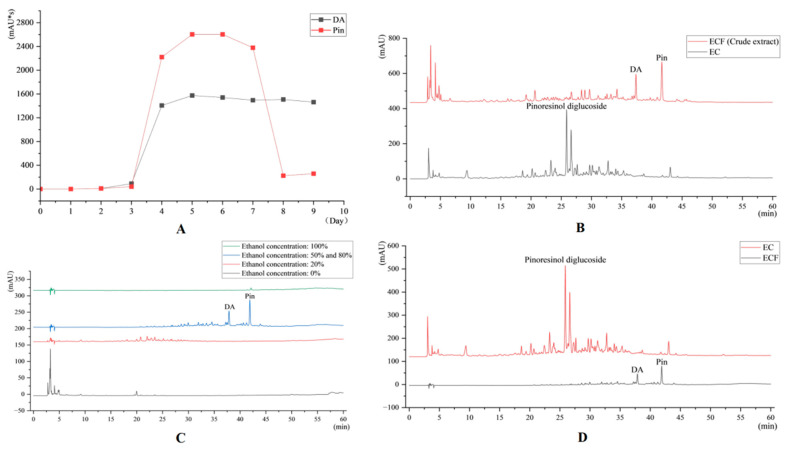 Figure 1
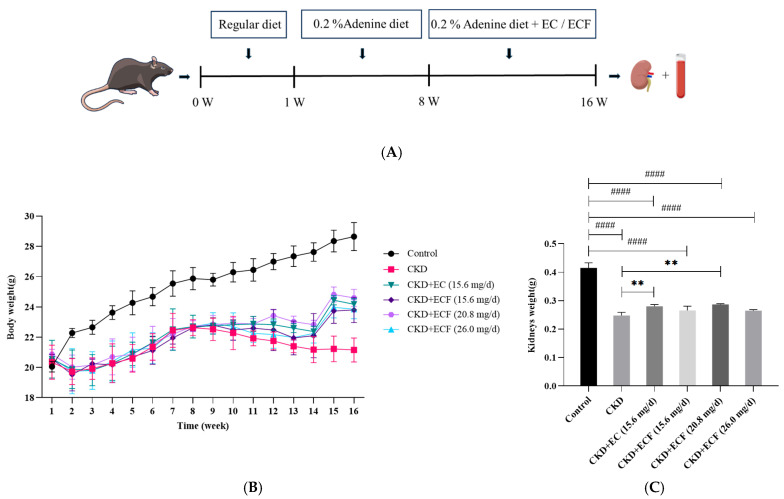 Figure 2
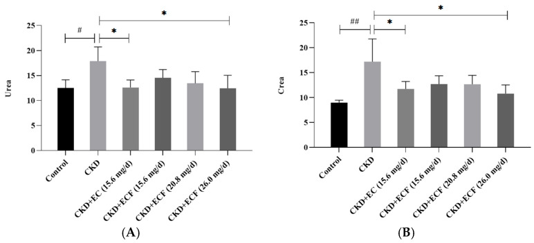 Figure 3
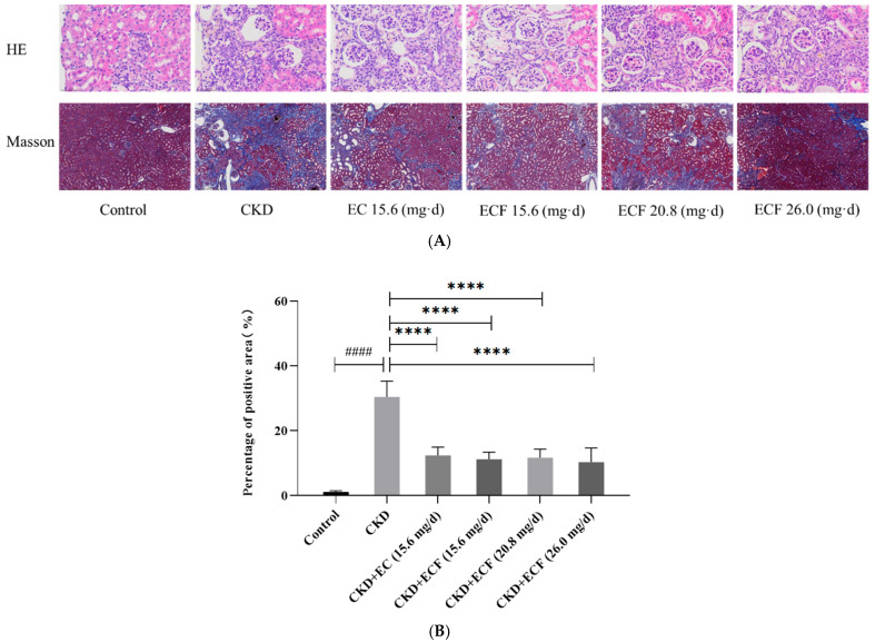 Figure 4
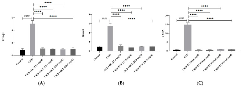 Figure 5
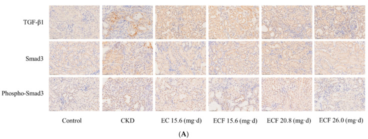 Figure 6
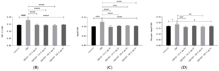 Figure 7
- —Sichuan Provincial Administration of Traditional Chinese Medicine face-to-face project
Peer Reviews
No public reviews on file for this paper yet. If you reviewed it on a platform where reviews are public (OpenReview, ICLR, NeurIPS, ICML), you can paste yours below so the community can read it here.
Videos
No videos yet. Explain this paper in a talk, walkthrough, or lecture? Add one.
Taxonomy
TopicsPhytochemical and Pharmacological Studies · Medicinal Plants and Bioactive Compounds · Phytochemistry and Biological Activities
1. Introduction
Renal fibrosis is a common pathophysiological feature of almost all progressive chronic kidney disease (CKD) [1]. Interstitial myofibroblasts are the primary effector cells in renal fibrosis, generating significant amounts of extracellular matrix (ECM), resulting in renal function loss and the concurrent degradation of renal structure [2]. The pathogenesis of renal fibrosis is intricate and complex, and experimental studies of Chinese medicines against renal fibrosis have made some progress, revealing the great potential of Chinese drugs in the prevention and treatment of renal fibrosis [3,4]. Numerous kidney diseases have been linked to the TGF-β1/Smad pathway. The process of renal fibrosis can be stopped and ECM deposition in renal tissues reversed by blocking the pathway’s activation [5]. Contemporary pharmacological research has shown numerous herbal remedies and monomer compounds to have antifibrotic properties. Natural medicine and its extracts may affect fibrosis by influencing the TGF-β1/Smad signal transduction pathway [6].
Eucommiae cortex (EC) is the dried bark of Eucommia ulmoides, a plant of the family Eucommiaceae [7], a precious economic tree species endemic to China. It is distributed in Sichuan, Guizhou, Yunnan, and other places and has a long history of medicinal use. It is now widely used in medicine, food processing, feed, industrial production, and other fields [8]. EC is rich in natural active ingredients, from which 246 compounds have been isolated and identified [9], mainly including lignans, iridoids, flavonoids, phenylpropanoids, and EC gum. Its pharmacological effects mainly include lowering blood pressure, enhancing immunity, regulating blood lipids, lowering blood sugar, protecting the liver, choleretic and diuretic activities, protecting nerve cells, regulating bone metabolism, tonifying and protecting the kidneys, and stabilizing the fetus [10].
EC has the effect of tonifying the liver and kidneys and strengthening muscles and bones. The lignans, iridoids, and phenylpropanoid constituents in EC are the main constituents and are the basis of medicinal substances in EC, which have a good anti-renal fibrosis effect [11]. The lignans in EC are protective against hypertensive renal damage, which may be related to inhibiting renal aldose reductase [12]. EC ameliorates hypertensive renal damage, diabetic nephropathy, and adenine renal injury-induced renal fibrosis in rats [13]. EC upregulates the expression of MMP-2 in the renal tissues of rats with unilateral ureteral obstruction (UUO), which slows down the disease progression of renal fibrosis [14]. EC’s decoction can improve patients’ renal function [15]. EC can inhibit the up-regulation of CTGF expression in the renal tubular epithelium of UUO rats, slowing the progression of tubular injury and interstitial fibrosis [16].
The microbial fermentation of traditional Chinese medicine has a long history. Microbial fermentation can release the active ingredients as soon as possible or produce new active ingredients with enhanced efficacy and reduced toxic side effects [17]. The microbial fermentation of EC can produce pinoresinol (Pin) and dehydrodiconiferyl alcohol (DA) [18]. Therefore, we studied the impact of EC and its microbial fermentation products on the TGF-β1/Smad pathway and renal fibrosis to inform clinical drug development.
2. Results
2.1. HPLC Results
The temporal profile of Pin and DA content during fermentation was analyzed (Figure 1A), and the highest content of both compounds was observed on the fifth day of fermentation. A comparative high-performance liquid chromatography (HPLC) analysis between EC extract and ECF crude extract was performed (Figure 1B). Different concentrations of eluent were collected, and based on the HPLC results, 50% ethanol eluent and 80% ethanol eluent were mixed, concentrated, and dried to obtain the final ECF drug formulation. The HPLC results of different concentration eluents of the EC crude extracts are shown in Figure 1C. The HPLC results of gavage preparation in the EC and ECF groups are shown in Figure 1D.
2.2. Effect of EC and ECF on Body Weight of Mice
The experimental protocol for the animal model is shown in Figure 2A. The CKD group exhibited the lowest body mass and the control group maintained the highest during the study period (Figure 2B). Statistical analysis demonstrated that the control group showed significantly higher body weight compared to other groups on the second week post-modeling. By the 15th week, the CKD group displayed considerably reduced body weight relative to all other groups. A comparative analysis of kidney tissue weights (Figure 2C) showed that the control group had significantly higher kidney weights, while the CKD group had significantly lower kidney weights. These results suggest that EC and ECF can improve weight loss and reduce renal tissue damage caused by adenine-induced renal fibrosis.
2.3. Effects of EC and ECF on Creatinine and Urea Nitrogen in Mice
The mice in the CKD group exhibited significantly elevated serum creatinine (Crea) and urea nitrogen (Urea) concentrations compared to the control group. Remarkably, both EC and ECF treatments attenuated these abnormalities, with serum Urea and Crea levels in the EC/ECF-treated groups observed to approximate those of the control cohort (Figure 3A,B). The CKD + ECF (26 mg/day) group demonstrated the most pronounced reduction in Urea and Crea. The findings showed that EC and ECF may effectively decrease CKD mice’s Urea and Crea serum levels.
2.4. Effects of EC and ECF on Histopathologic Changes in Mice with Chronic Kidney Disease
The results of the Masson and HE staining of renal tissue are displayed in Figure 4A. All groups except the control group had tubular proteins, brownish-yellow material, foci of mineralization, and varied degrees of peritubular depressions and interstitial fibrosis. The samples from each group varied in terms of the degree of mineralization: control group < CKD + EC (15.6 mg/d) group < CKD + ECF (20.8 mg/d) group < CKD + ECF (26 mg/d) group < CKD + ECF (15.6 mg/d) group < CKD group. The fibrosis severity of the samples in each group was as follows: control group < CKD + ECF (26 mg/d) group < CKD + ECF (15.6 mg/d) group < CKD + ECF (20.8 mg/d) group < CKD + EC (15.6 mg/d) group < CKD group. The visual field’s blue area per unit area was measured, and the significance analysis of the percentage area of positivity in each group is shown in the graph (Figure 4B). There was a significant decrease in all other groups compared to the CKD group. The results indicated that EC and ECF attenuated adenine-induced renal fibrosis.
2.5. qRT-PCR Measurement of TGF-β1, Smad3, α -SMA, and mRNA Expression
A relative quantitative analysis of target genes was performed using the control group to examine changes in the mRNA expression levels of target genes in kidney tissues compared to the control group. The TGF-β1, Smad3, and α-SMA expression levels were higher in the CKD group compared to the control group. As illustrated in Figure 5A–C, no significant differences were observed in TGF-β1, Smad3, and α-SMA expression levels between the EC, ECF, and control groups. The group with CKD + ECF (20.8 mg/d) had a more significant down-regulation of TGF-β1, while the group with CKD + ECF (15.6 mg/d) had a more substantial down-regulation of α-SMA and Smad3. The findings suggest that ECF and EC can reduce the expression of TGF-β1, Smad3, and α-SMA and attenuate the adenine-induced renal fibrosis.
2.6. Immunohistochemical Results
Each group’s renal tissues underwent immunohistochemical staining. The average optical density (OD) of the positive expression unit field of view was measured, and the results are shown in Figure 6A. The TGF-β1, Smad3, and phospho-Smad3 levels were considerably more significant in the CKD group than in the control group. They were significantly lower in the EC and ECF groups than in the CKD group, as illustrated in Figure 6B–D. TGF-β1 was considerably down-regulated in the CKD + ECF group (20.8 mg/d); Smad3 and phospho-Smad3 were significantly down-regulated in the CKD + EC group (15.6 mg/d). The findings showed that in the renal tissues of CKD mice, the ECF and EC groups dramatically decreased the expressions of TGF-β1, Smad3, and phospho-Smad3.
3. Discussion
Microbial fermentation can change the content and structure of existing compounds in plants, reduce toxic substances, and produce abundant secondary metabolites through various metabolic pathways [19]. For example, microbial fermentation promotes the release of active components such as phenolic compounds and flavonoids [20], and antioxidant activity is improved [21]. Combining fermentation with EC may lead to more possibilities. Focusing on the investigation and optimization of the fermentation performance of different oil-containing yeasts on Eucommia hydrolysate, the aim was to explore and evaluate the functions and effects of fermentation with mixed strains compared to single strains [22]. The degradation patterns of hemicellulose, cellulose, and lignin in raw materials by solid-state fermentation with three edible fungi were investigated using Eucommia bark and Eucommia leaf residue as substrates [23]. We studied the fermentation of Cordyceps militarisin order to increase the content of active ingredients in Eucommia and poplar flowers and then utilize the nutrients to promote the growth of Cordyceps militaris and increase the production of antibacterial and antiviral actives [24]. To explore the effect of probiotic fermentation on some active ingredients of EC, Mulberry leaves, and Gynostemma, the author used three mixed probiotic bacteria, including Bacillus subtilis, to start fermentation tests on three herbal decoctions and their decoction mixtures, respectively [25]. A kind of lactic acid bacteria was also used to ferment EC mushroom sauce [26]. EC is fermented by microorganisms to produce the fermentation products Pin and DA, so this experiment focused on the mitigating effects of EC and ECF on renal fibrosis.
The results of the study showed that exogenous supplementation with EC and ECF successfully reduced creatinine and urea nitrogen levels; down-regulated the expression levels of TGF-β1, α-SMA, Smad3, and phospho-Smad3 in the TGF-β1/Smad signaling pathway; and ameliorated renal fibrosis. As a result, both EC and ECF may have renoprotective effects and provide a reference for relevant clinical drug development. However, the underlying process of CKD is complex and includes many cytokines and signaling pathways. Whether ECF and EC can ameliorate CKD through other signaling pathways needs further exploration.
4. Materials and Methods
4.1. Reagents and Antibodies
Specific information is provided in Table 1. EC was purchased from Sichuan Zangxi tang Biotechnology Co., Ltd. (No. 230625, Guanghan, China); chemicals and HPLC-grade acetonitrile were purchased from Chengdu kelong chemical Co., Ltd. (Chengdu, China).
4.2. EC Extraction and Drug Preparation
The EC powder was subjected to methanol-assisted thermal reflux extraction under optimized conditions, with a material-to-solvent ratio of 1:20 (w/v). After ultrasonic extraction for 30 min, the filtrate was taken and the residue was extracted again using the above method, and then the two filtrates were combined and vacuum-filtrated through Whatman No. 1 filter paper. The pooled filtrates were concentrated via rotary evaporation (Yingyu R201D system, 40 °C, 0.09 MPa vacuum) and subsequently vacuum-dried to yield the EC extract. The EC extract was dissolved in methanol, analyzed by HPLC, and compared with the relevant regulations of EC in the Chinese Pharmacopoeia. Batches of acceptable quality were taken for subsequent experiments. The EC extract was dissolved in 0.05% carboxymethylcellulose sodium (CMC-Na) to make a suspension for gastrointestinal administration according to the EC dose recommended by the Chinese Pharmacopoeia and referring to the literature dose [27].
4.3. Microbial Fermentation Product Preparation
4.3.1. Optimal Fermentation Time
The fermentation broth containing EC extract was configured. Here, 1000 mL of fermentation solution contained 200 g of fresh potato, 20 g of analytically pure glucose, 1.5 g of analytically pure magnesium sulfate, 3 g of analytically pure potassium dihydrogen phosphate, and 140 mg of VB_1_. The EC extract was obtained by extracting 50 g of EC powder according to the method described in Section 4.2. The fermentation process employs traditional mucor [18] for microbial transformation. Fermentation medium aliquots (10 mL) were aseptically sampled at 24 h intervals (Days 1–9). Methanol (1:2 v/v) was added to the sample and sonicated (60 min). The solution was concentrated to near dryness using a rotary evaporator, dissolved in anhydrous methanol (AR grade), and filtered through a 0.22 μm nylon membrane before HPLC analysis.
4.3.2. Macroporous Resin Fractionation
The EC fermentation broth with optimal fermentation time was screened according to the HPLC results of different fermentation times. The fermentation broth was taken and added with methanol (1:2 v/v) and ultrasonicated (60 min). Then, the solution was concentrated to near dryness using a rotary evaporator and dried in a reduced-pressure drying oven to obtain the crude extract of ECF. AB-8 resin was pre-conditioned through sequential ethanol (95%, v/v) and deionized water (18.2 MΩ·cm) washing cycles. The ECF crude extract was loaded onto the adsorbent at 3 mL/min, and then eluted with ethanol using a stepwise gradient: 0% → 20% → 50% → 80% →100% at 2 mL/min under UV monitoring (280 nm). The eluted fractions were rotary-evaporated, lyophilized, and analyzed by HPLC.
4.3.3. ECF Drug Preparation
The ECF crude extract product of microbial fermentation of EC was taken at the optimum fermentation time. Gradient ethanol elution was performed using AB-8 macroporous adsorbent resin. The target fractions were collected, concentrated under reduced pressure (40 °C, 0.09 MPa), and lyophilized to obtain the ECF extract. Based on the daily dosage of EC specified in the Chinese Pharmacopoeia, the lyophilized extract was homogenized with 0.05% CMC-Na [28] to formulate a gastric suspension in the recommended dose.
4.4. Experimental Animals
Male C57BL/6 mice (21 ± 2 g body weight) were procured from Chengdu Dashuo Biotechnology Co., Ltd. (Animal Production License No. SCXK 200-030; Chengdu, China). The animal experimental protocols and operational procedures were based on national regulations on experimental animal welfare and ethics. They were examined by the Animal Welfare Ethics Committee of Chengdu Medical College (No. 014, Cheng Yi Dong Lun [2022]).
4.5. HPLC Conditions
Chromatographic conditions: a Unisil 5-120 C18 ultra column (5 μm, 4.6 mm × 250 mm), temperature maintained at 30 °C, with a mobile phase flow rate of 1 mL/min and detection wavelength at 227 nm. Detailed elution parameters are provided in Table 2.
4.6. Adenine-Diet-Induced Renal Fibrosis
After a week of acclimatization feeding, 36 male C57BL/6 mice (22 ± 2 g) were randomly divided into six groups (n = 6): control, CKD, CKD + EC (15.6 mg/d), CKD + ECF (15.6 mg/d), CKD + ECF (20.8 mg/d), and CKD + ECF (26 mg/d). Except for the control group, which was given a typical control diet (Wuxi Daitz Biotechnology Co., Ltd.), all groups were provided meals containing 0.2% adenine [29]. The blank and CKD groups were gavaged with 0.2 mL of CMC-Na solution daily, while the CKD + EC and CKD + ECF groups were gavaged once daily with 0.2 mL of 0.05% CMC-Na solution containing the relevant concentration of the drug. After 16 weeks, the mice were euthanized, and samples of blood and kidney tissue were gathered for additional research.
4.7. Renal Function Analysis
The blood samples were placed in an incubator at 37 °C for 30 min and then transferred to a centrifuge that had been pre-cooled to 4 °C. The supernatant was extracted for kidney function measurements following a 15 min [30] centrifugation at 3000 rpm. A fully automated biochemical analyzer (model: 3100) was used to measure renal function parameters such as Crea and Urea.
4.8. Analysis of Renal Histopathology
HE: For embedding, 4% paraformaldehyde-fixed tissue samples were rinsed under running water, trimmed, and placed into pathological embedding plastic baskets for gradient alcohol dehydration, xylene transparency, wax dipping, and embedding. Sectioning: tissues were cut into 5 µm thick slices using a Leica RM2235 slicer, spread in warm water, and fixed onto slides. Staining: sections were deparaffinized to water, stained with hematoxylin–eosin, dehydrated in gradient alcohol, made transparent using xylene, and sealed with neutral resin glue. Pathological changes in the liver and kidney were observed under the microscope. The entire sectioned area was observed, and photographic recordings were made using a microimaging system for each group of areas with apparent lesions. Masson staining: Sections were deparaffinized to water and stained with Weigert’s hematoxylin stain, Lichun red acidic magenta solution, 1% phosphomolybdic acid, 1% glacial acetic acid, aniline blue staining solution, and gradient alcohol dehydration followed by xylene transparency, and the sections were sealed with neutral resin glue. Renal tissues were photographed with a microimaging system. The area of positive expression in the pictures was measured using Image-Pro plus 6.0.
4.9. qRT-PCR Measurement of TGF-β1, Smad3, and α-SMA mRNA Expression
4.9.1. RNA Extraction and Concentration Determination
The procedure is as follows: Take an appropriate amount (about 100 mg) of tissue sample, add 1 mL of RNAiso Plus, homogenize, and leave at room temperature for 5 min; centrifuge at 12,000× g 4 °C for 5 min, take 800 μL of the supernatant, add 200 μL of chloroform, shake and mix well, and leave at room temperature for 5 min; centrifuge at 12,000× g 4 °C for 15 min; take the supernatant, add 500 μL of isopropanol and mix well, and leave at room temperature for 10 min; centrifuge at 12,000× g 4 °C for 10 min. After removing the supernatant, add 500 μL of isopropanol, mix thoroughly, and leave for 10 min at room temperature. Centrifuge at 12,000× g for 10 min at 4 °C, discard the supernatant, add 1 mL of 75% ethanol, mix well, centrifuge at 7500× g for 5 min at 4 °C, discard the supernatant, and dry the precipitate at room temperature. Add RNase-free water to dissolve the precipitate.
An ultraviolet spectrophotometer was used to determine the concentration of RNA. The concentration of the RNA sample was adjusted with RNase-free water.
4.9.2. cDNA Synthesis
Reverse transcription was used (refer to the UltraStart SYBR Green qPCR Master Mix Reverse Transcription Kit instruction manual). The reverse transcription system is shown in Table 3. After the system was made, all of the reagents underwent liquid sedimentation, 25 °C reaction for 10 min, 55 °C reaction for 15 min, and 85 °C response for 5 min. After denaturing the template–primer mixture, the reaction tube was immediately put on ice to terminate the reaction. Then, 80 μL of DEPC water was added for dilution to obtain cDNA.
4.9.3. qRT-PCR Assay
The qRT-PCR assay was performed according to the instructions for the StormstarSybrGreen qPCR Master Mix kit. A 20 μL system was used for the assay, and the assay system is shown in Table 4.
4.9.4. Primer Design and Synthesis
Primers were designed with Oligo7 software, and species-specific primer matching was performed using the NCBI Primer-Blast system. Primers were synthesized by the Chengdu Branch of Bioengineering (Shanghai) Co., Ltd., (Shanghai, China). The primer sequence information is shown in Table 5.
4.9.5. Data Processing
Data results were analyzed using the 2^−ΔΔCT^ method.
4.10. Immunohistochemistry Analysis
Protein expression levels were detected by immunohistochemistry. First, paraffin was removed and sliced, before being hydrated and dewaxed. Antigen repair, blocking, containment, incubation of primary and secondary antibodies, DAB color development, and hematoxylin re-staining were carried out, and gradient alcohol dehydration was followed by xylene transparency and neutral resin adhesive sealing of the film. The positive expression of the target was observed under a microscope.
4.11. Statistical Analysis
The data in this study were analyzed and plotted using GraphPad Prism 9.0 [31]. Significant differences between groups were tested using a one-way analysis of variance (ANOVA). p < 0.05 indicates statistical significance.
The reference list from the paper itself. Each links out to its DOI / PubMed record.
- 1Chen C. Feng C. Luo Q.L. Zeng Y. Yuan W. Cui Y. Tang Z. Zhang H. Li T. Peng J. CD 5L up-regulates the TGF-β signaling pathway and promotes renal fibrosis Life Sci.202435412294510.1016/j.lfs.2024.12294539127319 · doi ↗ · pubmed ↗
- 2Wang X.H. Liu X.H. Xu L.M. Li Y. Zheng B. Xia C. Wang J. Liu H. Targeted delivery of type I TGF-β receptor-mimicking peptide to fibrotic kidney for improving kidney fibrosis therapy via enhancing the inhibition of TGF-β1/Smad and p 38 MAPK pathways Int. Immunopharmacol.202413711248310.1016/j.intimp.2024.11248338880023 · doi ↗ · pubmed ↗
- 3Ye J.L. Li L.D. Yan Z.Y. Progress of experimental animal studies on Chinese herbal medicine against renal fibrosis Clin. J. Tradit. Chin. Med.2012248890
- 4Zhu J.H. Wang L. Ma Z.X. Duan J.A. Tao J.H. Rehmannia glutinosa Libosch and Cornus officinalis Sieb herb couple ameliorates renal interstitial fibrosis in CKD rats by inhibiting the TGF-β1/MAPK signaling pathway J. Ethnopharmacol.202431811703910.1016/j.jep.2023.11703937579922 · doi ↗ · pubmed ↗
- 5Zhang H. Luo J.H. Yang L. Tong Y.T. Cai H.L. Effects of astragalus polysaccharide modulating TGF-β1/Smad/AP-1 signalling pathway on renal fibrosis in rats with chronic renal failure Chin. J. Gerontol.20244440404044
- 6Wan Y.G. Che X.Y. Sun W. Huang Y.R. Meng X.J. Chen H.L. Shi X.M. Tu Y. Wu W. Liu Y.L. Low-dose of multi-glycoside of Tripterygium wilfordii Hook. f., a natural regulator of TGF-β1/Smad signaling activity improves adriamycin-induced glomerulosclerosis in vivo J. Ethnopharmacol.20141511079108910.1016/j.jep.2013.12.00524362077 · doi ↗ · pubmed ↗
- 7China Pharmacopoeia Committee Pharmacopoeia of the People’s Republic of China: Volume I, 2020 China Medical Science Press Beijing, China 2020172
- 8Zhang R.Z. Li Q. Pei J. Dang R. Yang Y.T. Wang C. Overview of the Chemical Components and Pharmacological Effects of Eucommia ulmoides J. Shaanxi Univ. Chin. Med.2024478791