Whispers in the Lungs: Small Extracellular Vesicles in Lung Cancer and COPD Crosstalk
Yetemwork Aleka, Fantahun Biadglegne, Ulrich Sack

TL;DR
This paper reviews how tiny cell particles called sEVs could help diagnose and monitor lung cancer and COPD, but more research is needed to make them clinically useful.
Contribution
Highlights the dual role of sEVs in lung cancer and COPD as potential biomarkers and emphasizes the need for large-scale studies.
Findings
sEVs from lung cancer carry onco-proteins like EGFR mutations and miR-21.
sEVs from COPD contain pro-inflammatory cytokines like IL-6 and TNF-α.
sEVs show promise as biomarkers but face challenges in clinical application due to extraction and analysis limitations.
Abstract
Lung cancer is a very serious disease, and it becomes worse when combined with chronic obstructive pulmonary disease [COPD], another chronic lung illness. Both are linked to smoking and long-term lung inflammation. Scientists are now focusing on tiny particles called small extracellular vesicles (sEVs), which are released by cells and carry important molecules. In lung cancer, sEVs often carry cancer-related signals, while in COPD, they carry inflammation-related ones. These signals could help doctors to diagnose and monitor the diseases more accurately. However, collecting and studying sEVs is still difficult, which limits their use in clinics. This review looks at the potential of sEVs as helpful tools in treating lung cancer and COPD and highlights the need for more research. Lung cancer is one of the deadliest forms of cancer. Its prognosis becomes even worse when it co-occurs with…
Genes, proteins, chemicals, diseases, species, mutations and cell lines named across the full text — each resolved to its canonical identifier and authoritative record.
Click any figure to enlarge with its caption.
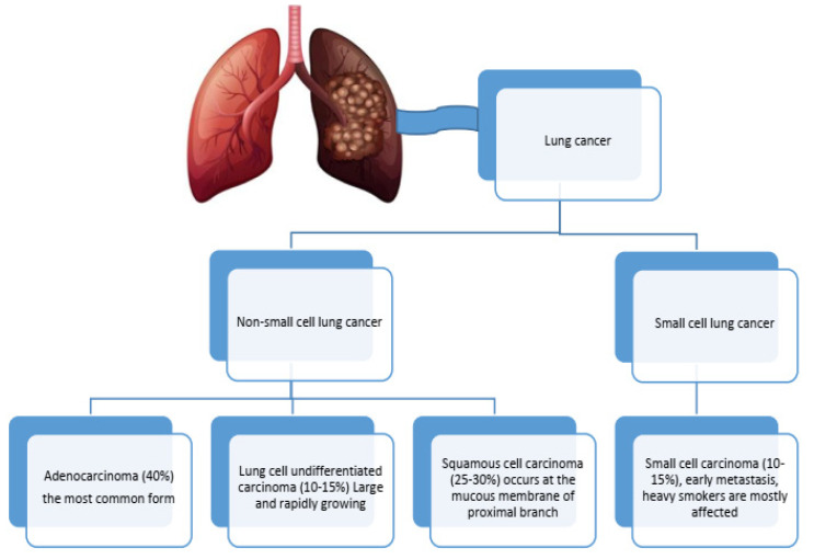 Figure 1
| Method | Advantage | Disadvantage | Reference | |
|---|---|---|---|---|
| Isolation | Ultracentrifugation | Large volumes processing | Time-consuming, costly equipment, non-EV contamination risk | [ |
| Density graident centrifugatin | Higher purity | Labor and time intensive | [ | |
| Immunoaffinity capture | High specificity | Potentia.antibody carryover, expensive | [ | |
| Polymer precipitation | Simple | Contaminants, co-precipitation | ||
| Size exclusion chromatography | Gentle on EVs | Limited sample volume, EV dilution | [ | |
| Microfluidic device | Rapid | Device.fabrication complexity; potential for clogging | [ | |
| Characterization | NTA | Provides size distribution and concentration, relatively quick | Limited sensitivity for small EVs, affected by sample purity | [ |
| Western blotting | Confirms presence of EVs marker | Labor and time consuming | [ | |
| Flowcytometry | Multiparametric analysis | Limited sensitivity for small EVs | [ | |
| TEM | Detailed, structural information visually | potential artifacts, not quantitative | [ | |
| Dynamic light scattering | Quick | Less accurate for polydisperse samples | [ | |
| SPR | Measures EV binding affinity | Complex data interpretation | [ |
- —German Academic Exchange Service (DAAD)
Peer Reviews
No public reviews on file for this paper yet. If you reviewed it on a platform where reviews are public (OpenReview, ICLR, NeurIPS, ICML), you can paste yours below so the community can read it here.
Videos
No videos yet. Explain this paper in a talk, walkthrough, or lecture? Add one.
Taxonomy
TopicsExtracellular vesicles in disease · Pulmonary Hypertension Research and Treatments · Circular RNAs in diseases
1. Introduction
As time elapses, the list of people dying from lung cancer continues to increase, owing to its rank as the second most fatal cancer type across the globe, with 2.5 million new cases and 1.8 million deaths annually. The development of lung cancer in conjunction with chronic obstructive pulmonary disease (COPD) differs by gender, age, and geographical location. The risk is approximately twofold for male COPD patients as opposed to females (5.09% versus 2.52%). However, the risk continues to grow with age, with more than 10% of younger lung cancer patients also having COPD. Geographically, the Western Pacific has the highest prevalence of COPD at 7.78%, followed by 3.25% in the Americas and 3.21% in Europe [1]. Smoking is the primary risk factor, alongside pollution and pre-existing lung conditions like COPD, which also contribute to high mortality rates. Late-stage diagnosis significantly affects survival, with 77% of cases being diagnosed at advanced stages [2]. Diagnosis involves using imaging techniques and biopsies to classify lung cancer into non-small-cell lung cancer (NSCLC) and small-cell lung cancer (SCLC) (Figure 1). Treatment varies by type and stage, including surgery [2], radiation, chemotherapy [3], and targeted therapies [4,5] based on genetic mutations. Recent advances in medicine, particularly using the immune system in treatment through immunotherapy [6], have shown immense possibility, especially concerning severe NSCLC cases. To improve the condition of the afflicted, the expansion of screening with the employment of new innovative biomarkers that aid in improving diagnosis and screening approaches is required. Efforts towards early detection screening have emerged to help lung cancer patients immensely. This is important in ensuring better outcomes for managing affected individuals. Scientists are searching for new biomarkers to aid in early lung cancer diagnosis and determine outcome prediction with or without other respiratory disease comorbidity [4,7,8,9,10]. The goal of this narrative review is to identify the roles, functions, and diagnostic or prognostic value of small EVs released from lung cancer or COPD patients using different original articles from peer-reviewed publishers such as Elsevier, Science Direct, PubMed, Scopus, Web of Science, and Google Scholar. It also analyzes the potential difficulties in distinguishing accurate small extracellular vesicle biomarkers considering the presence of comorbidities. We address the issues of interpreting biomarkers with concurrent pathologies and highlight important factors needed to refine accuracy in multi-faceted clinical situations.
Dual Battles: Immune Dynamics in COPD and Lung Cancer Progression
The immune system, which includes T cells, B cells, NK cells, macrophages, and dendritic cells, is essential in defending the body from cancerous cells. With lung cancer, the tumor microenvironment (TME) is a complex ecosystem that not only fosters tumor development but also shields it from immune response [11]. Cancer cells commonly use tactics to escape immune surveillance and strike, for instance, the downregulation of molecules responsible for antigen presentation [12], immunosuppressive cytokine secretion, inhibitory receptor expression, and recruitment of immunosuppressive cell types [13].
Furthermore, COPD is defined by persistent inflammation and constriction of the airways following the inhalation of harmful particulates or gases, especially cigarette smoke [14]. The response of the immune system is quite complicated and often overlaps with non-small-cell lung cancer, which is associated with a higher likelihood of lung cancer. Pro-inflammatory cytokines are released by macrophages and neutrophils in COPD, leading to tissue damage and mucus hypersecretion [15]. The cells that include Th1 and Th17 subsets activate macrophages and worsen inflammation. On the other hand, plasma cells differentiated from B cells are also active in inflammation [16]. The increased levels of inflammatory proteins and chemokines critically damage airway, cause inflammation and modify the environment. Tissue damage from oxidative balance in COPD is detrimental to the cells and increases inflammation. There are often such changes with persistent inflammation to the structure of the airways, which leads to further issues with the limitation of airflow and respiratory problems. This means that the region is subject to hyperinflammation, which may lead to a more persistent infection.
A combination of these two diseases can severely suppress immunity in the affected individual [17], making them experience elevated systemic oxidative stress and diminished levels of antioxidants and inflammatory cells [18]. Sustained lung inflammation due to irritants like cigarette smoke or neoplastic cells can be the cause of the priming of the immune system, shifting to the inflammatory state alongside tissue damage and further inflammation. COPD coupled with lung cancer can also increase the incidence and severity of infection due to impaired lung function and immunity while enhancing the chances of altering treatment selection and changing treatment response towards lung cancer. These two diseases can also lead to the compromising of the immune system, causing a shift towards a pro-inflammatory response and favoring tumor cell evasion [19]. The coexistence of these two conditions can also affect the selection and efficacy of lung cancer treatments, as well as increase susceptibility to respiratory infections due to compromised lung function and weakened immune response [20].
Therefore, early diagnosis and prognosis of treatment and its efficacy biomarkers is significant, as these biomolecules are expected to be accurate and precise. Recently, attention has shifted to extracellular vesicles (EVs) such as small EVs for their potential use in diagnostic and monitoring treatment purposes [10]. These biomolecules are noteworthy due to their small dimensions and originate from a unique process of production, namely an endocytic process, where they are substantially produced from parent cells. In the diseases of lung cancer and COPD, small EVs are known to participate in progression, immune modulation, and tissue remodeling, which goes beyond the activities of cells and includes tissue repair and fibrosis [21].
2. Small EVs as a Hidden Language
EVs are membrane-bound structures released by cells into bodily fluids that facilitate intercellular communication by transporting proteins, lipids, nucleic acids, and metabolites [22]. Varieties of these messengers are further explained in Table 1 and include small EVs (30–150 nm, located in multivesicular bodies) [23], microvesicles (100–1000 nm, from plasma membrane budding), and apoptotic bodies (500–2000 nm, released during cell death). EVs mediate development, physiological processes, and the regulation of immunity and have future potential in scientific investigation and medicine delivery. There are a few other subdivisions of EVs that are known, but for their indefinite overlap in size and biogenesis pathways, they are mostly included in one of the three main EV categories, as mentioned above. Certain EVs, however, enclose distinct molecular identities or functional roles in living systems, but within the context of current understanding, their classification will remain primarily based on their size. More studies must be conducted on the biogenesis, molecular composition, and functional attributes for EVs to aid in determining applicable classification strategies.
3. From Cells to Circulation
Small EVs facilitate intercellular communication, maintaining cellular balance and immune function [27]. They are secreted ubiquitously across cell types including γδ T lymphocytes, which demonstrate potential as cancer immune-therapeutics. Tumor-derived EVs function as antigen carriers, while immune cell-derived vesicles exhibit intrinsic antineoplastic activity via bioactive molecular cargo [28]. They promote cancer progression through angiogenesis [29], oncogenic molecule transfer, and metastatic site preparation [30]. EVs originate from tumor cells (promoting growth, angiogenesis, metastasis, immune evasion), immune cells (modulating anti-cancer responses), and respiratory cells. In lung cancer, tumor-derived EVs facilitate progression and metastasis [31]. During COPD, inflammation causes the immune system to be over activated and release pro-inflammatory mediators that damage tissues and remodel airways [32], while cigarette smoke-damaged epithelial cells release inflammatory EVs [14,33,34]. sEVs from patients suffering from COPD exhibited significantly higher levels of IL-1β, TNF-α, MMPs [35], and dysregulated miRNAs like miR-21 [34]. In a clinical setting, sEVs can be used as diagnostic and prognostic biomarkers for the detection of the disease in its early stages [36,37], disease monitoring [38,39], and even as therapeutic agents through the modulation of the sEV-mediated routes [40].
EV autoantibodies have been also identified as potential biomarkers in lung cancer, complementing their previously established role of EVs in biomarker identification. This finding expands the repertoire of molecular signatures that may facilitate the early detection and characterization of lung malignancies. The presence of these autoantibodies in patient serum provides an additional dimension to the multi-faceted approach of cancer diagnostics, potentially enhancing sensitivity and specificity when used in conjunction with EV-derived biomarkers [41].
4. From Sample to Signal: The Challenge of Isolation and Characterization
There is a significant lack of consensus regarding standardized protocols for EV isolation and characterization among researchers, representing a critical bottleneck in comparative studies of EVs with other molecular entities (Table 2). Despite numerous techniques being available, including ultracentrifugation [42], density gradient approaches [43], polymer-based precipitation [44], and size exclusion chromatography [45], methodological variations yield heterogeneous EV populations with differing purity profiles.
Similarly, characterization methods ranging from NTA [46], TEM [47], and flow cytometry to advanced techniques like fluorescence NTA [48] and ExoArc-SEC coupling [49] produce inconsistent results across laboratories. This methodological inconsistency impedes biomarker discovery efforts in lung cancer and COPD, where protein analysis workflows encompassing extraction, quantification [23], and profiling require rigorous standardization to establish clinically relevant markers [50].
Additional characterization includes lipid assays [51], functional assays, RNA expression analysis via qPCR [52], and Surface Plasmon Resonance (SPR) for binding kinetics [53]. On the other hand, most peripheral EVs are platelet-derived, creating significant challenges in developing EV-based diagnostic and prognostic markers. Platelets release abundant EVs during both normal physiology and sample processing, generating background noise that masks disease-specific signals. For reliable EV biomarker research, effective platelet removal protocols must be implemented [54]. Consequently, the field urgently requires guidelines establishing reproducible isolation and characterization protocols to facilitate meaningful cross-study comparisons and accelerate EV-based clinical applications.
5. Unlocking Cargos
Herein, we define small EVs possessing unique bioactive characteristics that allow the modulation of several cellular activities. The comprehensive protein composition of small EVs involves an array of proteins related to membrane transport, antigen capturing, and cell communication. Additionally, small EVs carry different species of RNAs such as messenger RNA (mRNA), microRNA (miRNA), and long non-coding RNA (lncRNA) that alter the expression of genes in each target cell. Furthermore, the small EVs comprise lipids to improve their structural stability due to their ability to bind with the cells of interest [56].
The identified biomarkers listed in Table 3 are diagnostically noted to be present in the context of lung cancer. However, the coexistence of these biomarkers may allow us to reasonably assume alterations to the downregulation or overexpression modifier. Their expression will be based on their relationship with lung cancer pathology; however, there is room for, and need for, further examination.
6. Bridging the Dual Treats: Expression Role in Comorbid Lung Cancer and COPD
While identifying disease-specific sEV markers in comorbid conditions remains difficult, some markers have been identified for various pathological states. The dominant pan-EV markers are tetraspanins (CD9, CD63, CD81), endosomal sorting complex required for transport (ESCR) components (TSG101, ALIX), and membrane proteins (flotillin-1, annexins). Furthermore, the conserved cargos across diverse cell-derived EVs are heat shock proteins (HSP70, HSP90) and cytoskeletal components (actin, tubulin), as demonstrated by the comparative analysis presented in Table 4. The presence of these markers facilitates the isolation and characterization of EVs, but their widespread presence is counterproductive when attempting to identify disease-specific vesicle populations. This highlights the need for further refined approaches to analyze the EV signatures of distinct diseases, especially with more complex pathological cases that have comorbidities [90].
7. Concluding the Promise of sEVs
The field of lung cancer sEV markers is developing quickly and has considerable prognostic and therapeutic prospects. The discovery of sEV-associated proteins, microRNA (miRNA), and DNA mutations elements enable non-invasive early diagnosis and treatment decisions. Yet, the application of these strategies in clinical practice needs to address standardization, secure large clinical studies, and resolve ethical issues.
The integration of sEV markers into general medical practice may improve the prognosis and quality of life of patients suffering both from lung cancer and COPD. We suggest the study of sEV markers during co-infections of COPD, allergies, and other inflammatory diseases to gain better insights and formulate new diagnostic and therapeutic strategies. To conclude, though the sEV markers in question are extremely promising, their clinical application is bound by the need for extensive proof of concept with supporting data. Their adoption and integration will reshape lung cancer management and care alongside other related diseases, ultimately improving patients’ health and quality of life.
The reference list from the paper itself. Each links out to its DOI / PubMed record.
- 1Luo G. Zhang Y. Rumgay H. Morgan E. Langselius O. Vignat J. Vignat J. Colombet M. Bray F. Estimated worldwide variation and trends in incidence of lung cancer by histological subtype in 2022 and over time: A population-based study Lancet Respir. Med.20251334836310.1016/S 2213-2600(24)00428-439914442 · doi ↗ · pubmed ↗
- 2Zhou C. Qin Y. Zhao W. Liang Z. Li M. Liu D. Bai L. Chen Y. Chen Y. Cheng Y. International expert consensus on diagnosis and treatment of lung cancer complicated by chronic obstructive pulmonary disease Transl. Lung Cancer Res.2023121661170110.21037/tlcr-23-33937691866 PMC 10483081 · doi ↗ · pubmed ↗
- 3Gessner P. Tessema B. Scholz M. Sack U. Boldt A. Kühnapfel A. Gessner C. The influence of anti-cancer therapies on lymphocyte subpopulations of lung cancer patients Front Immunol.202314123909710.3389/fimmu.2023.123909737701442 PMC 10493868 · doi ↗ · pubmed ↗
- 4Figarol S. Delahaye C. Gence R. Doussine A. Cerapio J.P. Brachais M. Tardy C. Béry N. Asslan R. Colinge J. Farnesyltransferase inhibition overcomes cancer adaptive resistance to targeted therapies Nat. Commun.202415534510.1038/s 41467-024-49360-438937474 PMC 11211478 · doi ↗ · pubmed ↗
- 5Di Noia V. D’Aveni A. D’Argento E. Rossi S. Ghirardelli P. Bortolotti L. Vavassori V. Bria E. Ceresoli G.L. Treating disease progression with osimertinib in EGFR-mutated non-small-cell lung cancer: Novel targeted agents and combination strategies ESMO Open 2021610028010.1016/j.esmoop.2021.10028034634633 PMC 8506968 · doi ↗ · pubmed ↗
- 6Dai X. Lu L. Deng S. Meng J. Wan C. Huang J. Sun Y. Hu Y. Wu B. Wu G. USP 7 targeting modulates anti-tumor immune response by reprogramming Tumor-associated Macrophages in Lung Cancer Theranostics 2020109332934710.7150/thno.4713732802195 PMC 7415808 · doi ↗ · pubmed ↗
- 7Han S. Jiang D. Zhang F. Li K. Jiao K. Hu J. Song H. Ma Q.Y. Wang J. A new immune signature for survival prediction and immune checkpoint molecules in non-small cell lung cancer Front. Oncol.202313109531310.3389/fonc.2023.109531336793597 PMC 9924230 · doi ↗ · pubmed ↗
- 8Gaga M. Chorostowska-Wynimko J. Horváth I. Tammemagi M.C. Shitrit D. Eisenberg V.H. Liang H. Stav D. Levy Faber D. Jansen M. Validation of Lung Epi Check, a novel methylation-based blood assay, for the detection of lung cancer in European and Chinese high-risk individuals Eur. Respir. J.202157200268210.1183/13993003.02682-202033122336 PMC 7806969 · doi ↗ · pubmed ↗