Automated Cell Counting in CSF Diagnostics Revisited—Friend or Foe?
Axel Haarmann, Jörg Schubert, Udo Steigerwald, Michael K. Schuhmann

TL;DR
This study compares manual and automated cell counting in cerebrospinal fluid and finds automated systems to be accurate and reliable, even at low cell counts.
Contribution
Demonstrates the diagnostic accuracy of automated cell counting systems in cerebrospinal fluid, especially at low cell counts.
Findings
Automated and manual cell counting showed strong correlation across all samples.
Automated systems were accurate and sensitive even at low leukocyte counts (<20 cells/µl).
Abstract
Background/Objectives: The gold standard for cerebrospinal fluid leukocyte counting is manual counting in a Fuchs–Rosenthal chamber. Recent advances in automated body-fluid-counting systems, offering a time- and labor-saving solution, are challenging this dogma. Yet, the equivalence of diagnostic accuracy is still debated in the community. Methods: We compared manual and automated cell counting of cerebrospinal fluid samples of lumbar punctures and extraventricular drains with both low and high leukocyte counts, shedding light on the variability of results between man and machine. Results: Automated and manual cell counting showed a strong correlation across all samples, particularly in the subgroup of patients with fewer than 20 cells/µl, where outliers could become especially clinically relevant. Conclusions: We found the automated counting system to be highly accurate and not lacking…
Genes, proteins, chemicals, diseases, species, mutations and cell lines named across the full text — each resolved to its canonical identifier and authoritative record.
Click any figure to enlarge with its caption.
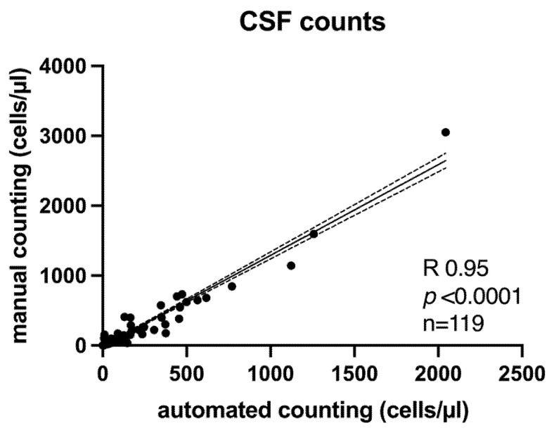 Figure 1
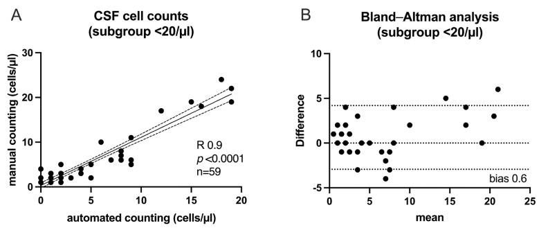 Figure 2
- —Open Access Publication Fund of the University of Wuerzburg
Peer Reviews
No public reviews on file for this paper yet. If you reviewed it on a platform where reviews are public (OpenReview, ICLR, NeurIPS, ICML), you can paste yours below so the community can read it here.
Videos
No videos yet. Explain this paper in a talk, walkthrough, or lecture? Add one.
Taxonomy
TopicsMultiple Sclerosis Research Studies · Sepsis Diagnosis and Treatment · Neuroinflammation and Neurodegeneration Mechanisms
1. Introduction
Cerebrospinal fluid (CSF) analysis is an essential backbone of the neurological diagnostic armamentarium. As a sensitive marker of intrathecal inflammation, the leukocyte count in particular is an important time-critical tool in acute diagnosis as well as in monitoring response to therapy. Since the beginning of the 20th century, the gold standard for counting CSF leukocytes has been manual counting in a Fuchs–Rosenthal counting chamber [1]. Automated cell-counting systems, well established in blood testing, are increasingly used in the analysis of many other body fluids. As cells pass through the detection system in an electrolyte solution, these machines use a combination of impedance-based counting and light-scattering techniques to quantify erythrocytes and platelets and identify different leukocyte types. Forward light scatter correlates with cell size, and side scatter correlates with granularity, providing information on cell structure and size. This allows for differentiation between mononuclear cells (lymphocytes, monocytes) and polymorphnuclear cells (such as neutrophils, eosinophils, and basophils) [2]. To date, automated CSF cell counting has only played a minor role in centers specializing in CSF diagnostics [3,4]. This is mainly due to a perceived lack of sensitivity at low event rates in both healthy and altered CSF [5,6,7,8]. On the contrary, these systems have the advantage of high availability and require less specialized staff to provide 24/7 diagnostics [9]. This is also important given the need for immediate processing of CSF samples, which can rapidly lose their diagnostic value if transport, storage, and analysis exceed a 2 h time frame [10]. For clinical practice, it is therefore necessary to clarify whether rapid automated counting is actually inferior to manual counting in all clinical scenarios (spinal tap, extraventricular drains, pure leukocytosis, and hemorrhagic CSF).
2. Materials and Methods
Between 25 July 2024 and 6 September 2024, we compared 119 CSF samples obtained as part of routine clinical examinations. These samples included 64 from lumbar punctures (LP) and 55 from an external ventricular drain (EVD). Samples (from the identical tube) were simultaneously analyzed (within 60 min), either by manual unstained counting in the Fuchs–Rosenthal chamber or by a Sysmex XN-9000 (Norderstedt, Germany) with body fluid mode. For manual counting, 20 µL of native CSF was added to the Fuchs–Rosenthal chamber and examined by an experienced technician using a Leica DM4B microscope (Wetzlar, Germany). If necessary, CSF was diluted with 0.9% NaCl (Fresenius Kabi, Bad Homburg, Germany) or with Türk’s solution (Sigma-Aldrich, Taufkirchen, Germany) prior to analysis.
For automated cell counting on the XN-9000 system, the measurement channel was first changed to body fluids mode. After flushing the system and carrying out a background measurement, it was ensured that there were no particles in the measurement channel that could lead to interference with the body fluids, which are generally low in cell counts. The measurement required 160 microlitres of CSF, of which 80 microlitres were used to measure the cell count.
The analysis was approved by the local ethics committee of the University of Würzburg, Germany (reference No 2024091801; 2024). Statistical testing was performed with GraphPad Prism (version 10.0, Boston, MA, USA).
3. Results
Overall, all samples (n = 119; LP = 64; EVD = 55) showed a high correlation of both counting methods (Figure 1) (R 0.95, p < 0.0001). There was a slight increase in deviation with increasing cell count but without systematic bias and within a range that did not affect clinical decisions.
Given the higher diagnostic impact, particularly in influencing treatment decisions such as whether to initiate anti-infective treatment, we then focused on a sub-group of patients with a cell count of less than 20/µL (n = 59; LP = 51; EVD = 8), showing the same strong relationship between both techniques (Figure 2A) (R 0.9, p < 0.0001). To evaluate how well the two different measurement methods agree with each other, we used a Bland–Altman plot (Figure 2B). We were able to rule out systematic bias and show good consistency between the two techniques, suggesting that both methods can be used interchangeably.
Of the 59 patients, the clinical threshold of 5 cells/µL was crossed in 2 pairs (2 vs. 5 and 5 vs. 2 cells). Yet, this diagnostic uncertainty cannot be attributed to any of the methods and underlines why an evaluation in the clinical context is necessary. The detailed data are presented in Table 1.
To validate the reliability of the automated counting, we also performed a dilution series (Supplementary Materials, Figure S1).
4. Discussion
The need for rapid analysis and skilled personnel for manual cell counting continues to drive efforts to automate CSF analysis. For example, colleagues have recently introduced a new method using a microchip-based automated image analysis device for this purpose [11]. In response to this clinical need, we examined CSF samples using the Sysmex system XN-9000 with body fluid mode, which is already used in our laboratory workflow for hematology diagnostics. We show a representative dataset of comparative analysis of both LP and EVD CSF samples that yield comparable results irrespective of manual or automated cell counting. Given the important role of standardized pre-analytical handling, particularly in CSF diagnostics, the analysis was performed from CSF of the same collecting tube within 60 min.
Looking at the two samples that differed enough to cross a diagnostic border (2->5; 5->2; bold in Table 1), achieving a concordance rate of >98%, the difference was of borderline clinical relevance. We decided to include EVD samples in our analysis because we believe that automated cell counting can excel in this area. In patients with subarachnoidal hemorrhage for example, CSF samples are often heavily contaminated with blood and require significant dilution for manual counting, which can increase the potential for error. In this context, the key diagnostic parameter is the trend in cell count over time not detailed cell morphology [12]. Simple longitudinal leukocyte and erythrocyte counts (combined with glucose and lactat levels) are also sufficient to monitor the risk of ventriculitis [13]. Automated cell counting also has the advantage of providing rapid differentiation of leukocytes into clinically relevant mononuclear and polymorphnuclear cells, which is more time-consuming manually and requires staff experienced in differentiating stained cells. Although this may be sufficient for most time-critical decisions, one should keep in mind that automated differentiation provides only a rough picture of the situation. In particular, automated CSF cell counting is of limited diagnostic value when morphological assessment is required, for example, to detect malignant cells or to identify specific cell types, such as erythrophages or siderophages [14]. This also applies to blasts in suspected lymphomatous meningitis or plasma cells in certain neuroinflammatory conditions. In addition, CSF may be contaminated by chrondroblasts or bone marrow cells that are not correctly identified in automated analysis.
Although markedly elevated leukocyte counts and a high proportion of polymorphonuclear cells should already raise strong suspicion for bacterial meningitis, manual microscopy offers the critical advantage in such cases of potentially enabling direct detection of bacteria. Thus, in these scenarios, automation may fail to capture subtle but diagnostically important features, making manual microscopy essential for accurate evaluation.
These limitations can be addressed through well-structured clinical workflows that include routine cytospin preparation. While particularly relevant for patients with suspected malignancy or subarachnoid hemorrhage, we recommend this practice routinely, as lumbar punctures are often performed early in the diagnostic process, when clinical assessment may still evolve. Routine cytospin preparation helps avoid repeat punctures, minimizing patient burden while preserving the option for retrospective morphological evaluation.
Furthermore, the interpretation of automated counts requires a plausibility check by the physician. Here, modern automated counters with body fluid modes can support diagnostics by detecting cells with markedly increased autofluorescence, a potential indicator of malignancy typically linked to large nuclei [15]. While this method lacks the sensitivity and specificity of microscopy, such findings should prompt mandatory manual review to confirm or rule out the presence of malignant cells [16].
In general, caution is warranted, as automated classification of cellular elements in CSF carries an inherent risk of misinterpretation, especially in the absence of morphological validation. The core issue is that automated analyzers rely on the above mentioned physical and biochemical surrogates, such as cell size, impedance, or fluorescence intensity, to identify and classify particles. This creates a diagnostic black box, where the system may report numerical values without reliably distinguishing true cells from artifacts. Strongly fluorescent particles may be reported as highly fluorescence cells, but this label cannot distinguish malignant cells from non-cellular elements like protein aggregates or debris. This highlights how reliance on automated measurements can lead to inaccurate conclusions if the results are interpreted without considering the underlying technical limitations. Thus, while results of automated counters can be diagnostically useful, they require cautious interpretation and confirmation by manual microscopy.
5. Conclusions
In summary, automated cell counting serves as a reliable and complementary diagnostic tool in the modern CSF laboratory. However, it cannot replace hands-on microscopy as soon as detailed cytological evaluation, such as identifying malignant cells, is required. Thus, its use is limited more by the clinician’s ability to select the appropriate method and critically interpret the results than by technical feasibility or reliability.
The reference list from the paper itself. Each links out to its DOI / PubMed record.
- 1Isenmann S. Strik H. Wick M. Gross C.C. Liquorzytologie: Methoden und Möglichkeiten Fortschr. Neurol. Psychiatr.20178561663010.1055/s-0043-11382329017200 · doi ↗ · pubmed ↗
- 2Alcaide Martin M.J. Altimira Queral L. Sahuquillo Frias L. Valina Amado L. Merino A. Garcia de Guadiana-Romualdo L. Automated cell count in body fluids: A review Adv. Lab. Med.2021214917710.1515/almed-2021-001137363326 PMC 10197423 · doi ↗ · pubmed ↗
- 3Aguadero V. Cano-Corres R. Berlanga E. Torra M. Evaluation of biological fluid analysis using the sysmex XN automatic hematology analyzer Cytom. B Clin. Cytom.20189468068810.1002/cyto.b.2158728834596 · doi ↗ · pubmed ↗
- 4Cho J. Oh J. Lee S.G. Lee Y.H. Song J. Kim J.H. Performance Evaluation of Body Fluid Cellular Analysis Using the Beckman Coulter Uni Cel Dx H 800, Sysmex XN-350, and UF-5000 Automated Cellular Analyzers Ann. Lab. Med.20204012213010.3343/alm.2020.40.2.12231650728 PMC 6822009 · doi ↗ · pubmed ↗
- 5Aune M.W. Becker J.L. Brugnara C. Canfield W. Dorfman D.M. Fiehn W. Fischer G. Fitzpatrick P. Flaming T.H. Henriksen H.K. Automated flow cytometric analysis of blood cells in cerebrospinal fluid: Analytic performance Am. J. Clin. Pathol.200412169070010.1309/EKFW 9E 3LLFXE 15X 915151209 · doi ↗ · pubmed ↗
- 6Van Acker J.T. Delanghe J.R. Langlois M.R. Taes Y.E. De Buyzere M.L. Verstraete A.G. Automated flow cytometric analysis of cerebrospinal fluid Clin. Chem.20014755656010.1093/clinchem/47.3.55611238311 · doi ↗ · pubmed ↗
- 7Wick M. Gross C.C. Tumani H. Wildemann B. Stangel M. On Behalf Of The German Society Of Csf D. Clinical Neurochemistry Dgln E.V. Automated Analysis of Cerebrospinal Fluid Cells Using Commercially Available Blood Cell Analysis Devices-A Critical Appraisal Cells 202110123210.3390/cells 1005123234069775 PMC 8157290 · doi ↗ · pubmed ↗
- 8Sandhaus L.M. Automated flow cytometric analysis of blood cells in cerebrospinal fluid Am. J. Clin. Pathol.200512315415762292 · pubmed ↗