Correction: Almutary et al. Development of 3D-Bioprinted Colitis-Mimicking Model to Assess Epithelial Barrier Function Using Albumin Nano-Encapsulated Anti-Inflammatory Drugs. Biomimetics 2023, 8, 41
Abdulmajeed G. Almutary, Abdullah M. Alnuqaydan, Saleh A. Almatroodi, Hamid A. Bakshi, Dinesh Kumar Chellappan, Murtaza M. Tambuwala

Abstract
Genes, proteins, chemicals, diseases, species, mutations and cell lines named across the full text — each resolved to its canonical identifier and authoritative record.
Click any figure to enlarge with its caption.
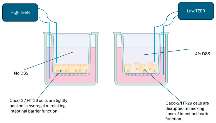 Figure 1
Peer Reviews
No public reviews on file for this paper yet. If you reviewed it on a platform where reviews are public (OpenReview, ICLR, NeurIPS, ICML), you can paste yours below so the community can read it here.
Videos
No videos yet. Explain this paper in a talk, walkthrough, or lecture? Add one.
Taxonomy
Topics3D Printing in Biomedical Research · Cancer Cells and Metastasis · Innovative Microfluidic and Catalytic Techniques Innovation
1. Error in Figure
In the original publication [1], there was a mistake in the published Figure 3, which had the following caption: “The manifestation in 3D-printed Caco-2 and HT-29 constructs. The 3D-printed Caco-2 and HT29 constructs were fixed in a 2% paraformaldehyde solution (10 mM calcium chloride, 50 mM sucrose in PBS) and paraffin embedded by using standard techniques. Using standard protocols, sections were stained with haematoxylin and eosin (H&E). (Scale bar: 20µm) Colitis manifestation was determined by viewing images under a microscope at 200×. (A,B) show untreated control 3D-printed CaCo-2 and HT-29 cell constructs, whereas (C,D) depict a DSS-induced colitis-like condition in 3D-printed Caco-2 and HT29 cellular constructs.” The figure mistakenly used a different source that was previously published. The corrected Figure 3 is provided below with the following caption: “Dextran sulphate sodium (DSS) on apical side of the cells disrupts epithelial cell barrier function, which, in turn, lower the resistance in the flow of electric current as measured using a TEER reader.”
2. Text Correction
There was an error in the original publication, particularly regarding the information provided in the histological images of Section 3.2. Also, Section 2.6, “Histology”, has been removed. The word “Measurements” was also added in Section 3.3, “Barrier Function Measurements of 3D-Printed Caco-2 and HT-29 Constructs”.
The following correction has been made to Paragraph 1 of Section 3.2:
3.2. Induction of Disease in a 3D-Printed Caco-2 and HT-29 Model
Caco-2 and HT-29 cells printed in a hydrogel using a 3D structure were treated with DSS 4% w/v for 24 h and showed a significant increase in epithelial disorganization/disfunction compared to the controls, which was assessed by the measurement of trans-epithelial electrical resistance (TEER), as depicted in Figure 3. This indicated that DSS induces a colitis-like condition in vitro [2].
3. References
Reference [2] has been newly added. Original ref. [20] was deleted. With this correction, the order of some references has been adjusted accordingly.
The authors state that the scientific conclusions are unaffected. This correction was approved by the Academic Editor. The original publication has also been updated.
The reference list from the paper itself. Each links out to its DOI / PubMed record.
- 1Almutary A.G. Alnuqaydan A.M. Almatroodi S.A. Bakshi H.A. Chellappan D.K. Tambuwala M.M. Development of 3D-Bioprinted Colitis-Mimicking Model to Assess Epithelial Barrier Function Using Albumin Nano-Encapsulated Anti-Inflammatory Drugs Biomimetics 202384110.3390/biomimetics 801004136810372 PMC 9944493 · doi ↗ · pubmed ↗
- 2Araki Y. Sugihara H. Hattori T. In vitro effects of dextran sulfate sodium on a Caco-2 cell line and plausible mechanisms for dextran sulfate sodium-induced colitis Oncol. Rep.2006161357136210.3892/or.16.6.135717089061 · doi ↗ · pubmed ↗