The genome sequence of the European ground squirrel, Spermophilus citellus (Linnaeus, 1766)
Dimitra-Lida Rammou, Dionisios Youlatos, Alexandros Triantafyllidis, Pawel Michalak, Gerrit Wehrenberg, Camila do Nascimento Moreira

TL;DR
This paper presents the genome sequence of the European ground squirrel, including a detailed assembly of its chromosomes and mitochondrial DNA.
Contribution
The study provides a high-quality genome assembly for Spermophilus citellus, including chromosomal pseudomolecules and the mitochondrial genome.
Findings
The genome assembly is 3,090.03 megabases long, with 95.47% scaffolded into 20 chromosomal pseudomolecules.
The mitochondrial genome is 16.45 kilobases in length and has been fully assembled.
Abstract
We present a genome assembly from a female Spermophilus citellus (European ground squirrel; Chordata; Mammalia; Rodentia; Sciuridae). The genome sequence has a total length of 3,090.03 megabases. Most of the assembly (95.47%) is scaffolded into 20 chromosomal pseudomolecules, including the X sex chromosome. The mitochondrial genome has also been assembled, with a length of 16.45 kilobases.
Genes, proteins, chemicals, diseases, species, mutations and cell lines named across the full text — each resolved to its canonical identifier and authoritative record.
Click any figure to enlarge with its caption.
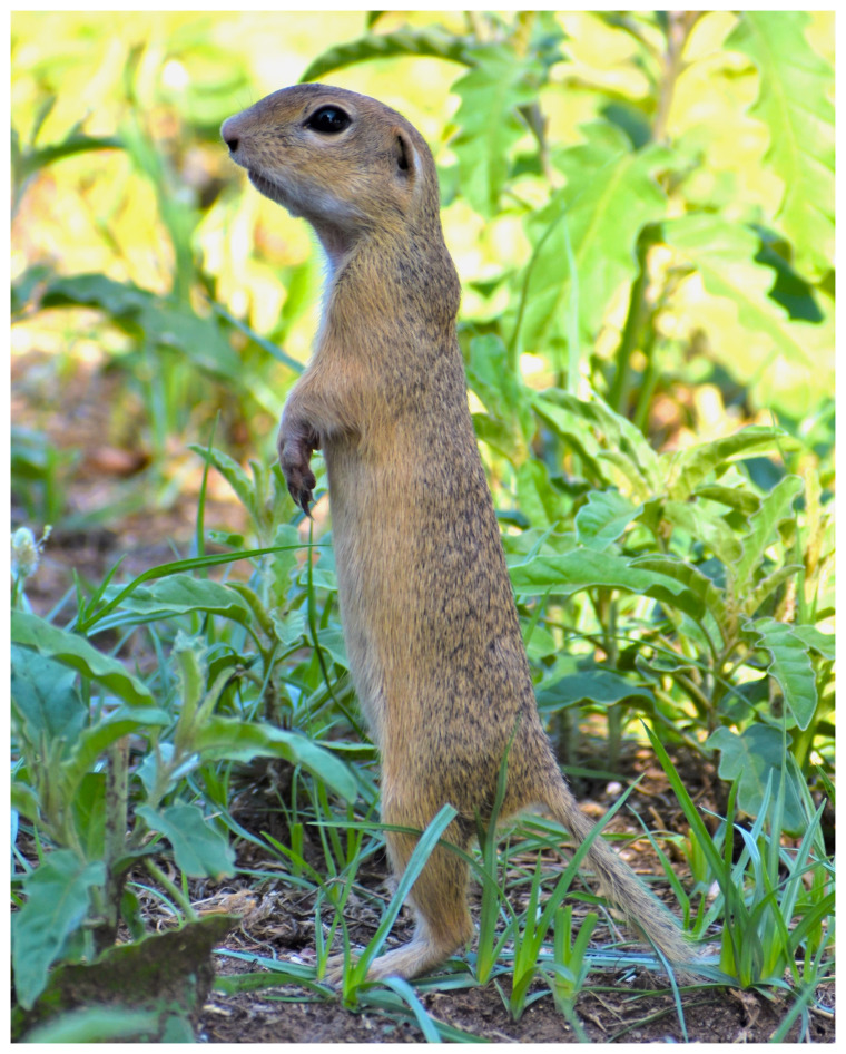 Figure 1
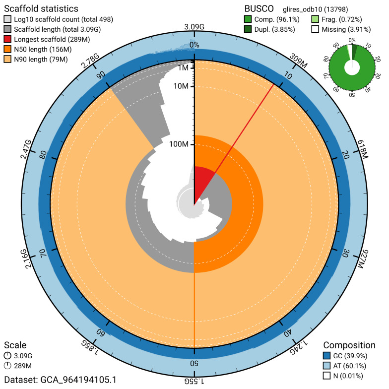 Figure 2
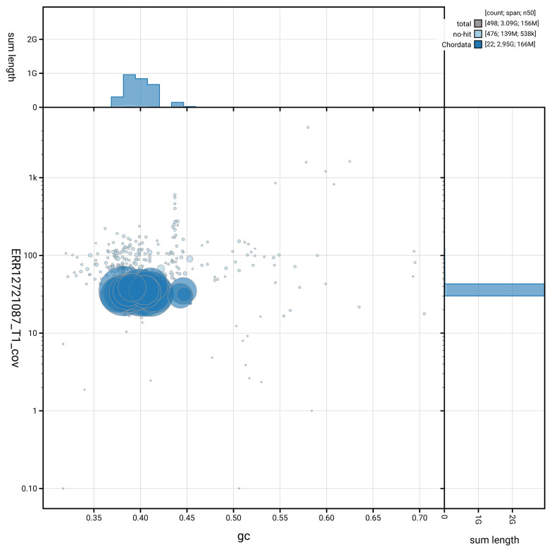 Figure 3
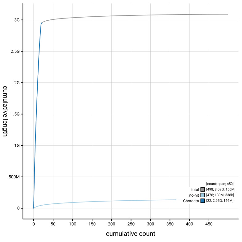 Figure 4
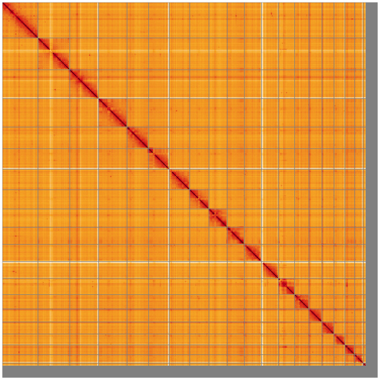 Figure 5
| Project information | |||
|---|---|---|---|
|
| Spermophilus citellus (European suslik) | ||
|
| PRJEB73447 | ||
|
|
| ||
|
| SAMEA10332752 | ||
|
| 9997 | ||
| Specimen information | |||
|
|
|
|
|
|
| mSpeCit3 | SAMEA10332755 | blood |
|
| mSpeCit2 | SAMEA10332754 | skin |
| Sequencing information | |||
|
|
|
|
|
|
| ERR12723499 | 3.13e+09 | 472.86 |
|
| ERR12721086 | 2.44e+06 | 31.51 |
|
| ERR12721087 | 7.57e+06 | 97.68 |
| Genome assembly | ||
|---|---|---|
| Assembly name | mSpeCit3.1 | |
| Assembly accession | GCA_964194105.1 | |
|
|
| |
| Assembly level for primary assembly | chromosome | |
| Span (Mb) | 3,090.03 | |
| Number of contigs | 1,933 | |
| Number of scaffolds | 497 | |
| Longest scaffold (Mb) | 288.97 | |
| Assembly metric | Measure |
|
| Contig N50 length | 3.45 Mb |
|
| Scaffold N50 length | 155.99 Mb |
|
| Consensus quality (QV) | Primary: 60.8; alternate: 58.9;
|
|
|
| Primary: 95.78%; alternate:
|
|
| BUSCO
| C:96.1%[S:92.2%,D:3.8%],
|
|
| Percentage of assembly mapped to
| 95.48% |
|
| Sex chromosomes | X |
|
| Organelles | Mitochondrial genome: 16.45 kb |
|
| INSDC accession | Name | Length (Mb) | GC% |
|---|---|---|---|
| 1 | 288.97 | 38 | |
| 2 | 258.54 | 40 | |
| 3 | 232.93 | 41 | |
| 4 | 231.55 | 40.5 | |
| 5 | 175.33 | 39 | |
| 6 | 166.8 | 41.5 | |
| 7 | 166.25 | 38.5 | |
| 8 | 155.99 | 38 | |
| 9 | 139.52 | 41.5 | |
| 10 | 137.97 | 39.5 | |
| 11 | 136.59 | 38.5 | |
| 12 | 131.62 | 41 | |
| 13 | 116.84 | 40.5 | |
| 14 | 105.9 | 38.5 | |
| 15 | 97.87 | 40.5 | |
| 16 | 87.8 | 39 | |
| 17 | 78.99 | 44.5 | |
| 18 | 69.43 | 44.5 | |
| 19 | 22.32 | 44.5 | |
| X | 149.01 | 37.5 | |
| MT | 0.02 | 35.5 |
| Software
| Version | Source |
|---|---|---|
| BEDTools | 2.30.0 |
|
| BLAST | 2.14.0 |
|
| BlobToolKit | 4.3.9 |
|
| BUSCO | 5.5.0 |
|
| bwa-mem2 | 2.2.1 |
|
| Cooler | 0.8.11 |
|
| DIAMOND | 2.1.8 |
|
| fasta_
| 0.2.4 |
|
| FastK | 666652151335353eef2fcd58880bcef5bc2928e1 |
|
| Gfastats | 1.3.6 |
|
| GoaT CLI | 0.2.5 |
|
| Hifiasm | 0.19.8-r603 |
|
| HiGlass | 44086069ee7d4d3f6f3f0012569789ec138f42b8
|
|
| MerquryFK | d00d98157618f4e8d1a9190026b19b471055b
|
|
| Minimap2 | 2.24-r1122 |
|
| MitoHiFi | 3 |
|
| MultiQC | 1.14, 1.17, and 1.18 |
|
| Nextflow | 23.10.0 |
|
| PretextView | 0.2.5 |
|
| samtools | 1.19.2 |
|
| sanger-tol/
| - |
|
| sanger-tol/
| 0.5.1 |
|
| Seqtk | 1.3 |
|
| Singularity | 3.9.0 |
|
| TreeVal | 1.2.0 |
|
| YaHS | 1.2a.2 |
|
- —Wellcome Trust
Peer Reviews
No public reviews on file for this paper yet. If you reviewed it on a platform where reviews are public (OpenReview, ICLR, NeurIPS, ICML), you can paste yours below so the community can read it here.
Videos
No videos yet. Explain this paper in a talk, walkthrough, or lecture? Add one.
Taxonomy
TopicsBat Biology and Ecology Studies · Animal Ecology and Behavior Studies · Genetic diversity and population structure
Species taxonomy
Eukaryota; Opisthokonta; Metazoa; Eumetazoa; Bilateria; Deuterostomia; Chordata; Craniata; Vertebrata; Gnathostomata; Teleostomi; Euteleostomi; Sarcopterygii; Dipnotetrapodomorpha; Tetrapoda; Amniota; Mammalia; Theria; Eutheria; Boreoeutheria; Euarchontoglires; Glires; Rodentia; Sciuromorpha; Sciuridae; Xerinae; Marmotini; Spermophilus; Spermophilus citellus (Linnaeus, 1766) (NCBI:txid9997)
Background
Spermophilus citellus ( Figure 1), the European ground squirrel, is a semifossorial rodent with an elongated body between 18–24 cm, short tail measuring 20–40% of head-body length, grayish-yellow fur, and small ears. Its body mass varies from 145 g to 520 g, depending on age, sex, life cycle and environment ( Matějů, 2008; Ramos-Lara et al., 2014; Ružić, 1978). It inhabits grasslands and agricultural ecosystems up to 2,500 m, forming colonies and constructing burrows for hibernation, reproduction, and shelter. As a keystone species, it supports predators, enhances vegetation diversity and soil structure, and provides habitat for reptiles, birds, and arthropods in abandoned burrows.
A juvenile Spermophilus citellus from Thessaloniki, Greece (photo by Dionisios Youlatos) (not the specimen used for genome sequencing).
Spermophilus citellus is endemic to Central and Southeastern Europe, with its distribution divided by the Carpathian Mountains ( Kryštufek et al., 2009; Říčanová et al., 2013). Its northwestern range includes Czechia, Austria, Poland, Slovakia, Hungary, northern Serbia, and western Romania, while the southeastern range spans southern Serbia, North Macedonia, Bulgaria, southern Romania, Greece, Turkish Thrace, Moldova, and Ukraine ( Ćosić et al., 2024). Classified as Endangered [A3c ver 3.1], it is protected under the Bern Convention (Appendix II) and the EU Habitats and Species Directive (Annexes II and IV) ( Ćosić et al., 2024). Its populations are in continuous decline, primarily due to habitat degradation and loss, threatening its long-term survival.
The recent sequencing project represents the first high-profile genomic initiative for this species, providing a crucial resource for investigating adaptive responses to environmental changes and human pressures. The broad latitudinal and altitudinal distribution of the target species offers a unique opportunity to examine adaptation to shifting environmental conditions, particularly in the context of climate change. Additionally, improving tools for evidence-based conservation management is essential. Integrating genetic diversity data into biodiversity conservation strategies remains a gap for this species, necessitating the development of efficient indicators to assess the adaptive potential of the species populations.
Genome sequence report
Sequencing data
The genome of a specimen of Spermophilus citellus was sequenced using Pacific Biosciences single-molecule HiFi long reads, generating 129.19 Gb (gigbases) from 10.02 million reads. GenomeScope analysis of the PacBio HiFi data estimated the haploid genome size at 3,115.01 Mb, with a heterozygosity of 0.18% and repeat content of 31.24%. These values provide an initial assessment of genome complexity and the challenges anticipated during assembly. Based on this estimated genome size, the sequencing data provided approximately 30.0x coverage of the genome. Chromosome conformation Hi-C sequencing produced 472.86 Gb from 3,131.52 million reads. Table 1 summarises the specimen and sequencing information.
Table 1.: Specimen and sequencing data for Spermophilus citellus.
Assembly statistics
The primary haplotype was assembled, and contigs corresponding to an alternate haplotype were also deposited in INSDC databases. The assembly was improved by manual curation, which corrected 218 misjoins or missing joins and removed 12 haplotypic duplications. These interventions reduced the total assembly length by 0.84%, decreased the scaffold count by 26.66%, and increased the scaffold N50 by 13.24%. The final assembly has a total length of 3,090.03 Mb in 497 scaffolds, with 1,436 gaps, and a scaffold N50 of 155.99 Mb ( Table 2).
Table 2.: Genome assembly data for Spermophilus citellus.
The snail plot in Figure 2 provides a summary of the assembly statistics, indicating the distribution of scaffold lengths and other assembly metrics. Figure 3 shows the distribution of scaffolds by GC proportion and coverage. Figure 4 presents a cumulative assembly plot, with separate curves representing different scaffold subsets assigned to various phyla, illustrating the completeness of the assembly.
Genome assembly of Spermophilus citellus, mSpeCit3.1: metrics. The BlobToolKit snail plot provides an overview of assembly metrics and BUSCO gene completeness. The circumference represents the length of the whole genome sequence, and the main plot is divided into 1,000 bins around the circumference. The outermost blue tracks display the distribution of GC, AT, and N percentages across the bins. Scaffolds are arranged clockwise from longest to shortest and are depicted in dark grey. The longest scaffold is indicated by the red arc, and the deeper orange and pale orange arcs represent the N50 and N90 lengths. A light grey spiral at the centre shows the cumulative scaffold count on a logarithmic scale. A summary of complete, fragmented, duplicated, and missing BUSCO genes in the glires_odb10 set is presented at the top right. An interactive version of this figure is available at https://blobtoolkit.genomehubs.org/view/GCA_964194105.1/dataset/GCA_964194105.1/snail.
Genome assembly of Spermophilus citellus, mSpeCit3.1: BlobToolKit GC-coverage plot.Blob plot showing sequence coverage (vertical axis) and GC content (horizontal axis). The circles represent scaffolds, with the size proportional to scaffold length and the colour representing phylum membership. The histograms along the axes display the total length of sequences distributed across different levels of coverage and GC content. An interactive version of this figure is available at https://blobtoolkit.genomehubs.org/view/GCA_964194105.1/blob.
Genome assembly of Spermophilus citellus, mSpeCit3.1: BlobToolKit cumulative sequence plot.The grey line shows cumulative length for all scaffolds. Coloured lines show cumulative lengths of scaffolds assigned to each phylum using the buscogenes taxrule. An interactive version of this figure is available at https://blobtoolkit.genomehubs.org/view/GCA_964194105.1/dataset/GCA_964194105.1/cumulative.
Most of the assembly sequence (95.48%) was assigned to 20 chromosomal-level scaffolds, representing 19 autosomes and the X sex chromosome. These chromosome-level scaffolds, confirmed by Hi-C data, are named according to size ( Figure 5; Table 3). During curation, it was noted that the order and orientation of contigs in Chromosome 12 between approximately 23.6–68.8Mb are uncertain. Chromosome X was identified by alignment to the genomes of Sciurus vulgaris (GCA_902686455.2) ( Mead et al., 2020b) and Sciurus carolinensis (GCA_902686445.2) ( Mead et al., 2020a).
Genome assembly of Spermophilus citellus: Hi-C contact map of the mSpeCit3.1 assembly, visualised using HiGlass.Chromosomes are shown in order of size from left to right and top to bottom. An interactive version of this figure may be viewed at https://genome-note-higlass.tol.sanger.ac.uk/l/?d=c65KyKOKQG60ig0sTFmtoQ.
Table 3.: Chromosomal pseudomolecules in the genome assembly of Spermophilus citellus, mSpeCit3.
The mitochondrial genome was also assembled. This sequence is included as a contig in the multifasta file of the genome submission and as a standalone record.
Assembly quality metrics
The estimated Quality Value (QV) and k-mer completeness metrics, along with BUSCO completeness scores, were calculated for each haplotype and the combined assembly. The QV reflects the base-level accuracy of the assembly, while k-mer completeness indicates the proportion of expected k-mers identified in the assembly. BUSCO scores provide a measure of completeness based on benchmarking universal single-copy orthologues.
The combined primary and alternate assemblies achieve an estimated QV of 59.9. The k-mer recovery for the primary haplotype is 95.78%, and for the alternate haplotype 62.61%; the combined primary and alternate assemblies have a k-mer recovery of 99.41%. BUSCO analysis using the glires_odb10 reference set ( n = 13,798) identified 96.1% of the expected gene set (single = 92.2%, duplicated = 3.8%).
Table 2 provides assembly metric benchmarks adapted from Rhie et al. (2021) and the Earth BioGenome Project Report on Assembly Standards September 2024. The assembly achieves the EBP reference standard of 6.C.Q59.
Methods
Sample acquisition
On September 1, 2021, four samples were collected from two living Spermophilus citellus specimens belonging to a single population in Thessaloniki, Greece (latitude: N 23.00, longitude: E 40.53, elevation: 25 m). Due to the species’ endangered and protected status, only non-lethal sampling was performed. The first specimen (ID: ERGA_AT_GR_011, SAMEA10332751, ToLID: mSpeCit2) was an adult female from which skin tissue (SAMEA10332754) and blood (SAMEA10332753) were collected. The second specimen (ID: ERGA_AT_GR_012, SAMEA10332752, ToLID: mSpeCit3) was a juvenile female from which skin tissue (SAMEA10332756) and blood (SAMEA10332755) were obtained. Immediately after collection, all samples were placed in Eppendorf tubes, transferred to a liquid nitrogen container, and subsequently stored in a –80°C freezer for preservation. Following sampling, both individuals were released back into their natural habitat. The species identification and sample collection were conducted by Dimitra-Lida Rammou, Dionisios Youlatos, and Anastasia Diakou.
The specimen with ID ERGA_A_GR_012 (ToLID mSpeCit3) was used for PacBio HiFi sequencing, from which the genome was assembled. The specimen with ID ERGA_AT_GR_011, ToLID mSpeCit2) was used for Hi-C scaffolding of the assembly.
Nucleic acid extraction
The workflow for high molecular weight (HMW) DNA extraction at the Wellcome Sanger Institute (WSI) Tree of Life Core Laboratory includes a sequence of procedures: sample preparation and homogenisation, DNA extraction, fragmentation and purification. Detailed protocols are available on protocols.io ( Denton et al., 2023b). The mSpeCit3 sample was prepared for DNA extraction on dry ice ( Jay et al., 2023). The blood sample was homogenised using a PowerMasher II tissue disruptor ( Denton et al., 2023a). HMW DNA was extracted using the Manual MagAttract v1 protocol ( Strickland et al., 2023b). DNA was sheared into an average fragment size of 12–20 kb in a Megaruptor 3 system ( Todorovic et al., 2023). Sheared DNA was purified by solid-phase reversible immobilisation, using AMPure PB beads to eliminate shorter fragments and concentrate the DNA ( Strickland et al., 2023a). The concentration of the sheared and purified DNA was assessed using a Nanodrop spectrophotometer and Qubit Fluorometer using the Qubit dsDNA High Sensitivity Assay kit. Fragment size distribution was evaluated by running the sample on the FemtoPulse system.
Hi-C sample preparation
Tissue from the skin of the mSpeCit2 sample was processed for Hi-C sequencing at the WSI Scientific Operations core, using the Arima-HiC v2 kit. In brief, 20–50 mg of frozen tissue (stored at –80 °C) was fixed, and the DNA crosslinked using a TC buffer with 22% formaldehyde concentration. After crosslinking, the tissue was homogenised using the Diagnocine Power Masher-II and BioMasher-II tubes and pestles. Following the Arima-HiC v2 kit manufacturer's instructions, crosslinked DNA was digested using a restriction enzyme master mix. The 5’-overhangs were filled in and labelled with biotinylated nucleotides and proximally ligated. An overnight incubation was carried out for enzymes to digest remaining proteins and for crosslinks to reverse. A clean up was performed with SPRIselect beads prior to library preparation. Additionally, the biotinylation percentage was estimated using the Qubit Fluorometer v4.0 (Thermo Fisher Scientific) and Qubit HS Assay Kit and Arima-HiC v2 QC beads.
Library preparation and sequencing
Library preparation and sequencing were performed at the WSI Scientific Operations core.
** PacBio HiFi **
At a minimum, samples were required to have an average fragment size exceeding 8 kb and a total mass over 400 ng to proceed to the low input SMRTbell Prep Kit 3.0 protocol (Pacific Biosciences, California, USA), depending on genome size and sequencing depth required. Libraries were prepared using the SMRTbell Prep Kit 3.0 (Pacific Biosciences, California, USA) as per the manufacturer's instructions. The kit includes the reagents required for end repair/A-tailing, adapter ligation, post-ligation SMRTbell bead cleanup, and nuclease treatment. Following the manufacturer’s instructions, size selection and clean up was carried out using diluted AMPure PB beads (Pacific Biosciences, California, USA). DNA concentration was quantified using the Qubit Fluorometer v4.0 (Thermo Fisher Scientific) with Qubit 1X dsDNA HS assay kit and the final library fragment size analysis was carried out using the Agilent Femto Pulse Automated Pulsed Field CE Instrument (Agilent Technologies) and gDNA 55kb BAC analysis kit.
Samples were sequenced on a Revio instrument (Pacific Biosciences, California, USA). Prepared libraries were normalised to 2 nM, and 15 μL was used for making complexes. Primers were annealed and polymerases were hybridised to create circularised complexes according to manufacturer’s instructions. The complexes were purified with the 1.2X clean up with SMRTbell beads. The purified complexes were then diluted to the Revio loading concentration (in the range 200–300 pM), and spiked with a Revio sequencing internal control. Samples were sequenced on Revio 25M SMRT cells (Pacific Biosciences, California, USA). The SMRT link software, a PacBio web-based end-to-end workflow manager, was used to set-up and monitor the run, as well as perform primary and secondary analysis of the data upon completion.
** Hi-C **
For Hi-C library preparation, DNA was fragmented using the Covaris E220 sonicator (Covaris) and size selected using SPRISelect beads to 400 to 600 bp. The DNA was then enriched using the Arima-HiC v2 kit Enrichment beads. Using the NEBNext Ultra II DNA Library Prep Kit (New England Biolabs) for end repair, a-tailing, and adapter ligation. This uses a custom protocol which resembles the standard NEBNext Ultra II DNA Library Prep protocol but where library preparation occurs while DNA is bound to the Enrichment beads. For library amplification, 10 to 16 PCR cycles were required, determined by the sample biotinylation percentage. The Hi-C sequencing was performed using paired-end sequencing with a read length of 150 bp on an Illumina NovaSeq 6000 instrument.
Genome assembly, curation and evaluation
** Assembly **
Prior to assembly of the PacBio HiFi reads, a database of k-mer counts ( k = 31) was generated from the filtered reads using FastK. GenomeScope2 ( Ranallo-Benavidez et al., 2020) was used to analyse the k-mer frequency distributions, providing estimates of genome size, heterozygosity, and repeat content.
The HiFi reads were first assembled using Hifiasm ( Cheng et al., 2021) with the --primary option. The Hi-C reads were mapped to the primary contigs using bwa-mem2 ( Vasimuddin et al., 2019). The contigs were further scaffolded using the provided Hi-C data ( Rao et al., 2014) in YaHS ( Zhou et al., 2023) using the --break option for handling potential misassemblies. The scaffolded assemblies were evaluated using Gfastats ( Formenti et al., 2022), BUSCO ( Manni et al., 2021) and MERQURY.FK ( Rhie et al., 2020).
The mitochondrial genome was assembled using MitoHiFi ( Uliano-Silva et al., 2023), which runs MitoFinder ( Allio et al., 2020) and uses these annotations to select the final mitochondrial contig and to ensure the general quality of the sequence.
** Assembly curation **
The assembly was decontaminated using the Assembly Screen for Cobionts and Contaminants (ASCC) pipeline. Flat files and maps used in curation were generated via the TreeVal pipeline ( Pointon et al., 2023). Manual curation was conducted primarily in PretextView ( Harry, 2022) and HiGlass ( Kerpedjiev et al., 2018), with additional insights provided by JBrowse2 ( Diesh et al., 2023). Scaffolds were visually inspected and corrected as described by Howe et al. (2021). Any identified contamination, missed joins, and mis-joins were amended, and duplicate sequences were tagged and removed. The curation process is documented at https://gitlab.com/wtsi-grit/rapid-curation.
** Assembly quality assessment **
The Merqury.FK tool ( Rhie et al., 2020), run in a Singularity container ( Kurtzer et al., 2017), was used to evaluate k-mer completeness and assembly quality for the primary and alternate haplotypes using the k-mer databases ( k = 31) that were computed prior to genome assembly. The analysis outputs included assembly QV scores and completeness statistics.
A Hi-C contact map was produced for the final version of the assembly. The Hi-C reads were aligned using bwa-mem2 ( Vasimuddin et al., 2019) and the alignment files were combined using SAMtools ( Danecek et al., 2021). The Hi-C alignments were converted into a contact map using BEDTools ( Quinlan & Hall, 2010) and the Cooler tool suite ( Abdennur & Mirny, 2020). The contact map was visualised in HiGlass ( Kerpedjiev et al., 2018).
The blobtoolkit pipeline is a Nextflow port of the previous Snakemake Blobtoolkit pipeline ( Challis et al., 2020). It aligns the PacBio reads in SAMtools and minimap2 ( Li, 2018) and generates coverage tracks for regions of fixed size. In parallel, it queries the GoaT database ( Challis et al., 2023) to identify all matching BUSCO lineages to run BUSCO ( Manni et al., 2021). For the three domain-level BUSCO lineages, the pipeline aligns the BUSCO genes to the UniProt Reference Proteomes database ( Bateman et al., 2023) with DIAMOND blastp ( Buchfink et al., 2021). The genome is also divided into chunks according to the density of the BUSCO genes from the closest taxonomic lineage, and each chunk is aligned to the UniProt Reference Proteomes database using DIAMOND blastx. Genome sequences without a hit are chunked using seqtk and aligned to the NT database with blastn ( Altschul et al., 1990). The blobtools suite combines all these outputs into a blobdir for visualisation.
The blobtoolkit pipeline was developed using nf-core tooling ( Ewels et al., 2020) and MultiQC ( Ewels et al., 2016), relying on the Conda package manager, the Bioconda initiative ( Grüning et al., 2018), the Biocontainers infrastructure ( da Veiga Leprevost et al., 2017), as well as the Docker ( Merkel, 2014) and Singularity ( Kurtzer et al., 2017) containerisation solutions.
Table 4 contains a list of relevant software tool versions and sources.
Wellcome Sanger Institute – Legal and Governance
The materials that have contributed to this genome note have been supplied by a Tree of Life collaborator. The Wellcome Sanger Institute employs a process whereby due diligence is carried out proportionate to the nature of the materials themselves, and the circumstances under which they have been/are to be collected and provided for use. The purpose of this is to address and mitigate any potential legal and/or ethical implications of receipt and use of the materials as part of the research project, and to ensure that in doing so we align with best practice wherever possible.
The overarching areas of consideration are:
Ethical review of provenance and sourcing of the materialLegality of collection, transfer and use (national and international)
Each transfer of samples is undertaken according to a Research Collaboration Agreement or Material Transfer Agreement entered into by the Tree of Life collaborator, Genome Research Limited (operating as the Wellcome Sanger Institute) and in some circumstances other Tree of Life collaborators.
The reference list from the paper itself. Each links out to its DOI / PubMed record.
- 1Abdennur N Mirny LA : Cooler: scalable storage for Hi-C data and other genomically labeled arrays. Bioinformatics. 2020;36(1):311–316. 10.1093/bioinformatics/btz 540 31290943 PMC 8205516 · doi ↗ · pubmed ↗
- 2Allio R Schomaker-Bastos A Romiguier J : Mito Finder: efficient automated large-scale extraction of mitogenomic data in target enrichment phylogenomics. Mol Ecol Resour. 2020;20(4):892–905. 10.1111/1755-0998.13160 32243090 PMC 7497042 · doi ↗ · pubmed ↗
- 3Altschul SF Gish W Miller W : Basic Local Alignment Search Tool. J Mol Biol. 1990;215(3):403–410. 10.1016/S 0022-2836(05)80360-2 2231712 · doi ↗ · pubmed ↗
- 4Bateman A Martin MJ Orchard S : Uni Prot: the universal protein knowledgebase in 2023. Nucleic Acids Res. 2023;51(D 1):D 523–D 531. 10.1093/nar/gkac 1052 36408920 PMC 9825514 · doi ↗ · pubmed ↗
- 5Buchfink B Reuter K Drost HG : Sensitive protein alignments at Tree-of-Life scale using DIAMOND. Nat Methods. 2021;18(4):366–368. 10.1038/s 41592-021-01101-x 33828273 PMC 8026399 · doi ↗ · pubmed ↗
- 6Challis R Kumar S Sotero-Caio C : Genomes on a Tree (Goa T): a versatile, scalable search engine for genomic and sequencing project metadata across the eukaryotic Tree of Life [version 1; peer review: 2 approved]. Wellcome Open Res. 2023;8:24. 10.12688/wellcomeopenres.18658.1 36864925 PMC 9971660 · doi ↗ · pubmed ↗
- 7Challis R Richards E Rajan J : Blob Tool Kit – interactive quality assessment of genome assemblies. G 3 (Bethesda). 2020;10(4):1361–1374. 10.1534/g 3.119.400908 32071071 PMC 7144090 · doi ↗ · pubmed ↗
- 8Cheng H Concepcion GT Feng X : Haplotype-resolved de novo assembly using phased assembly graphs with hifiasm. Nat Methods. 2021;18(2):170–175. 10.1038/s 41592-020-01056-5 33526886 PMC 7961889 · doi ↗ · pubmed ↗