Intravenous Misplacement of the Nephrostomy Catheter Into the Renal Vein Following Percutaneous Nephrolithotomy (PCNL): A Case Report and Literature Review
Shuah Ullah, Hammad Ur Rehman Shamsi, Ayesha Kamran, Abdullah Ayub, Dania Masood

TL;DR
A rare case of a nephrostomy catheter being misplaced into a renal vein after kidney stone surgery is reported and reviewed.
Contribution
This case report adds to the limited literature on intravenous nephrostomy tube misplacement following PCNL.
Findings
The nephrostomy catheter tip was found in the left renal vein after PCNL in a 63-year-old male.
Conservative management was successful without the need for surgical intervention.
Good imaging is critical to assess and manage such rare complications safely.
Abstract
Background: After percutaneous nephrolithotomy (PCNL), intravenous misplacement of a nephrostomy tube is a very rare clinical occurrence. This report summarizes the characteristics and management of intravenous misplacement of a nephrostomy tube. Case Presentation: We present a rare case of intravenous nephrostomy catheter misplacement after PCNL in a 63 years old male. The tip of the tube was located in the left renal vein. The patient was managed conservatively and treated safely. Conclusion: Intravenous nephrostomy tube misplacement is a rare PCNL complication. Good Imaging can rule out through and through renal vein perforation and thus patients can be safely managed using conservative approach.
Genes, proteins, chemicals, diseases, species, mutations and cell lines named across the full text — each resolved to its canonical identifier and authoritative record.
Click any figure to enlarge with its caption.
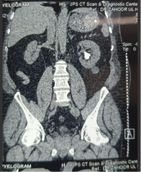 Figure 1
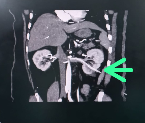 Figure 2
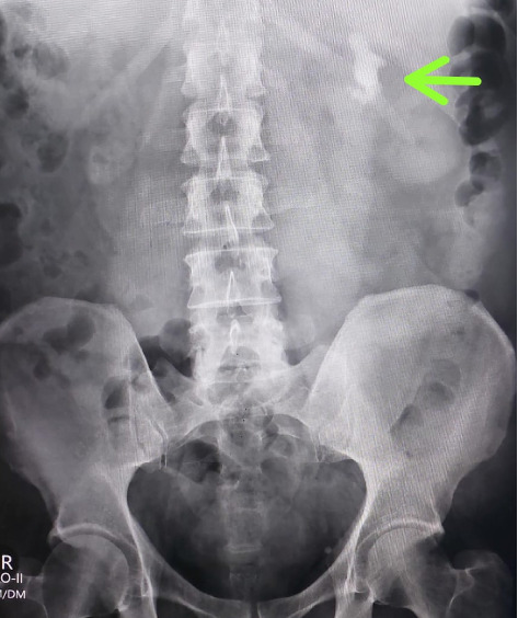 Figure 3
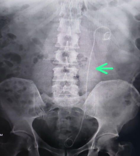 Figure 4
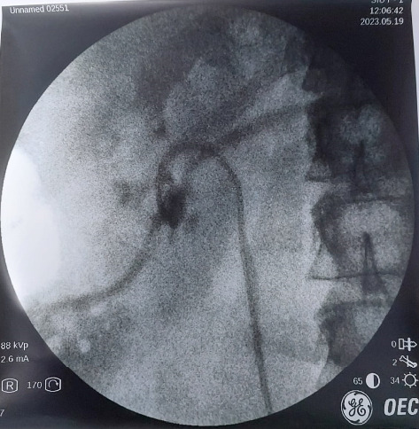 Figure 5
Peer Reviews
No public reviews on file for this paper yet. If you reviewed it on a platform where reviews are public (OpenReview, ICLR, NeurIPS, ICML), you can paste yours below so the community can read it here.
Videos
No videos yet. Explain this paper in a talk, walkthrough, or lecture? Add one.
Taxonomy
TopicsKidney Stones and Urolithiasis Treatments · Pediatric Urology and Nephrology Studies · Dialysis and Renal Disease Management
1. Introduction
Percutaneous nephrolithotomy (PCNL) was first introduced by Fernstrom and Johansson in 1976 [1]. It has become as a method to temporarily alleviate the obstruction, infection, and dilation of the kidney and is also the treatment of choice for renal calculi [2]. There are two mandatory steps of PCNL, i.e., puncturing and dilation. Rarely, during dilatation, dilators can cause direct injury to the main renal vein or its tributaries [3]. A nephrostomy tube is routinely placed after PCNL in the renal pelvis in order to control bleeding and drain the collecting system. Although it is a well-established procedure but complication rates of up to 7% have been reported [4]. Intraoperative complications include bleeding, damage to visceral organs, injury to the renal collecting system, pulmonary complications, extra-renal migration of stone fragments, thromboembolic disorders, and incorrect placement of the nephrostomy tube. On the other hand, postoperative complications include infection, persistent urinary fistula, sepsis, bleeding, infundibular stenosis, and, in severe cases, nephrectomy and patient mortality [4]. Therefore, the mechanism and cause of injury should be investigated properly. We report a case of misplacement of nephrostomy tube, who underwent PCNL.
2. Case Presentation
A 63-year-old male with no known comorbidities presented with a six-month history of left flank pain and lower urinary tract symptoms. Radiological investigations revealed renal calculi in the left kidney as shown in Figures 1, 2 and 3 indicating a need for PCNL. The procedure was performed, and a DJ stent was placed for postoperative drainage as shown in Figure 4. Additionally, a nephrostomy tube was inserted for drainage purposes.
The patient experienced intense bleeding after 5 h following the nephrostomy tube insertion, which subsequently resulted in hypotension later on. He was transferred to the High Dependency Unit (HDU), for resuscitation. Four units of blood along with fresh frozen plasma (FFP) were transfused, stabilizing the patient's hemodynamics. On further radiological imaging, specifically a CT angiogram, showed that the nephrostomy tube had accidently traversed the lower pole calyces of the left kidney, reaching the left renal vein, with its tip positioned only 2 mm away from the inferior vena cava (IVC) ostium (Figure 5). Patient was kept stabilized through fluid resuscitation and by keeping the PCN tube clamped.
The patient was taken back to the operating theatre to address the cause of bleeding on the second postoperative day. The nephrostomy tube was withdrawn from the renal vein under fluoroscopic guidance, and the plan was to keep it in the kidney pelvicalyceal system. However, due to a nonhydronephrotic system, it came out completely from the kidney, so it was kept as a drain for 24 h. The patient remained vitally stable afterward, and minimal hematuria was observed in the Foley's catheter, which resolved with hydration alone. Serial ultrasounds were performed at intervals of 6 h, all of which were unremarkable. Subsequent monitoring showed stable hemodynamics as shown in Tables 1 and 2, and no further bleeding episodes occurred.
3. Discussion
PCNL is now considered a standard procedure for patients with renal stones. The common complications after PCNL include sepsis, iatrogenic injury to adjacent structures (such as the liver, spleen, and bowel), failed renal access, perforation of the renal pelvis, pleural effusion, and pneumothorax [5]. The most significant complication associated with this procedure is hemorrhage. Severe bleeding can occur due to needle puncture, tract dilation, or nephrostomy tube placement [3]. The close association of the renal veins with the renal pelvis and major posterior calyces makes them susceptible to damage during the steps of PCNL [6].
In this case, PCNL was performed for renal calculi, and the nephrostomy tube was inserted for drainage of the collecting system. A DJ stent was placed to maintain the patency of the ureter after the procedure. Five hours after the placement of the nephrostomy tube, the tube was unclamped to observe urine output and the passage of residual stones. Upon unclamping, gross hematuria of approximately 500–1000 mL was observed. At that time, the patient was stable, so the PCN tube was kept clamped, and the patient was closely monitored alongside fluid replacement. Upon unclamping the PCN tube for the second time, similar gross hematuria with a blood loss of 500–1000 mL was observed. The patient subsequently became hemodynamically unstable and was shifted to the HDU, where 4 units of packed cell volume (PCV) and FFP were administered to maintain hemodynamics. Investigations were performed to identify the possible complications. An ultrasound was performed, but no significant findings were observed. On CT angiography, the nephrostomy tube was found to be in the renal vein, with its tip only 2 mm away from the IVC (as shown in Figure 5).
There are two possible ways to manage a misplaced nephrostomy tube: conservative and surgical. In our case, the patient was successfully managed using conservative treatment. In cases involving misplacement of a nephrostomy tube, depending on the postoperative time elapsed, nephrostomy tube transposition is advised under fluoroscopic guidance, and the surgical team should be prepared for emergency surgery if required [7]. Using fluoroscopic guidance with contrast, the nephrostomy tube was completely withdrawn, as the patient was hemodynamically and vitally stable. Repeated blood tests (Tables 1 and 2) and ultrasounds were performed. The patient developed slight hematuria, which was detected through the Foley's catheter, but it was managed by hydrating the patient under the cover of a diuretic. The patient was kept under observation for 48 h to monitor for any possible recurrent hematuria, but none occurred. The patient was discharged successfully. The data from these and other similar publications are summarized in Table 3.
The reference list from the paper itself. Each links out to its DOI / PubMed record.
- 1Fernström I. Johansson B. Percutaneous Pyelolithotomy. A New Extraction Technique Scandinavian Journal of Urology and Nephrology 197610325725910.1080/21681805.1976.118820842-s 2.0-00170468721006190 · doi ↗ · pubmed ↗
- 2Ganpule A. P. Vijayakumar M. Malpani A. Desai M. R. Percutaneous Nephrolithotomy (PCNL) a Critical Review International Journal of Surgery 201636 Pt D 66066410.1016/j.ijsu.2016.11.0282-s 2.0-8500701067527856356 · doi ↗ · pubmed ↗
- 3Mahmood S. N. Toffeq H. M. Renal Vein Injury During Percutaneous Nephrolithotomy Procedure Journal of Endourology Case Reports 20162114815110.1089/cren.2016.008927704054 PMC 5035830 · doi ↗ · pubmed ↗
- 4Lingeman J. E. Campbell-walsh Urology Urinary lithiasis and endourology 2007214311525
- 5Srivastava A. Singh K. J. Suri A. Vascular Complications after Percutaneous Nephrolithotomy: Are There Any Predictive Factors? Urology 2005661384010.1016/j.urology.2005.02.0102-s 2.0-2234445397015992882 · doi ↗ · pubmed ↗
- 6Kotb A. F. Elabbady A. Mohamed K. R. Atta M. A. Percutaneous Silicon Catheter Insertion into the IVC, Following Percutaneous Nephrostomy (PCN) Exchange Canadian Urological Association Journal 201377-8E 505E 50710.5489/cuaj.14032-s 2.0-8487977581023914270 PMC 3713152 · doi ↗ · pubmed ↗
- 7Mazzucchi E. Mitre A. Brito A. Arap M. Murta C. Srougi M. Intravenous Misplacement of the Nephrostomy Catheter Following Percutaneous Nephrostolithotomy: Two Case Reports Clinics 2009641697010.1590/S 1807-593220090001000132-s 2.0-5884910000119142555 PMC 2671967 · doi ↗ · pubmed ↗
- 8Dias-Filho A. C. Coaracy G. A. Borges W. Right Atrial Migration of Nephrostomy Catheter International Brazil Journal Urology 200531547047110.1590/s 1677-553820050005000092-s 2.0-2804444008716255794 · doi ↗ · pubmed ↗